2011 Vol.23(2)
Display Mode: |
2011, 23(2): 79-91.
doi: 10.1007/s11670-011-0079-2
 Abstract
 FullText HTML
 PDF 1039KB
Abstract:
ObjectiveA number of studies have reported the association of “XPA”, “XPC”, “XPD/ERCC2” gene polymorphisms with lung cancer risk. However, the results were conflict. To clarify the impact of polymorphisms of “XPA”, “XPC”, “XPD/ERCC2”, on lung cancer risk, a meta-analysis was performed in this study. MethodsThe electronic databases PubMed and Embase were retrieved for studies included in this meta-analysis by “XPA”, “XPC”, “XPD/ERCC2”, “lung”, “cancer/neoplasm/tumor/carcinoma”, “polymorphism” (An upper date limit of October, 31, 2009). A meta-analysis was performed to evaluate the relationship among XPA, XPC and XPD polymorphism and lung cancer risks. ResultsA total of 31 publications retrieved from Pubmed and Embase included in this study. XPC A939C CC genotype increased lung cancer risk in total population (recessive genetic model: OR=1.23, 95% CI:1.05-1.44; homozygote comparison: OR=1.21,95%CI:1.02-1.43and CC vs. CA contrast: OR=1.25,95%CI:1.06-1.48), except in Asians. XPD A751C, 751C allele and CC genotype also increased lung cancer risk in total population and in Caucasians (recessive genetic model: Total population: OR=1.20, 95%CI:1.07-1.35). No significant correlation was found between XPD A751C and lung cancer risk in Asians and African Americans. XPD G312A AA genotype increased lung cancer risk in total population, in Asians and Caucasians(recessive genetic model: Total population: OR=1.20, 95%CI: 1.06-1.36). No significant association was found between XPA G23A, XPC C499T, XPD C156A and lung cancer risk. ConclusionOur results suggest that the polymorphisms in XPC and XPD involve in lung cancer risks. XPA polymorphisms is less related to lung cancer risk.
2011, 23(2): 92-98.
doi: 10.1007/s11670-011-0092-5
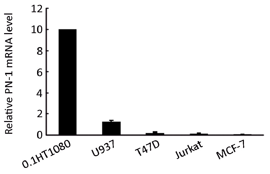
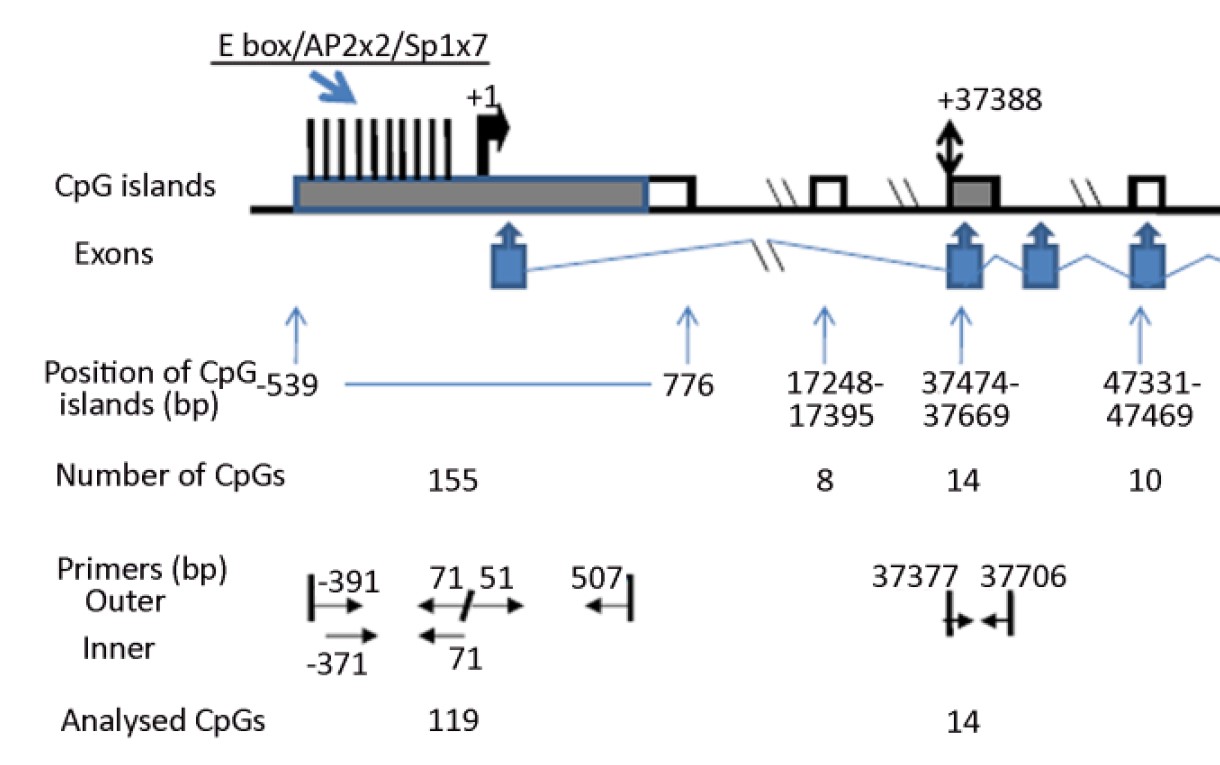
Abstract:
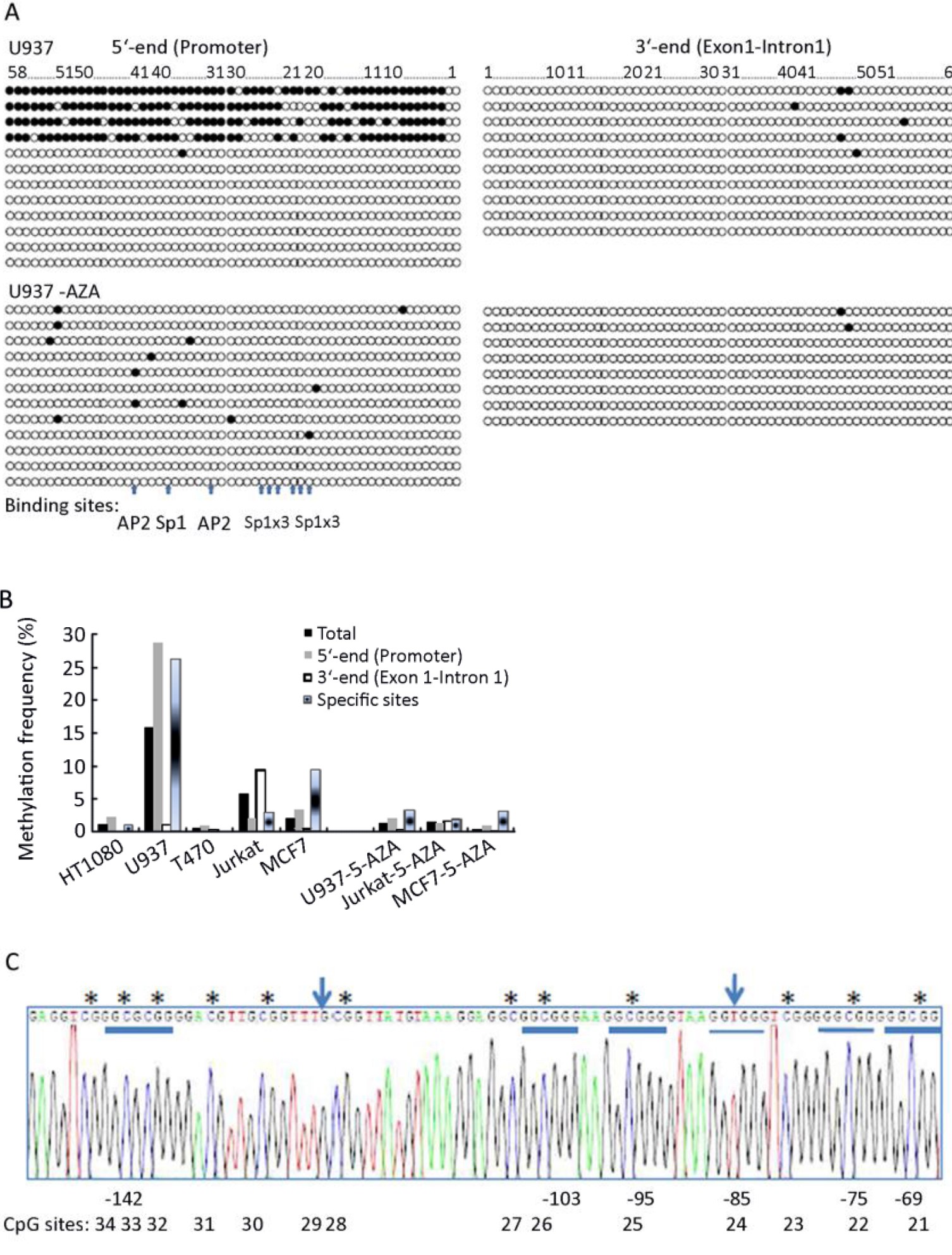
ObjectiveTo investigated whether epigenetic mechanisms contribute to the variable expression of variable protease nexin1(PN-1) encoded by the SERPINE2 gene in different cell types. MethodsWorking with 5 human cell lines, we determined the CpG methylation status within two CpG islands in the SERPINE2 gene by bisulphate sequencing and the PN-1 mRNA level by Q-RT PCR. ResultsA CpG island spanning the transcription initiation site showed little methylation in 3 of the cell lines and substantial methylation in 2 of the cell lines. A CpG island covering the translation starting site showed full methylation in all investigated cell lines. Methylation within the CpG island was not randomly distributed, but showed accumulation at specific sites. However, we were not able to distinguish any patterns which related the methylation frequency to the gene expression level. Inhibition of CpG methylation with 5-aza-2’-deoxycytidine led to a several fold increase in PN-1 mRNA levels, but based on the results on CpG methylation in the CpG island spanning the transcript, the effect is most likely indirect. ConclusionWe have carefully mapped the CpG methylation pattern in two CpG islands in the 5’ part of the SERPINE2 gene without finding any obvious inverse correlation between methylation frequency and expression level.

2011, 23(2): 99-106.
doi: 10.1007/s11670-011-0099-y
Abstract:
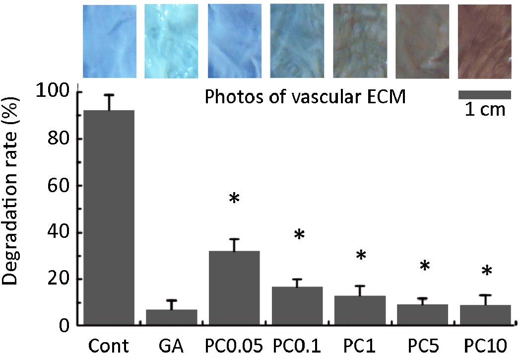
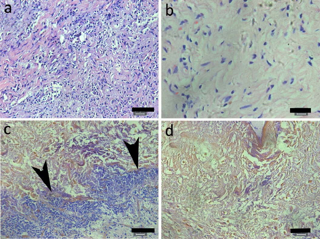
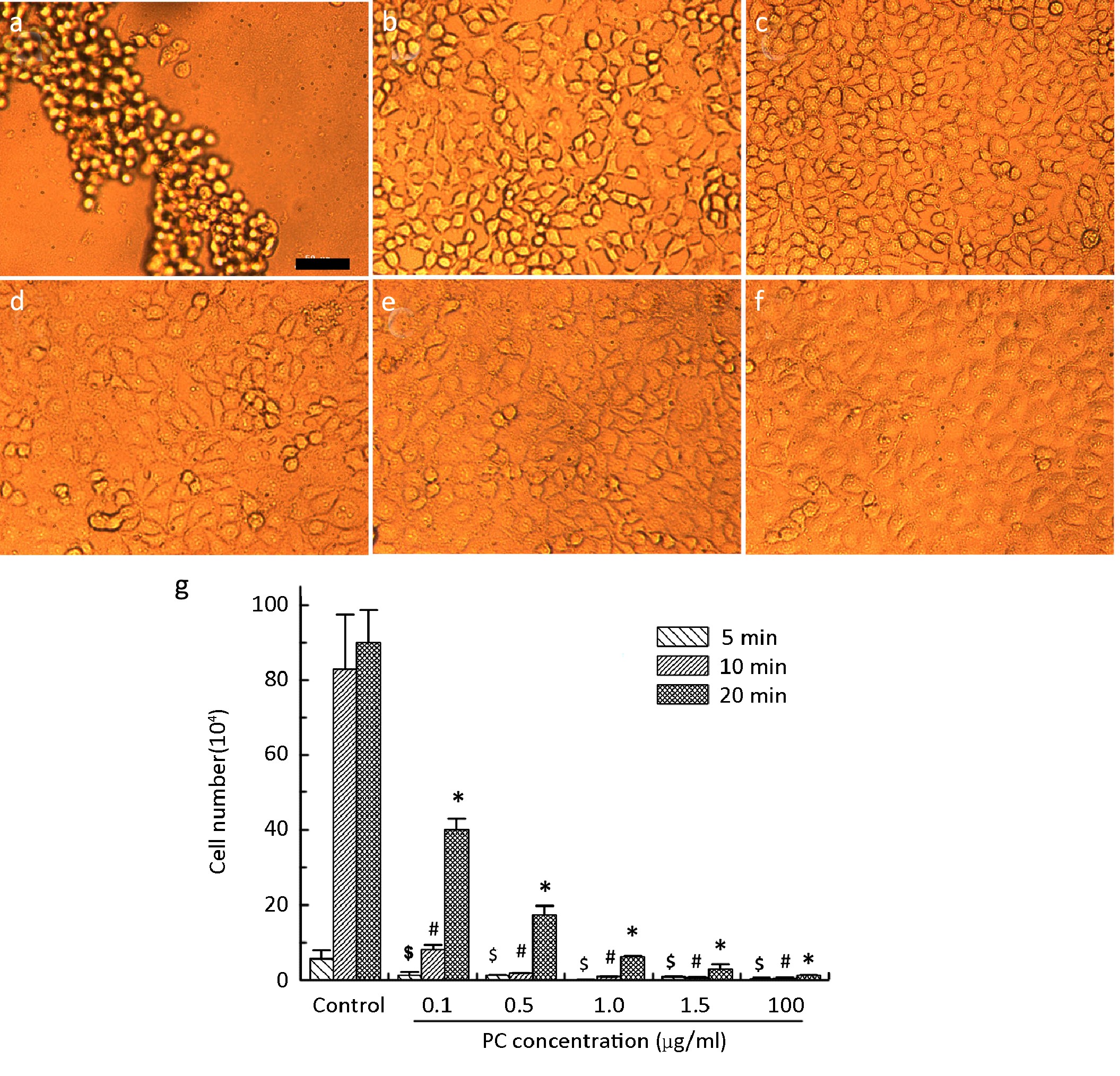
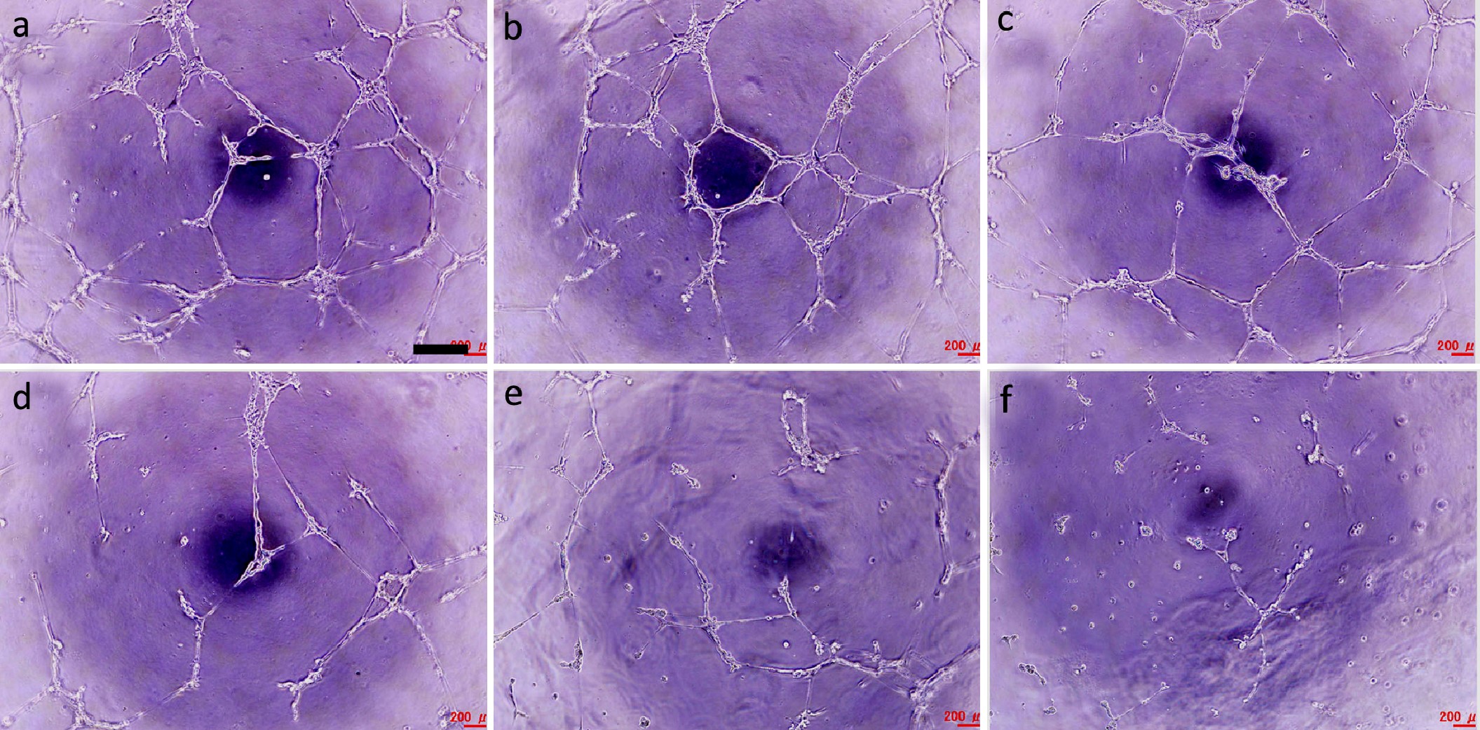
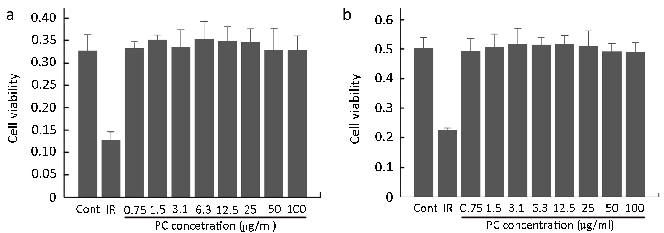
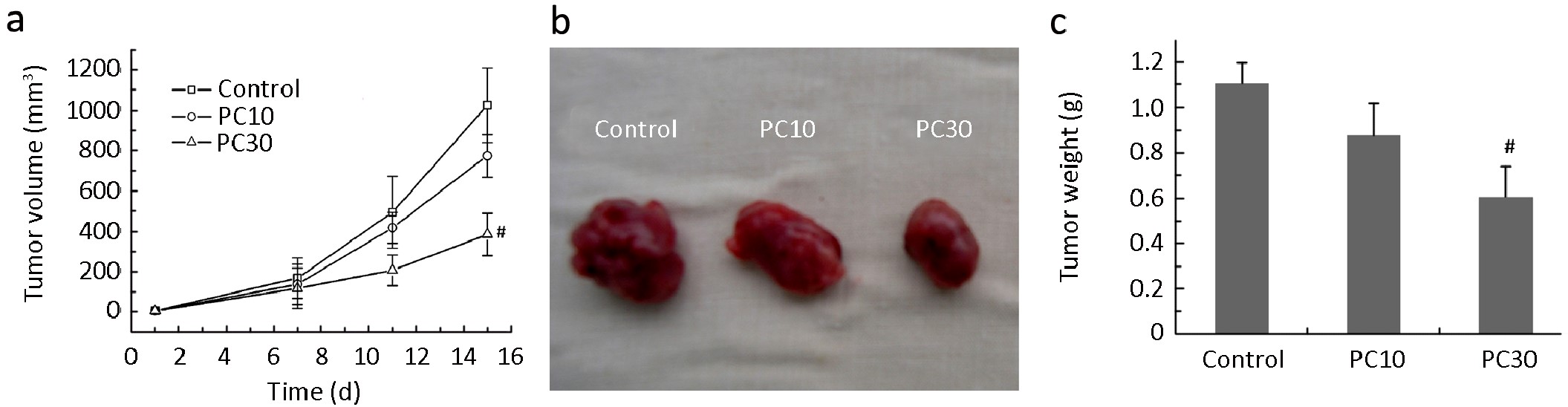
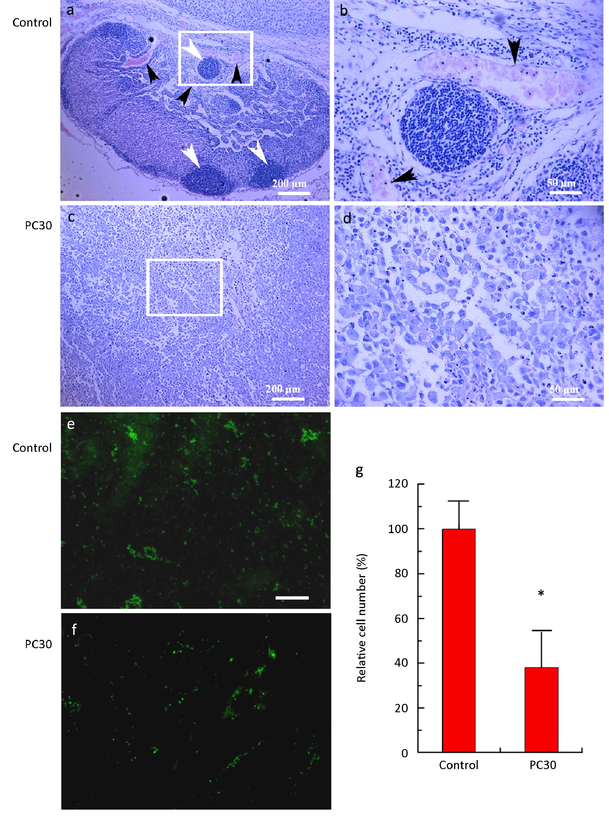
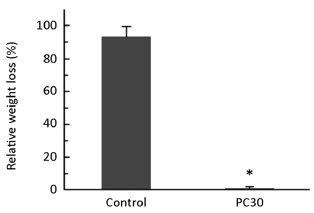
ObjectiveProcyanidins (PC) are widely available natural polyphenols. The present study is designed to investigate if PC can inhibit angiogenesis in lung adenocarcinoma xenografts through crosslinking vascular extracellular matrix (ECM) and preventing proteolysis by matrix metalloproteinases (MMPs). MethodsUsing the in vitro MMP-2 proteolysis and in vivo subcutaneous implantation models, we investigated if PC crosslinking inhibits MMP-mediated proteolysis. Using a cultured cell detachment assay, an in vitro angiogenesis assay, and a cell proliferation assay, we investigated if PC inhibits MMP-2-mediated endothelial cell detachment, angiogenesis, and cell proliferation, respectively. Using tumor xenografts, we evaluated if PC can inhibit growth of lung adenocarcinoma. ResultsPC crosslink vascular ECM proteins, protecting them against proteolysis by MMPs in vitro and in vivo, protecting cultured human umbilical vein endothelial cells from detachment by MMP-2, and inhibiting in vitro angiogenesis. However, PC (0.75-100 µg/ml) did not inhibit vascular and tumor cells proliferation. PC injections (30 mg PC/kg bodyweight) in situ had anticancer effects on xenografts of lung adenocarcinoma, most likely by inhibiting angiogenesis during ECM proteolysis by MMPs. ConclusionThe results suggest that PC may be important MMP inhibitors that can be used as therapeutic anticancer agents.
2011, 23(2): 107-111.
doi: 10.1007/s11670-011-0107-2
Abstract:
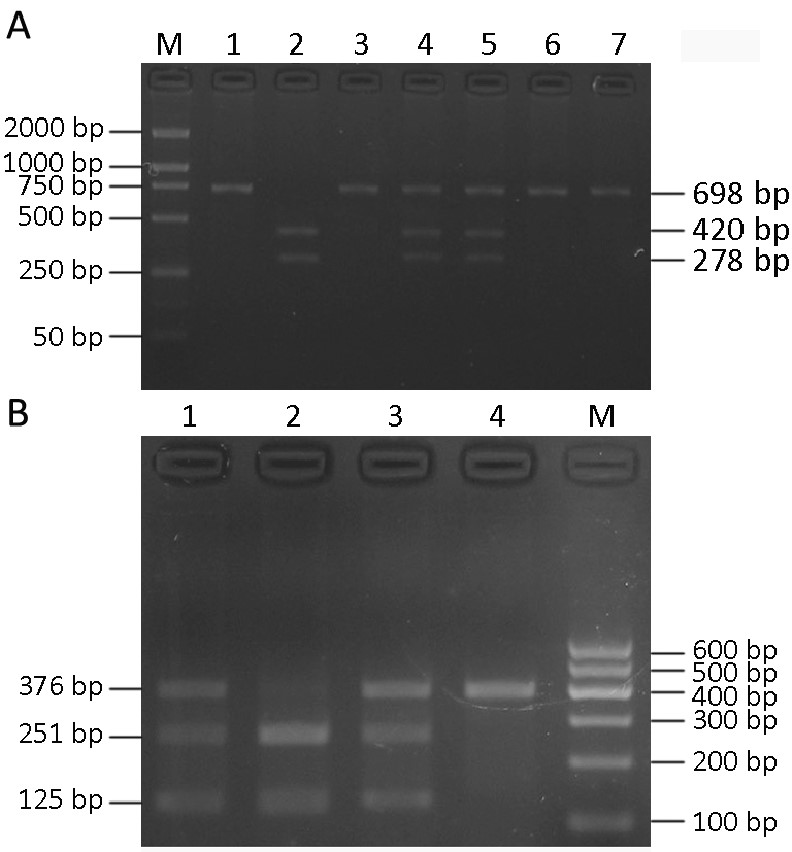
ObjectiveTo explore the relationship between cytochrome P450 2E1 (CYP2E1) RsaI/PstI and DraI polymorphism and lung cancer susceptibility in Mongolian and Han population in Inner Mongolia of China. MethodsCYP2E1 RsaI/PstI and DraI polymorphisms were detected by polymerase chain reaction-restriction fragment length polymorphism in 64 lung cancer patients, 150 healthy Mongolian and 150 healthy Han individuals. The distribution of genotype and allele frequencies of CYP2E1 RsaI/PstI and DraI polymorphisms were studied. ResultsThe risk of lung cancer was increased in individuals with CYP2E1 (cl/cl) and CYP2E1 (DD) with OR values of 2.431 (95%CI=1.082-5.460) and 2.778 (95%CI=1.358-5.683) respectively (P<0.05). When CYP2E1 RsaI/PstI and DraI polymorphisms were combined, the risk of lung cancer was reduced in individuals with CYP2E1 (cl/c2+c2/c2 and DD+CC) with OR values of 0.233 (95%CI=0.088-0.615, P<0.05). In smokers, the susceptibility to lung cancer was higher in the individuals with CYP2E1 (c1/c1) and CYP2E1 (DD) than in the individuals with c2 and C allele (P<0.05, OR=2.643 and 4.308 respectively). There was no significant difference in distribution of CYP2E1 genotype frequency between healthy Mongolian, Han population and lung cancer patients, healthy controls in Inner Mongolia. ConclusionCYP2E1 (c1/c1) and CYP2E1 (DD) are predisposing factors of lung cancer in population in Inner Mongolia. CYP2E1 (c2﹢C) co-mutation may decrease the risk of lung cancer. Smoking exerts synergetic effect with CYP2E1 (c1/c1) and CYP2E1 (DD) on the occurrence of lung cancer.
2011, 23(2): 112-117.
doi: 10.1007/s11670-011-0112-5
Abstract:
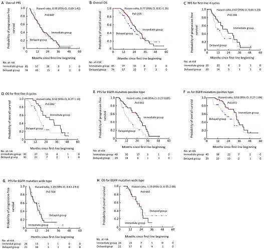
ObjectiveTo analyze the outcomes of patients who received TKI immediately after the first-line without progression as maintenance treatment (immediate group) vs. those received delayed treatment upon disease progression as second-line therapy (delayed group). MethodsThe study included 159 no-small-cell lung cancer (NSCLC) patients who received gefitinib or erlotinib as maintenance treatment in the immediate group (85 patients) or as second-line therapy in the delayed group (74 patients). The primary end point was progression-free survival (PFS). EGFR mutation status was detected using denaturing high-performance liquid chromatography (DHPLC). ResultsPFS was 17.3 and 16.4 months in the immediate and delayed groups, respectively (hazard ratio [HR], 0.99; 95% Confidence Interval [CI]: 0.69-1.42; P=0.947). In a subgroup analysis that included only patients with EGFR mutation, however, PFS was significantly longer in the immediate group than in the delayed group (HR, 0.48; 95% CI: 0.27-0.85; P=0.012). In patients with wild type EGFR, the risk for disease progression was comparable between the two groups (HR, 1.23; 95% CI: 0.61-2.51; P=0.564). No significant difference was demonstrated between the immediate and delayed group in terms of the overall survival (OS) (26.1 months vs. 21.6 months, respectively; HR=0.53; 95% CI: 0.27 to 1.06; P=0.072). There was also no difference in the incidence of adverse events between the two groups. ConclusionsEGFR TKI maintenance improves PFS in patients with EGFR mutation. Prospectively designed clinical studies that compare TKI immediate vs. delayed treatment after first-line chemotherapy upon disease progression are needed.
2011, 23(2): 118-122.
doi: 10.1007/s11670-011-0118-z
Abstract:
ObjectiveTo investigate the validity of CT perfusion in assessing angiogenic activity of lung cancer. MethodsFifty-six patients with lung cancer scheduled for elective surgical resection received 16-slice helical CT perfusion imaging. Time-density curve (TDC), blood flow (BF), blood volume (BV), mean transmit time (MTT) and permeability surface area product (PS) were calculated. 18F-deoxyglucose-positron emission tomography (FGD-PET) was carried out in 14 out of the 56 patients to calculate standardized uptake values (SUVs). Tumor microvessel density (MVD) was examined using CD34 immunohistochemical staining of the resected tumor tissue. Pearson’s correlation analysis was used to evaluate potential correlation between CT perfusion parameters and MVD or SUV. ResultsAverage time to peak height (TPH) of the TDCs (including two types of TDC) was 24.38±5.69 seconds. Average BF, BV, MTT and PS were 93.42±53.45 ml/100g/min,93.42±53.45 ml/100g,6.83±4.51 s and 31.92±18.73 ml/100g/min, respectively. Average MVD was 62.04±29.06/HPF. The mean SUV was 6.33±3.26. BF was positively correlated with MVD (r=0.620,P<0.01) and SUV (r=0.891, P<0.01). PS was also positively correlated with SUV (r=0.720, P<0.05). A positive correlation was also observed between tumor MVD and SUV (r=0.915, P<0.01). ConclusionsCT perfusion imaging is a reliable tool to evaluate the tumor neovascularity of lung cancer.

2011, 23(2): 123-128.
doi: 10.1007/s11670-011-0123-2
Abstract:
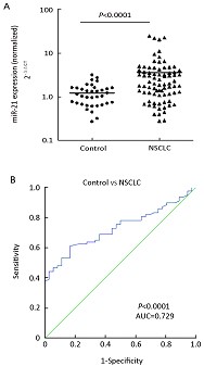
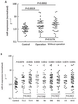
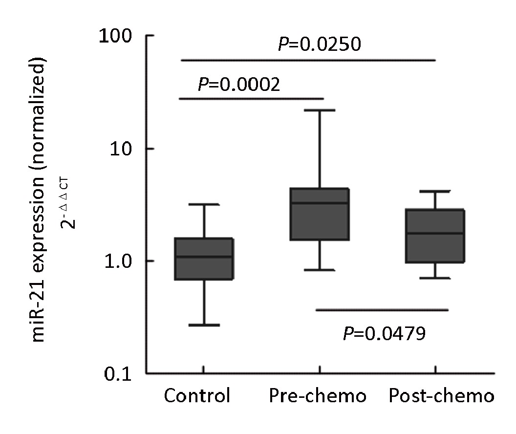
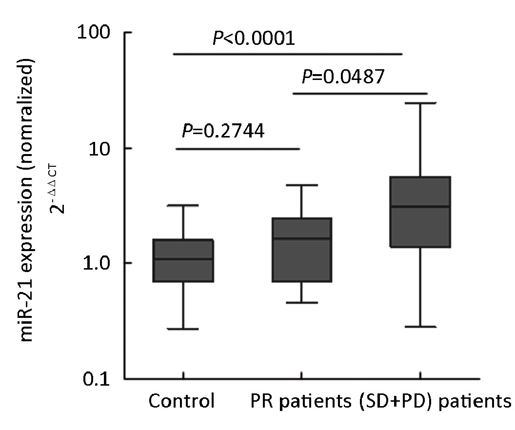
ObjectiveTo examine plasma microRNA-21 (miR-21) level in patients with non-small cell lung cancer (NSCLC) and its potential correlation with chemotherapeutic response. Methods77 NSCLC patients and 36 age and sex-matched healthy controls were included. Plasma miR-21 concentration was examined using a quantitative real-time reverse transcription polymerase chain reaction assay (qRT-PCR). Potential correlation between plasma mir-21 concentrations with chemotherapeutic responses was analyzed in 35 patients with advanced NSCLC (stages IIIB and IV). ResultsPlasma miR-21 was significantly higher in NSCLC patients relative to the healthy controls (P<0.0001). As a biomarker, plasma mir-21 had a receiver operating characteristic (ROC) curve area of 0.729 with 61.04% sensitivity and 83.33% specificity. Chemotherapeutic response in the 35 patients with advanced NSCLC (stages IIIB and IV) included partial response (PR) (n=11), stable disease and progression disease (SD+PD) (n=24). The overall response rate (CR+PR) was 31.4%. Plasma miR-21 in patients who achieved PR was significantly lower than those who did not respond (SD+PD) (P=0.0487), and comparable to that of the healthy controls (P=0.2744). ConclusionPlasma miR-21 is a good biomarker for NSCLC, and could be used to predict responses to chemotherapy.
2011, 23(2): 129-133.
doi: 10.1007/s11670-011-0129-9
Abstract:
ObjectiveTo evaluate the maximum tolerated dose (MTD) of docetaxel (DCT) and cisplatin (DDP) concurrently with three dimensional (3D) conformal radiotherapy or IMRT for patients with locally advanced non-small cell lung cancer (stage IIIa and IIIb) after 2–4 cycles of induction chemotherapy. MethodsFourteen patients with histological/cytological proven stage III non–small-cell lung cancer were eligible. 3D or IMRT radiotherapy (60-70Gy in 30-35 fractions, 6-7weeks, 2 Gy/fraction) was delivered concurrently with cisplatin and docetaxel, 2 cycles during concurrent chemoradiotherapy (CCRT). The level I dosage was composed of 56 mg/m2 DCT, on day 1 and 28mg/m2 DDP, on day 1 and day 2. The level II was composed of 60 mg/m2 DCT, on day 1 and 30 mg/ m2 DDP, on day 1 and day 2. The level III was composed of 64 mg/m2 DCT, on day 1 and 32 mg/ m2 DDP, on day 1 and day 2. ResultsFourteen patients were allocated and finished concurrent chemoradiotherapy. The dose-limiting neutropenia was at the dose Level III (64 mg/m2) and occurred in 2 of 5 patients. No dose limiting non-hematologic or hematologic toxicity occurred in the other patients. ConclusionsPatients with locally advanced non-small cell lung cancer may tolerate 60mg/m2 docetaxel and 60mg/m2 cisplatin for 2 cycles during concurrent radiotherapy after 2-3 cycles of induction chemotherapy.
2011, 23(2): 140-146.
doi: 10.1007/s11670-011-0140-1
Abstract:
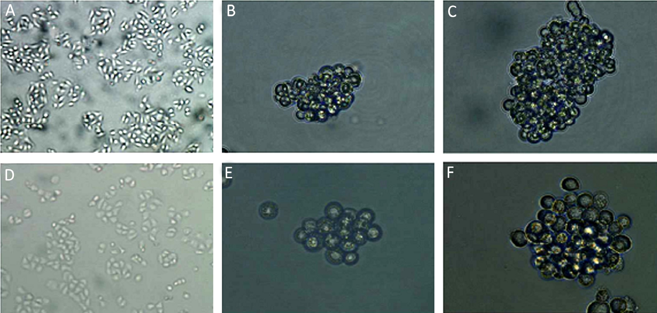
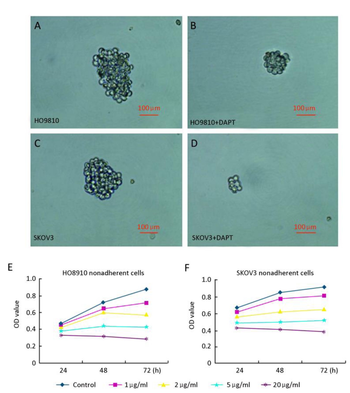
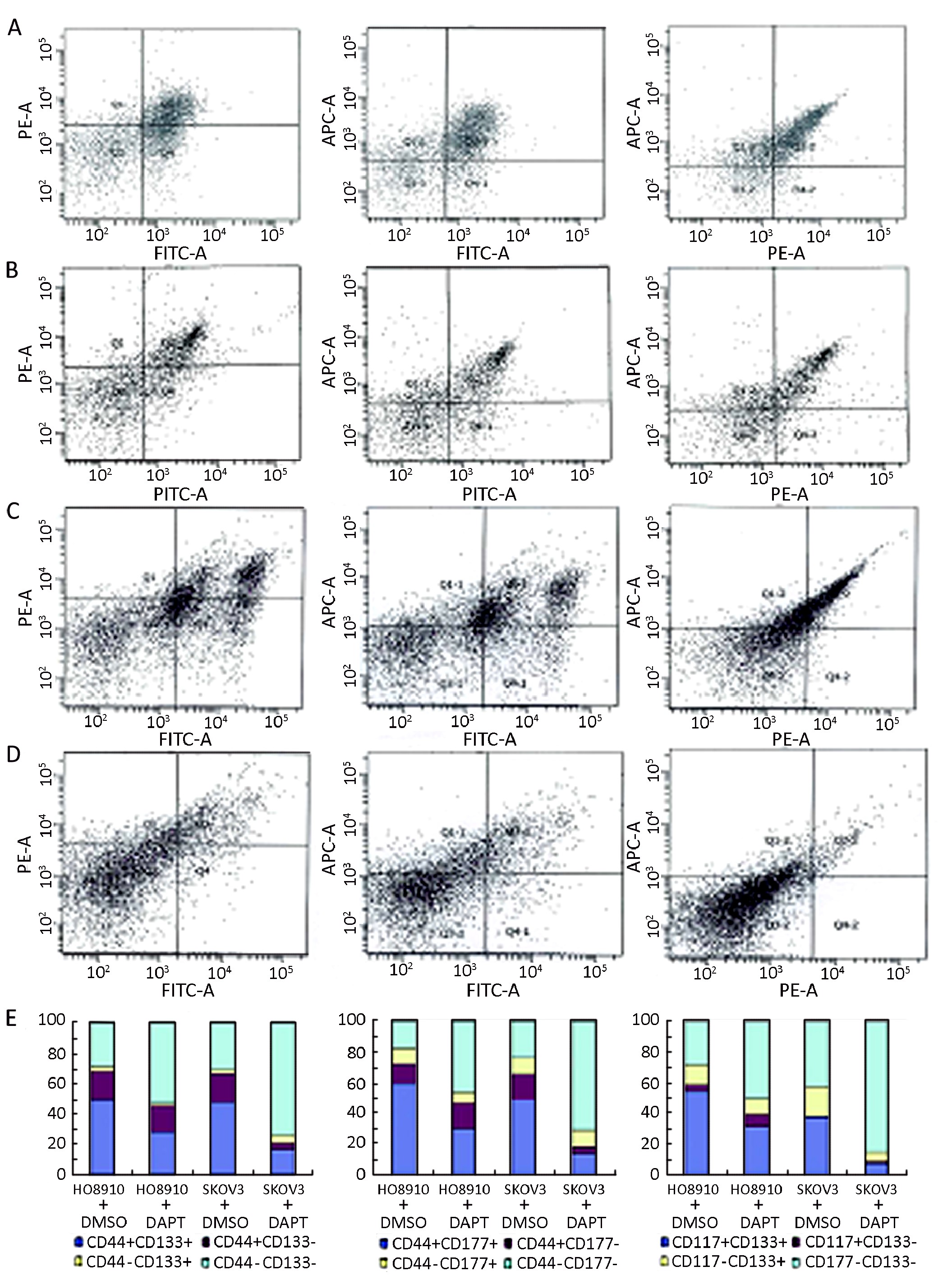
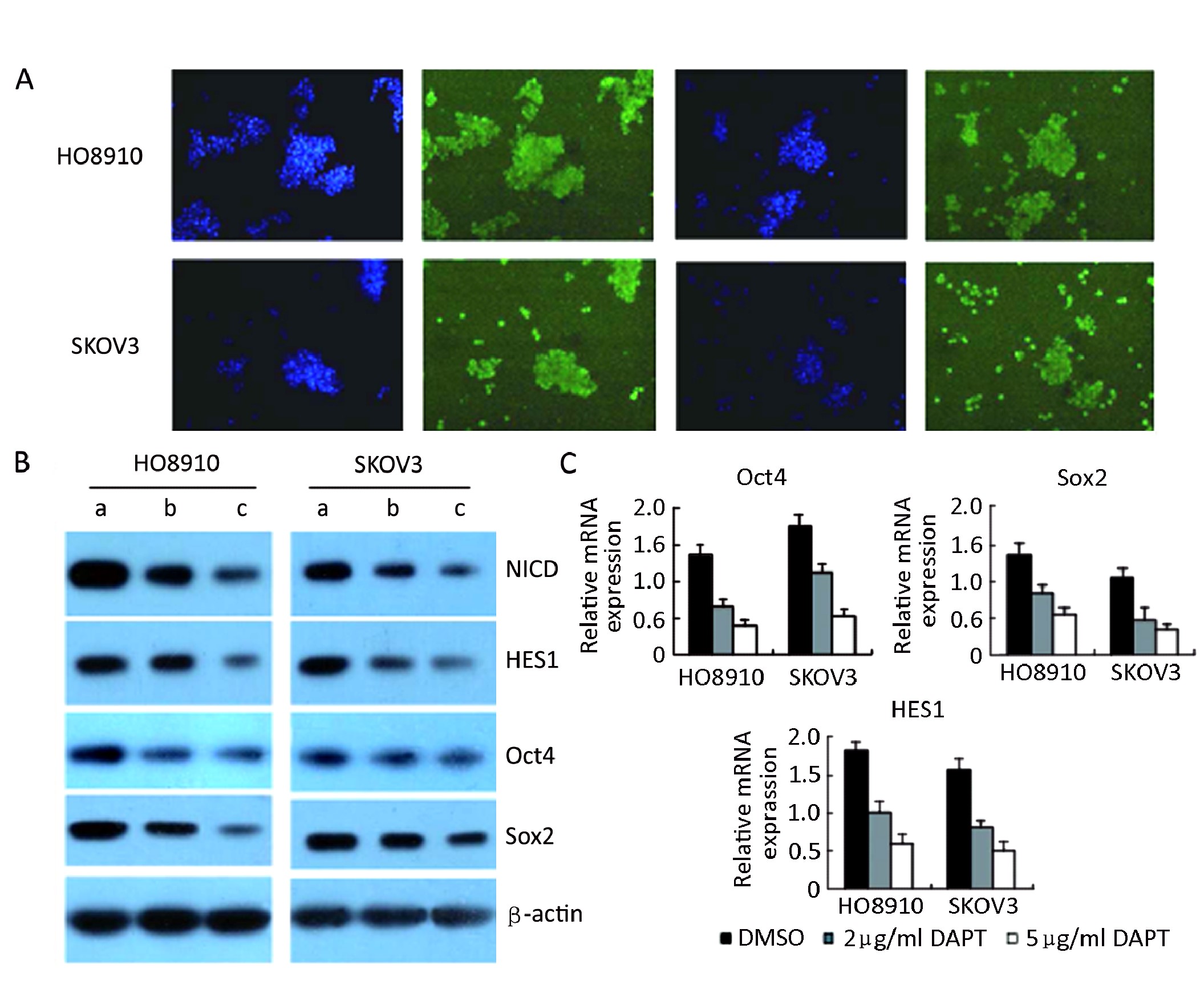
ObjectiveThe Notch signaling pathway plays an important role in the stem cell signaling network and contributes to tumorigenesis. However, the functions of Notch signaling in ovarian cancer stem cells (OCSCs) are not well understood. We aimed to investigate the effects of Notch blockade on self-renewal and stemness maintenance of OCSCs. MethodsOvarian cancer stem-like cells were enriched from ovarian cancer cell lines in serum-free medium. A γ-secretase inhibitor, (DAPT), was used to block Notch signaling. MTT assays were performed to assess self-renewal and proliferation inhibition, flow cytometry was performed to analyze cell surface marker and immunofluorescence, Western Blot and Real-time RT-PCR assays were performed to detect Oct4 and Sox2 protein and mRNA expression of the Ovarian cancer stem-like cells treated with DAPT. ResultsNotch blockade markedly inhibits self-renewal and proliferation of ovarian cancer stem-like cells, significantly downregulates the expression of OCSCs-specific surface markers, and reduces protein and mRNA expression of Oct4 and Sox2 in OCSC-like cells. ConclusionOur results suggest that Notch signaling is not only critical for the self-renewal and proliferation of OCSCs, but also for the stemness maintenance of OCSCs. The γ-secretase inhibitor is a promising treatment targeting OCSCs.
2011, 23(2): 147-152.
doi: 10.1007/s11670-011-0147-7
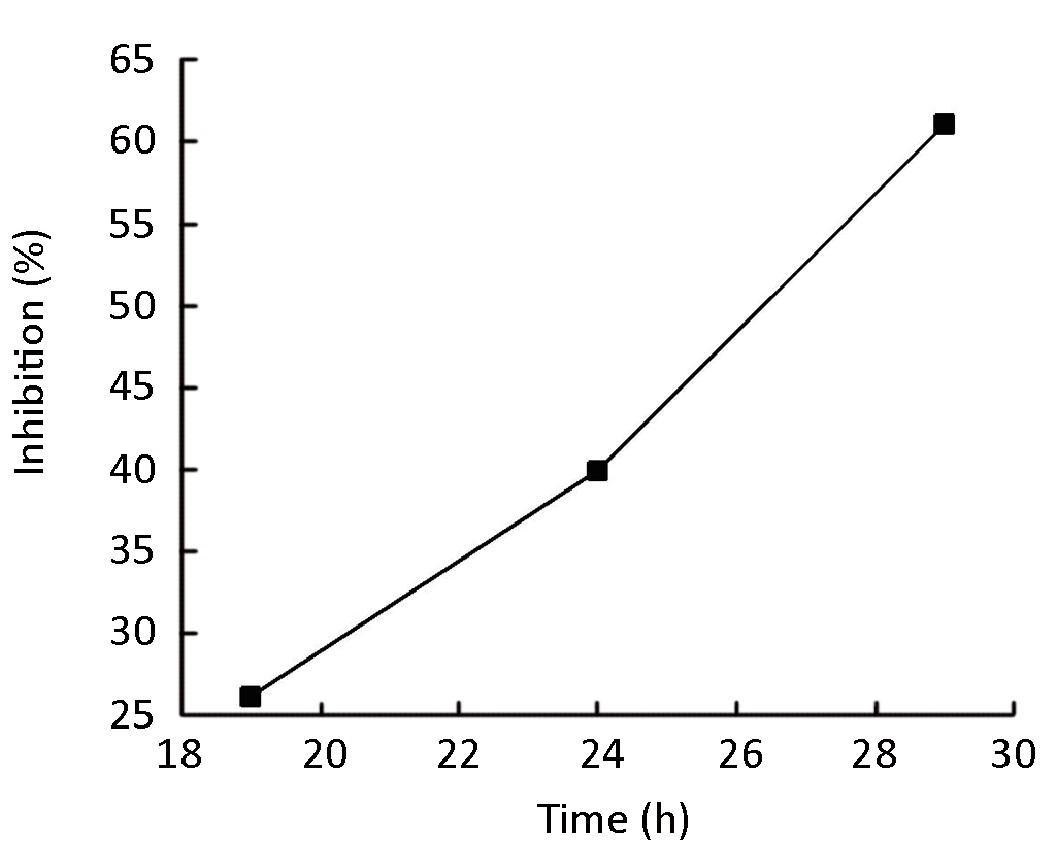
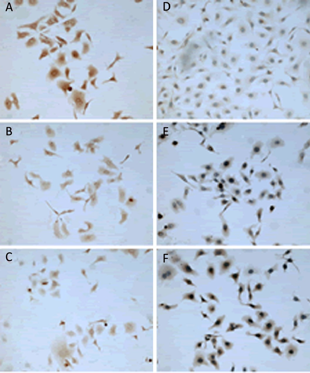
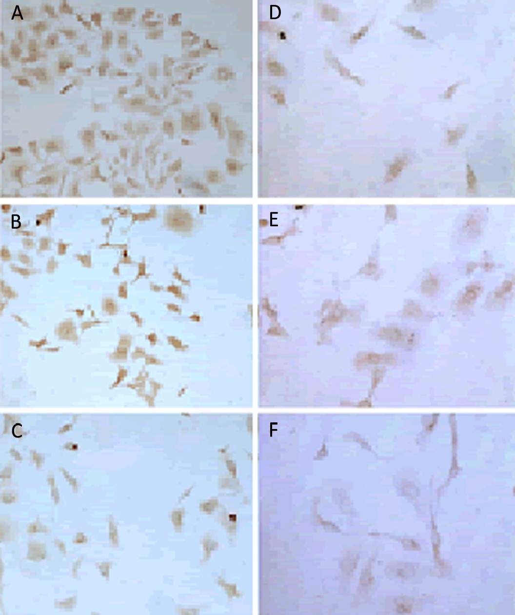
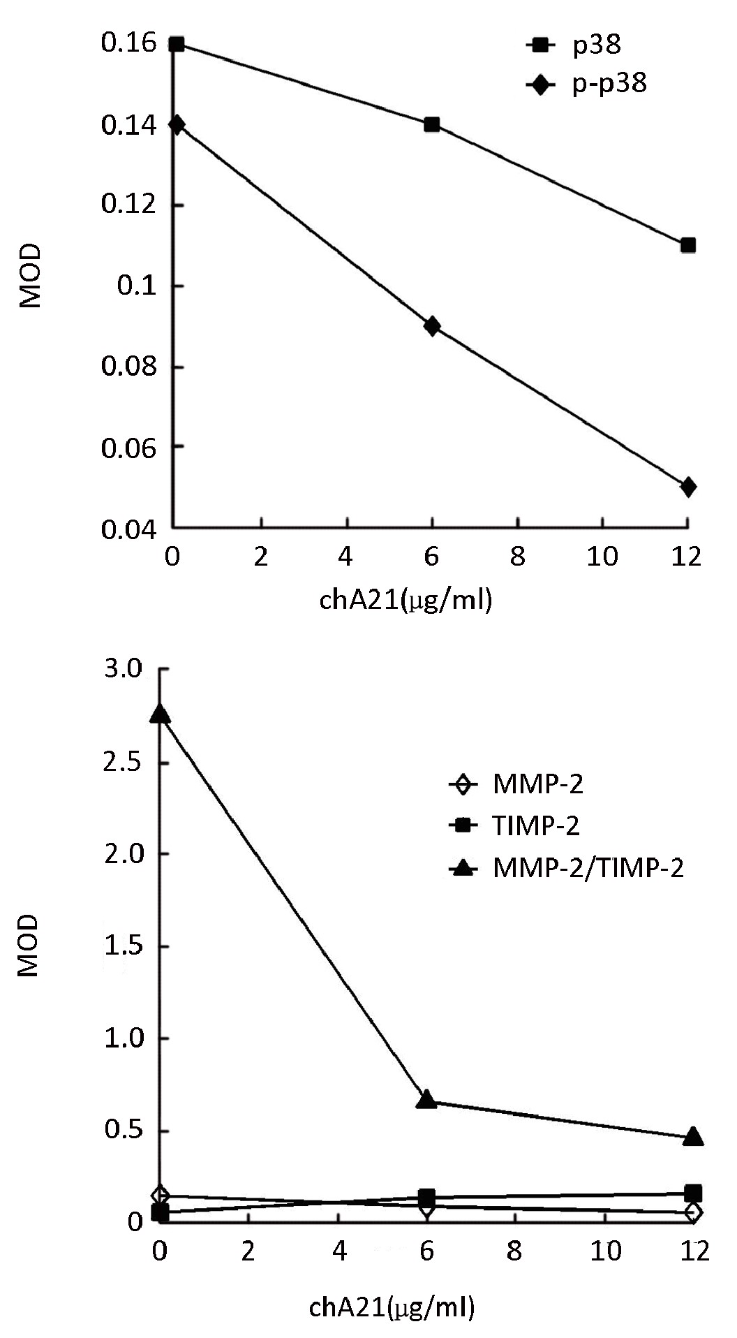
Abstract:
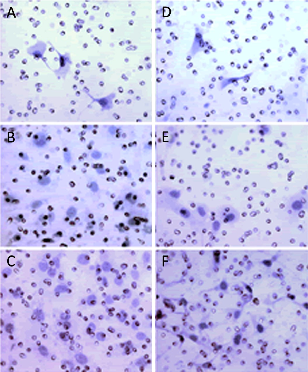
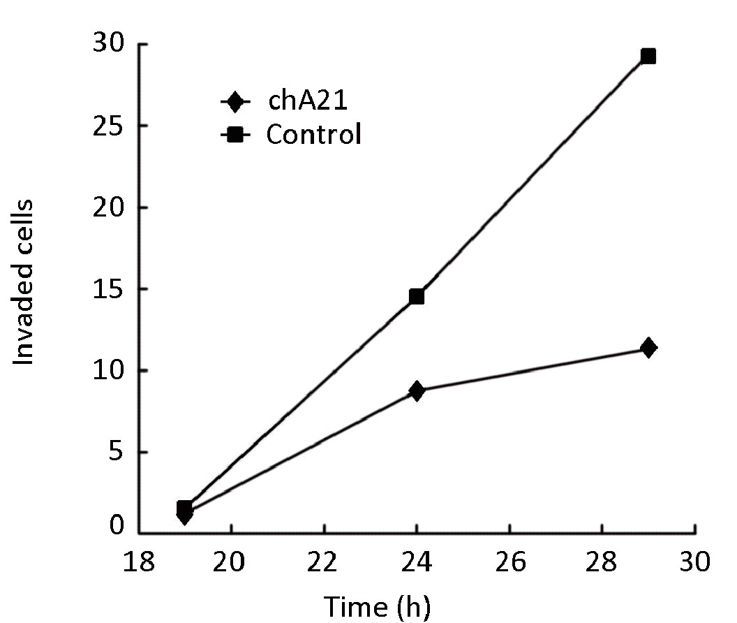
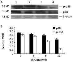
ObjectiveHER-2 plays an important role in the development and progression of ovarian carcinoma. A number of monoclonal antibodies (MAbs) and engineered antibody fragments (such as scFvs) against the subdomain II or IV of HER-2 extracellular domain (ECD) have been developed. We investigated the effect of chA21, an engineered anti-HER-2 antibody that bind primarily to subdomain I, on ovarian carcinoma cell invasion in vitro, and explored its possible mechanisms. MethodsGrowth inhibition of SK-OV-3 cells was assessed using a Methyl thiazolyl tetrazolium (MTT) assay. The invasion ability of SK-OV-3 was determined by a Transwell invasion assay. The expression of matrix metalloproteinase-2 (MMP-2) and its tissue inhibitors (TIMP-2) was detected by immunocytochemical staining, and the expression of p38 and the phosphorylation of p38 were assayed by both immunocytochemistry and Western blot. ResultsAfter treatment with chA21, the invasion of human ovarian cancer SK-OV-3 cells was inhibited in dose- and time-dependent manners. Simultaneously the expression of p38, phospho-p38, MMP-2 and the MMP-2/TIMP-2 ratio decreased, while TIMP-2 expression increased. Additionally, the decrease in phospho-p38 was much greater than that of p38. ConclusionchA21 may inhibit SK-OV-3 cell invasion via the signal transduction pathway involving MMP-2, TIMP-2, p38 and the activation of p38MAPK.


2011, 23(2): 153-159.
doi: 10.1007/s11670-011-0153-9
Abstract:
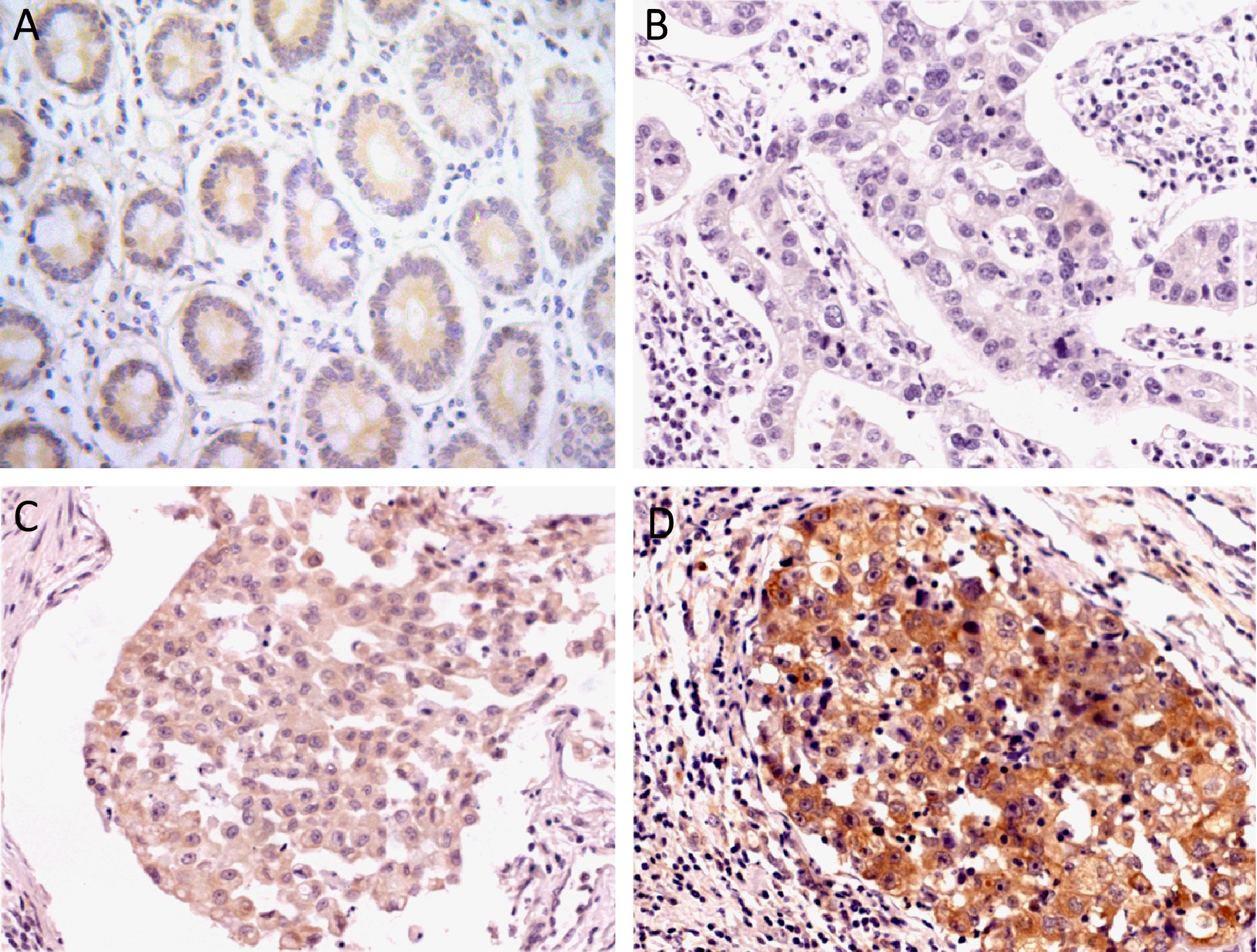
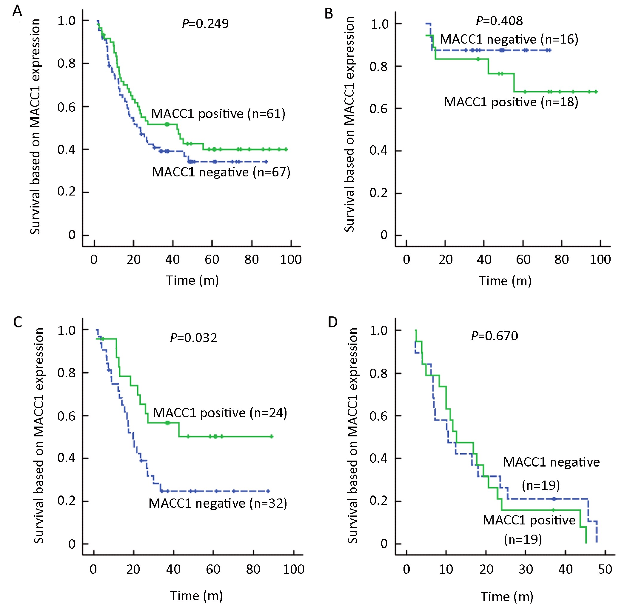
ObjectiveThe aim of this study was to detect metastasis-associated in colon cancer-1 (MACC1) expression in Chinese gastric cancer and analyze the relationship between MACC1 expression and postoperative survival. MethodsThe expression of MACC1 and c-MET protein in a sample of 128 gastric cancer tissues was detected by immunohistochemistry. A retrospective cohort study on the prognosis was carried out and data were collected from medical records. ResultsThe positive rate of MACC1 protein expression in gastric cancer was 47.66%, higher than that in adjacent noncancerous mucosa (P<0.001). MACC1 protein expression was not related to the clinicopathological variables involved. Kaplan-Meier analysis revealed that the survival of MACC1 positive group tended to be better than that of MACC1 negative group, particularly in patients with stage III carcinoma (P=0.032). Cox regression analysis revealed that MACC1 protein over-expression in gastric cancer tended to be a protective factor with hazard ratio of 0.621 (P=0.057). Immunohistochemical analysis showed that the positive rate of c-MET protein expression was much higher in cases with positive MACC1 expression in gastric cancer (P=0.002), but P53 expression was not associated with MACC1 expression. ConclusionMACC1 over-expression implies better survival and may be an independent prognostic factor for gastric cancer in Chinese patients.

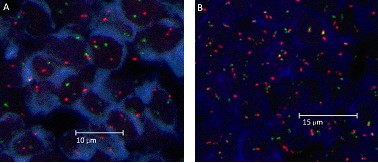
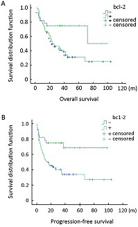
2011, 23(2): 160-164.
doi: 10.1007/s11670-011-0160-x
Abstract:
ObjectiveRecent studies have suggested that t(14;18) is present in a significant proportion of diffuse large B-cell lymphomas (DLBCLs). However, the prognostic significance of this translocation and its relationship with BCL-2 protein expression remains controversial. Our study aimed to investigate the predictive power of t(14;18) and BCL-2 protein expression in the prognosis of DLBCLs. MethodsBiopsy specimens from 106 DLBCLs were analyzed using interphase fluorescence in situ hybridization (FISH). Immunophenotypic analysis of CD20, CD3, CD10, BCL-6, MUM1 and BCL-2 was performed by immunohistochemistry. SPSS 13.0 software was used for statistical analysis. ResultsThe t(14;18) was identified in 27 of 106 cases (25.5%). The percentages of tumor cells expressing CD10, BCL-6, MUM1 and BCL-2 were 21.7%, 26.4%, 56.6% and 73.6%, respectively. The presence of this translocation was significantly correlated with the expression of CD10 and immunophenotypic subtype (p<0.001). No association was observed between BCL-2 protein expression and the presence of t(14;18). Multivariate analysis confirmed that both t(14;18) and BCL-2 expression were significantly associated with survival. Moreover, patients with t(14;18) had worse prognosis, compared with those with BCL-2 expression (for overall survival: hazard ratio, 4.235; 95%CI, 2.153-8.329, p<0.001 vs. hazard ration, 2.743; 95%CI, 1.262-5.962, p=0.011). ConclusionsThe t(14;18) is a useful prognostic tool for the evaluation of DLBCL immunophenotype and prognosis. The prognosis of GCB (germinal centre-like B cell) DLBCL patients should be made with the consideration of the presence of this translocation, and the detection of t(14;18) should be included as a routine diagnostic test in these cases.