2011 Vol.23(4)
Display Mode: |
2011, 23(4): 245-253.
doi: 10.1007/s11670-011-0245-6
 Abstract
 FullText HTML
 PDF 405KB
Abstract:
Following the classification of hepatocellular nodules by the International Working Party in 1995 and further elaboration by the International Consensus Group for Hepatocellular Neoplasia in 2009, entities under the spectrum of hepatocellular nodules have been better characterized. Research work hence has been done to answer questions such as distinguishing high-grade dysplastic nodules from early hepatocellular carcinoma (HCC), delineating the tumor cell origin of HCC, identifying its prognostic markers, and subtyping hepatocellular adenomas. As a result, a copious amount of data at immunohistochemical and molecular levels has emerged. A panel of immunohistochemical markers including glypican-3, heat shock protein 70 and glutamine synthetase has been found to be of use in the diagnosis of small, well differentiated hepatocellular tumors and particularly of HCC. The use of liver fatty acid binding protein (L-FABP), β-catenin, glutamine synthetase, serum amyloid protein and C-reactive protein is found to be helpful in the subtyping of hepatocellular adenomas. The role of tissue biomarkers for prognostication in HCC and the use of biomarkers in subclassifying HCC based on tumor cell origin are also discussed.
Following the classification of hepatocellular nodules by the International Working Party in 1995 and further elaboration by the International Consensus Group for Hepatocellular Neoplasia in 2009, entities under the spectrum of hepatocellular nodules have been better characterized. Research work hence has been done to answer questions such as distinguishing high-grade dysplastic nodules from early hepatocellular carcinoma (HCC), delineating the tumor cell origin of HCC, identifying its prognostic markers, and subtyping hepatocellular adenomas. As a result, a copious amount of data at immunohistochemical and molecular levels has emerged. A panel of immunohistochemical markers including glypican-3, heat shock protein 70 and glutamine synthetase has been found to be of use in the diagnosis of small, well differentiated hepatocellular tumors and particularly of HCC. The use of liver fatty acid binding protein (L-FABP), β-catenin, glutamine synthetase, serum amyloid protein and C-reactive protein is found to be helpful in the subtyping of hepatocellular adenomas. The role of tissue biomarkers for prognostication in HCC and the use of biomarkers in subclassifying HCC based on tumor cell origin are also discussed.


2011, 23(4): 254-258.
doi: 10.1007/s11670-011-0254-5
Abstract:
Nowadays, advanced non-small cell lung cancer (NSCLC) is still an incurable disease. However, recent researches on maintenance therapy have led to considerable progress. Recently, pemetrexed and erlotinib have been approved for maintenance chemotherapy by both the U.S. Food and Drug Administration and European Medicines Agency. However, there are not adequate data to support the maintenance therapy as the standard treatment for advanced NSCLC and there has been no conclusive predictor of who will get benefit from maintenance chemotherapy and what type of maintenance, continuation or switch, is preferred. This article reviews the main studies on maintenance therapy of advanced NSCLC and discusses the results available to date.
Nowadays, advanced non-small cell lung cancer (NSCLC) is still an incurable disease. However, recent researches on maintenance therapy have led to considerable progress. Recently, pemetrexed and erlotinib have been approved for maintenance chemotherapy by both the U.S. Food and Drug Administration and European Medicines Agency. However, there are not adequate data to support the maintenance therapy as the standard treatment for advanced NSCLC and there has been no conclusive predictor of who will get benefit from maintenance chemotherapy and what type of maintenance, continuation or switch, is preferred. This article reviews the main studies on maintenance therapy of advanced NSCLC and discusses the results available to date.
2011, 23(4): 265-270.
doi: 10.1007/s11670-011-0265-2
Abstract:
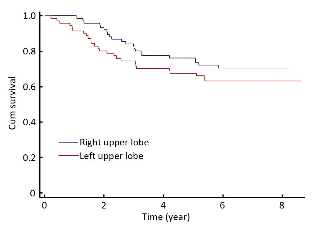
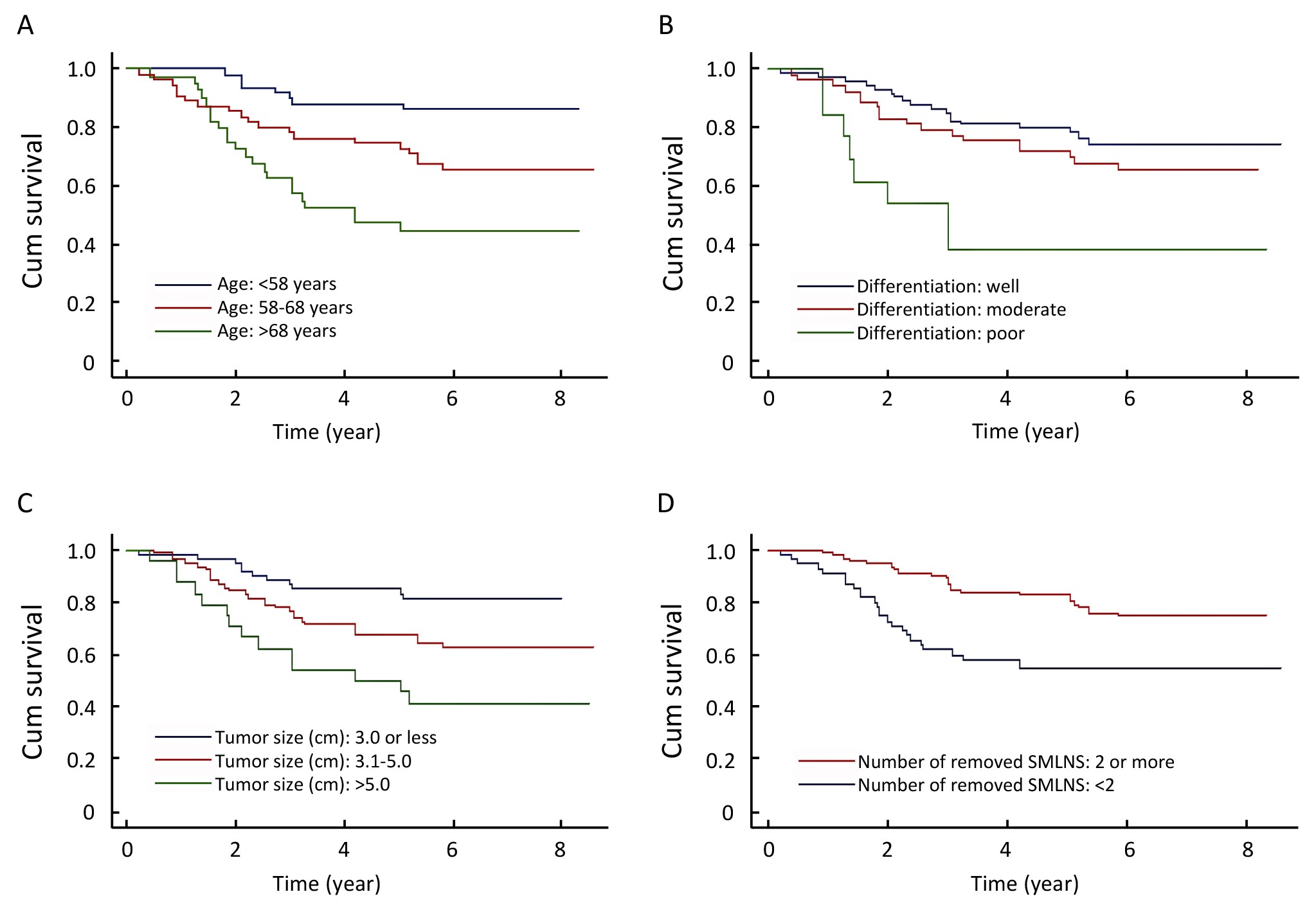
ObjectiveTo identify clinical and pathologic factors that were associated with the survival of stage IB upper lobe non-small cell lung cancer (NSCLC) patients. MethodsA retrospective study of 147 subjects who had undergone curative resection for stage IB upper lobe NSCLC was performed. Patients who had received any adjuvant or neo-adjuvant chemotherapy were excluded. Survival function curves were estimated using the Kaplan-Meier procedure. Crude and adjusted hazard ratios (HRs) of potential prognostic factors were estimated using Cox proportional hazards models. ResultsFive factors, including age, tumor size, histologic grade of differentiation, number of removed superior mediastinal lymph node stations and presence of visceral pleura invasion, were significantly and independently associated with mortality risk. Adjusted HRs were 2.6 [95% confidence interval (95% CI): 1.1-6.5] and 4.6 (95% CI: 1.9-11) for those aged 58−68 years and those >68 years, respectively, relative to those aged <58 years. HRs for those with poorly and moderately differentiated tumors were 6.4 (95% CI: 2.3-18) and 1.4 (95% CI: 0.7-2.8), respectively. HRs for those with tumor size 3.1−5 cm and >5 cm (vs≤3.0 cm) were 2.3 (95% CI: 1.1-4.9) and 4.3 (95% CI: 1.9-10), respectively. The presence of visceral pleura invasion also increased the risk of mortality (HR=4.0, 95% CI: 1.3-12). ConclusionAdvanced age, larger tumor size, poorly differentiated histology, smaller number of removed superior mediastinal lymph node stations, and presence of visceral pleura invasion were associated with poor survival of surgically treated stage IB upper lobe NSCLC patients.
2011, 23(4): 271-275.
doi: 10.1007/s11670-011-0271-4
Abstract:
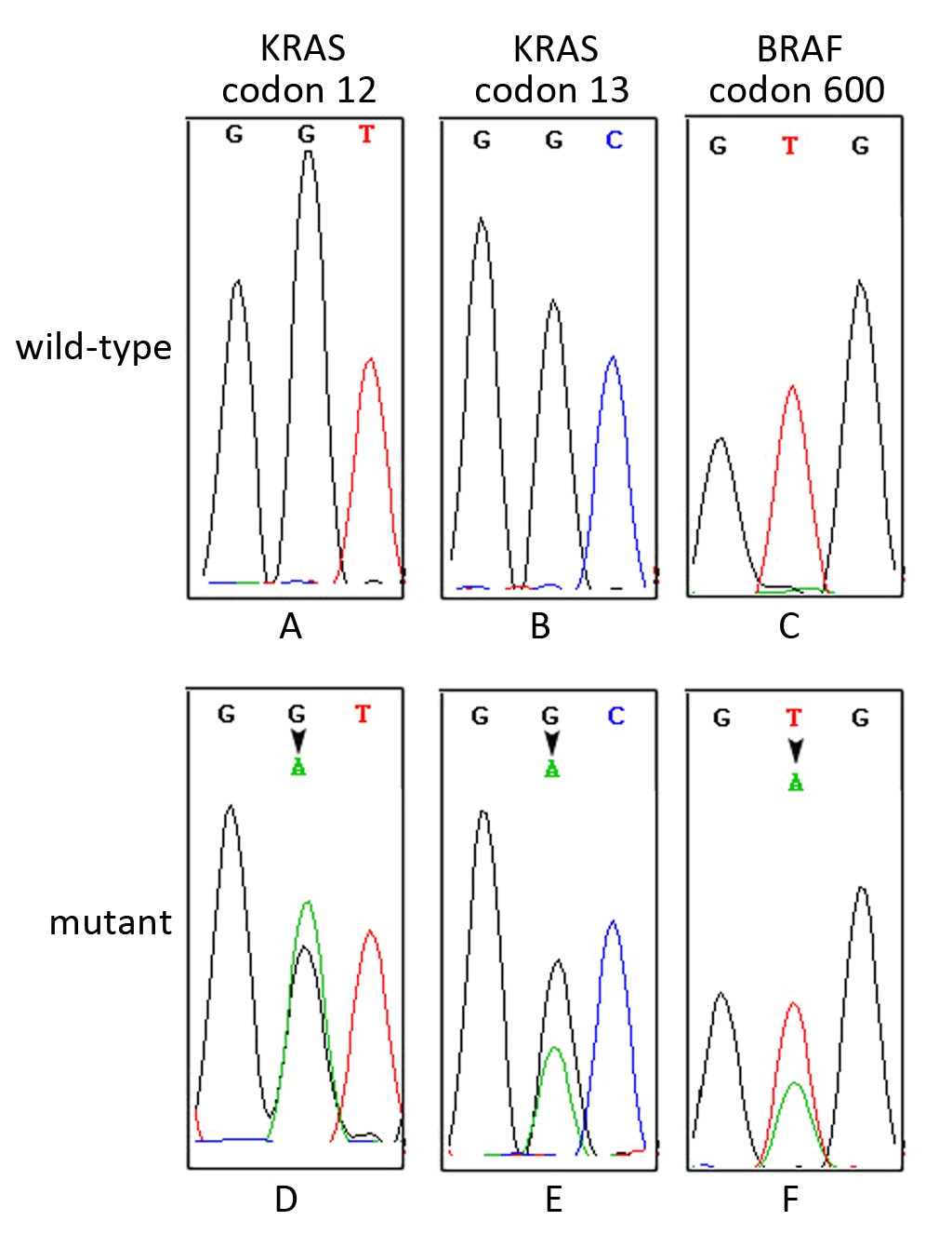
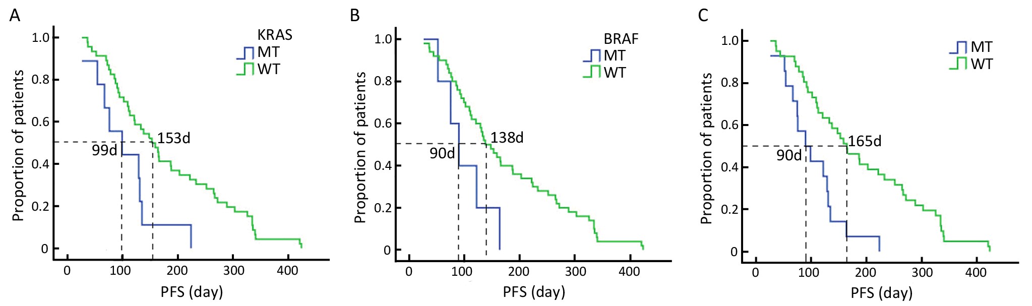
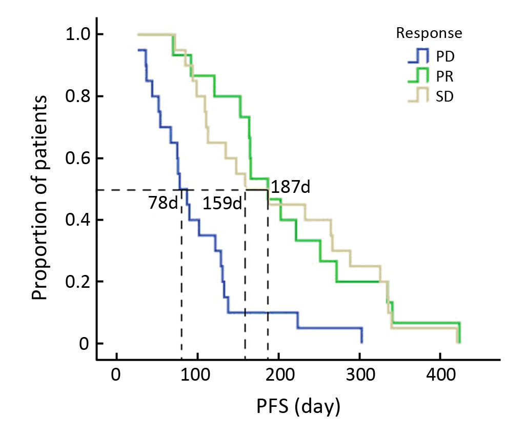
ObjectiveTo analyze the relationship between KRAS, BRAF mutations and the response toCetuximab in Chinese colorectal cancer patients. MethodsA total of273 Chinese colorectal cancer patients were evaluated for KRAS and BRAF mutations by Sanger sequencing. Among them, 59 patients with metastatic colorectal cancer (mCRC) were treated with Cetuximab in combination with chemotherapy from August 2005 to July 2009. Statistical analysis was conducted to assess the relationship between KRAS, BRAF mutations and the response or survival of 59 mCRC patients. ResultsKRAS and BRAF mutation rates were 38.5% (105/273) and 5.1% (14/273), respectively, and KRAS/BRAF mutations were mutually exclusive. Among 59 patients treated with Cetuximab plus chemotherapy, KRAS and BRAF mutations were identified in 11and 5 patients, respectively. The response rates and median progression-free survivals (PFS) in KRAS wild-type and mutant patients were 35.4% (17/48) vs. 9.1% (1/11) (P=0.054) and 153 days vs. 99 days (P=0.01), respectively.Also, the response rates and median PFS in BRAF wild-type and mutant patients were 37.2% (16/43) vs. 20% (1/5) (P=0.016) and 138 days vs. 90 days (P=0.036), respectively. ConclusionBesides KRAS, assessing BRAF mutation should also be required to select patients eligible for Cetuximab. Further prospective evaluation in large samples should be performed to confirm these preliminary findings.
2011, 23(4): 276-282.
doi: 10.1007/s11670-011-0276-z
Abstract:
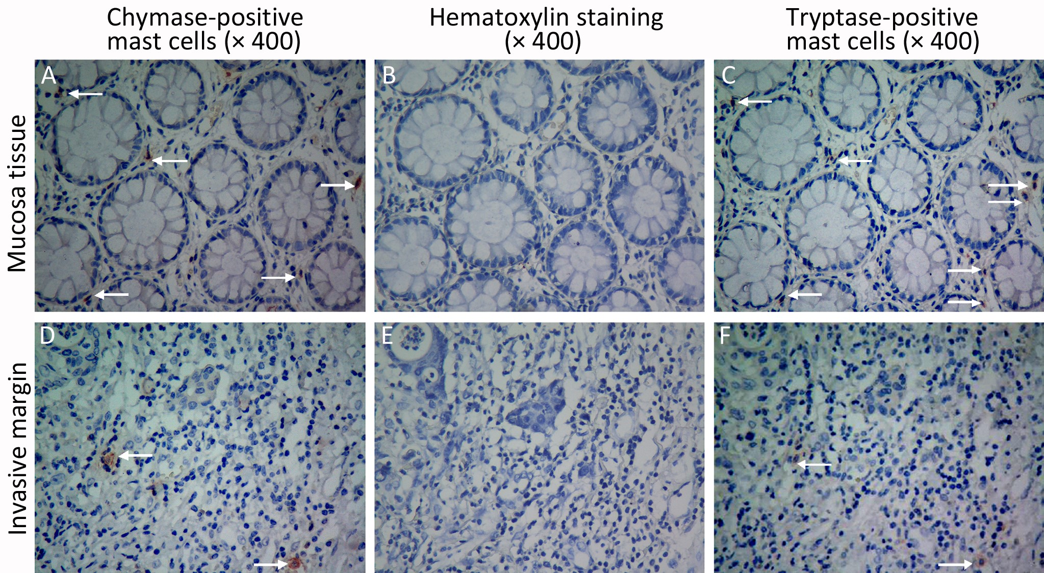
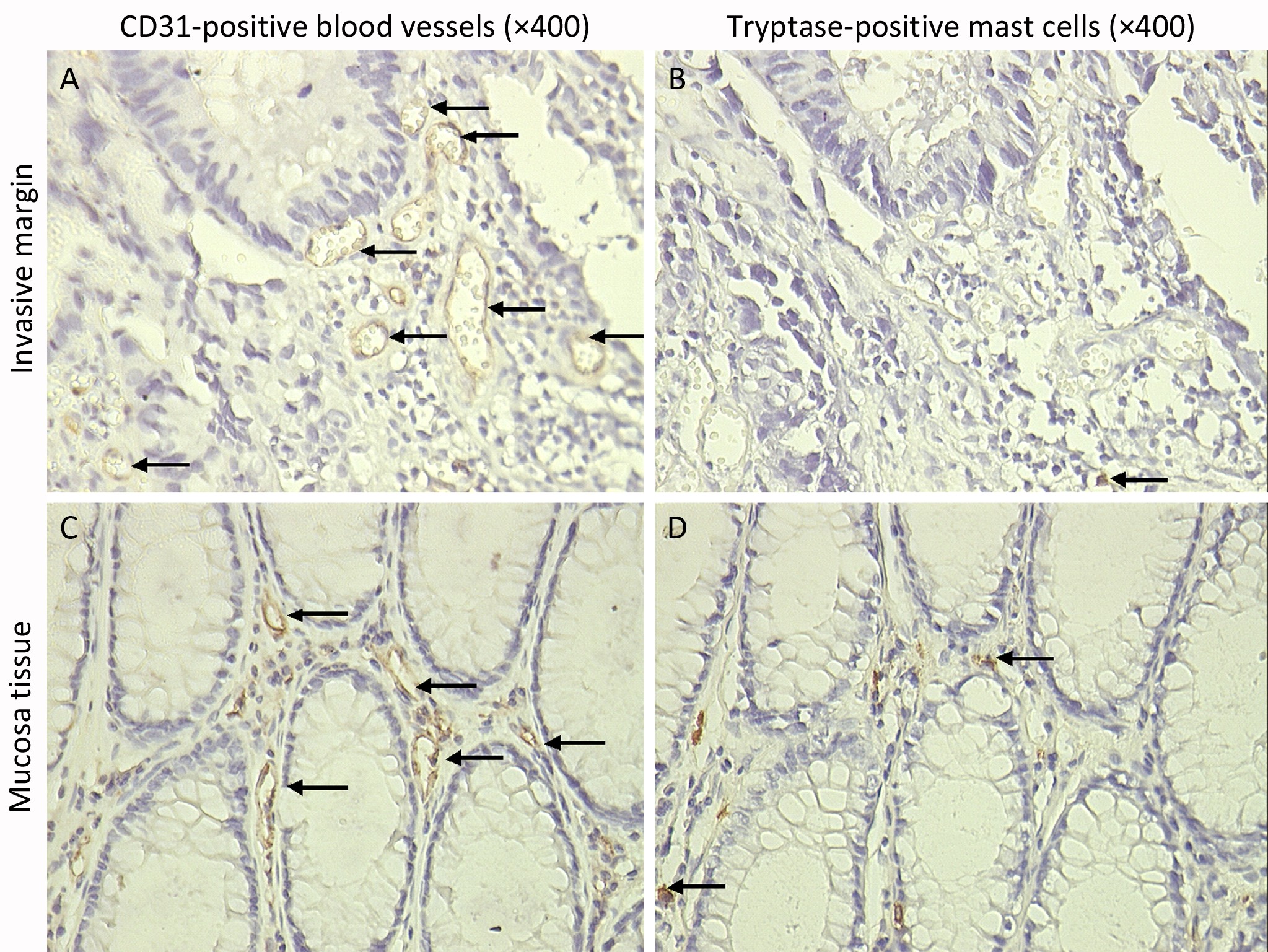
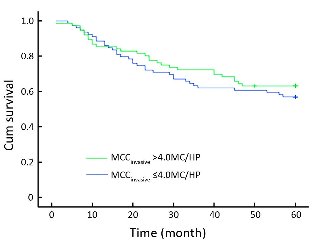
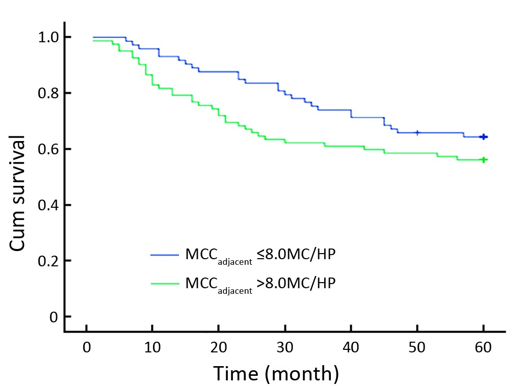
ObjectiveMast cells (MC) reside in the mucosa of the digestive tract as the first line against bacteria and toxins. Clinical evidence has implied that the infiltration of mast cells in colorectal cancers is related to malignant phenotypes and a poor prognosis. This study compared the role of mast cells in adjacent normal colon mucosa and in the invasive margin during the progression of colon cancer. MethodsSpecimens were obtained from 39 patients with colon adenomas and 155 patients with colon cancers treated at the Sun Yat-sen University Cancer Center between January 1999 and July 2004. The density of mast cells was scored by an immunohistochemical assay. The pattern of mast cell distribution and its relationship with clinicopathologic parameters and 5-year survival were analyzed. ResultsThe majority of mast cells were located in the adjacent normal colon mucosa, followed by the invasive margin and least in the cancer stroma. Mast cell count in adjacent normal colon mucosa (MCCadjacent) was associated with pathologic classification, distant metastases and hepatic metastases, although it was not a prognostic factor. In contrast, mast cell count in the invasive margin (MCCinvasive) was associated with neither the clinicopathlogic parameters nor overall survival. ConclusionMast cells in the adjacent normal colon mucosa were related to the progression of colon cancer, suggesting that mast cells might modulate tumor progression via a long-distance mechanism.
2011, 23(4): 283-287.
doi: 10.1007/s11670-011-0283-0
Abstract:
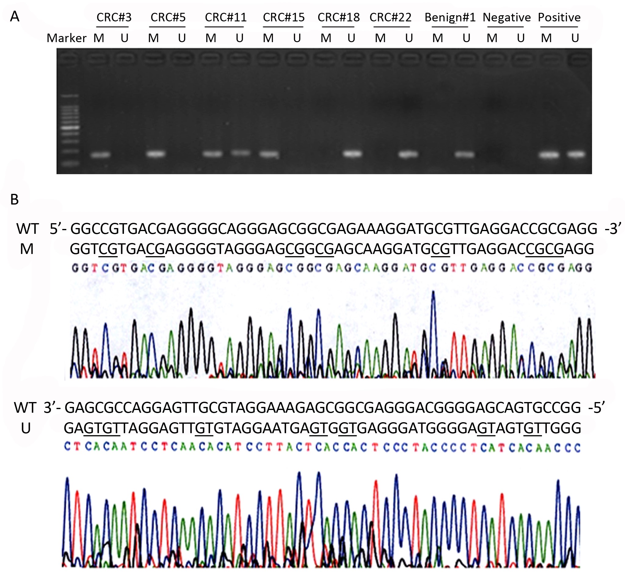
ObjectiveDeleted in liver cancer 1 (DLC1) is a new candidate tumor suppressor gene, whose down-regulation or even silence will result from promoter hypermethylation in various human cancers including colorectal cancer (CRC). The aim of this study is toevaluate the diagnostic role of DLC1gene methylationin the serum DNA from CRC patients. MethodsThis study enrolled 85 CRC patients and 45 patients with benign colorectal diseases. Methylation-specific polymerase chain reaction (MSP) was used to determine the promoter methylation status of DLC1 gene in serum DNA. The combination of DLC1 methylation and conventional tumor markers was further analyzed. ResultsHypermethylation of DLC1 was detected in 42.4% (36/85) of CRC serums, while seldom in the benign controls(8.9%, 4/45) (P<0.001). The aberrant DLC1 methylation in serum DNA was not associated with patients’ clinicopathological features and elevated CEA/CA19-9 levels. Furthermore, the combinational analysis of CEA, CA19-9 and DLC1 methylation showed a higher sensitivity and no reduced diagnostic specificity than CEA and CA19-9 combination for CRC diagnosis. ConclusionThe serum DLC1 methylation may be a promising biomarker for the early detection of CRC, which will further increase the diagnostic efficiency in combination with CEA and CA19-9.
2011, 23(4): 288-294.
doi: 10.1007/s11670-011-0288-8
Abstract:
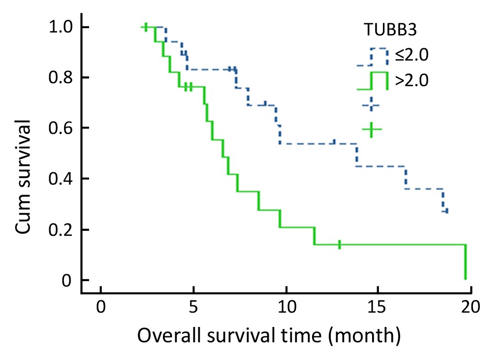
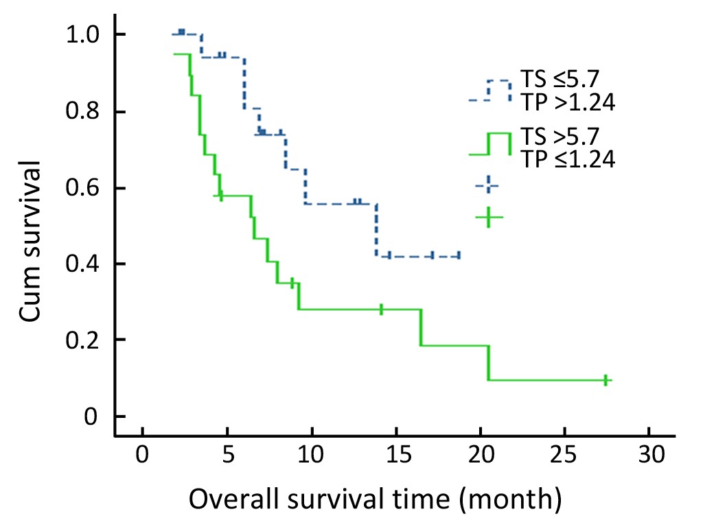
ObjectiveToevaluate the role of class III β-tubulin (TUBB3), thymidylate synthase (TS), thymidine phosphorylase (TP), and excision repair cross-complementing group 1 (ERCC1) in clinical outcome of advanced gastric cancer patients receiving capecitabine plus paclitaxel or cisplatin. MethodsThe clinical data and tumor specimens from 57 advanced gastric cancer patients receiving first-line capecitabine plus paclitaxel (cohort 1, n=36) and capecitabine plus cisplatin (cohort 2, n=21) were retrospectively collected, and TUBB3, TS, TP, and ERCC1 expressions were detected by real-time quantitative PCR. The associations between expressions of biomarkers and response or survival were analyzed statistically. ResultsThe median age of 57 patients was 57 years (range: 27–75 years) with 38 males and 19 females. Of all patients, the response rates of patients with high TP, low TP and high TS, low TS expressions were 57.1%, 27.6% (P=0.024), and 55.2%, 28.6% (P=0.042), respectively. Among cohort 1, the response rates and median overall survivals of patients with low and high TUBB3 expressions were 61.1% vs. 33.3% (P=0.095) and 13.8 months vs. 6.6 months (P=0.019), respectively; the response rate (87.5%) of patients with low TUBB3 and high TP expressions was higher than that (14.3%) of patients with high TUBB3 and low TP expressions (P=0.01). Among cohort 2, the response rates of patients with low ERCC1 and high ERCC1 expressions were 45.5% and 20.0% respectively (P=0.361). ConclusionTUBB3, TS and TP expressions could predict the response of advanced gastric cancer patients receiving capecitabine-based and paclitaxel-based chemotherapy. These results will be further confirmed in future large samples.
2011, 23(4): 295-300.
doi: 10.1007/s11670-011-0295-9
Abstract:
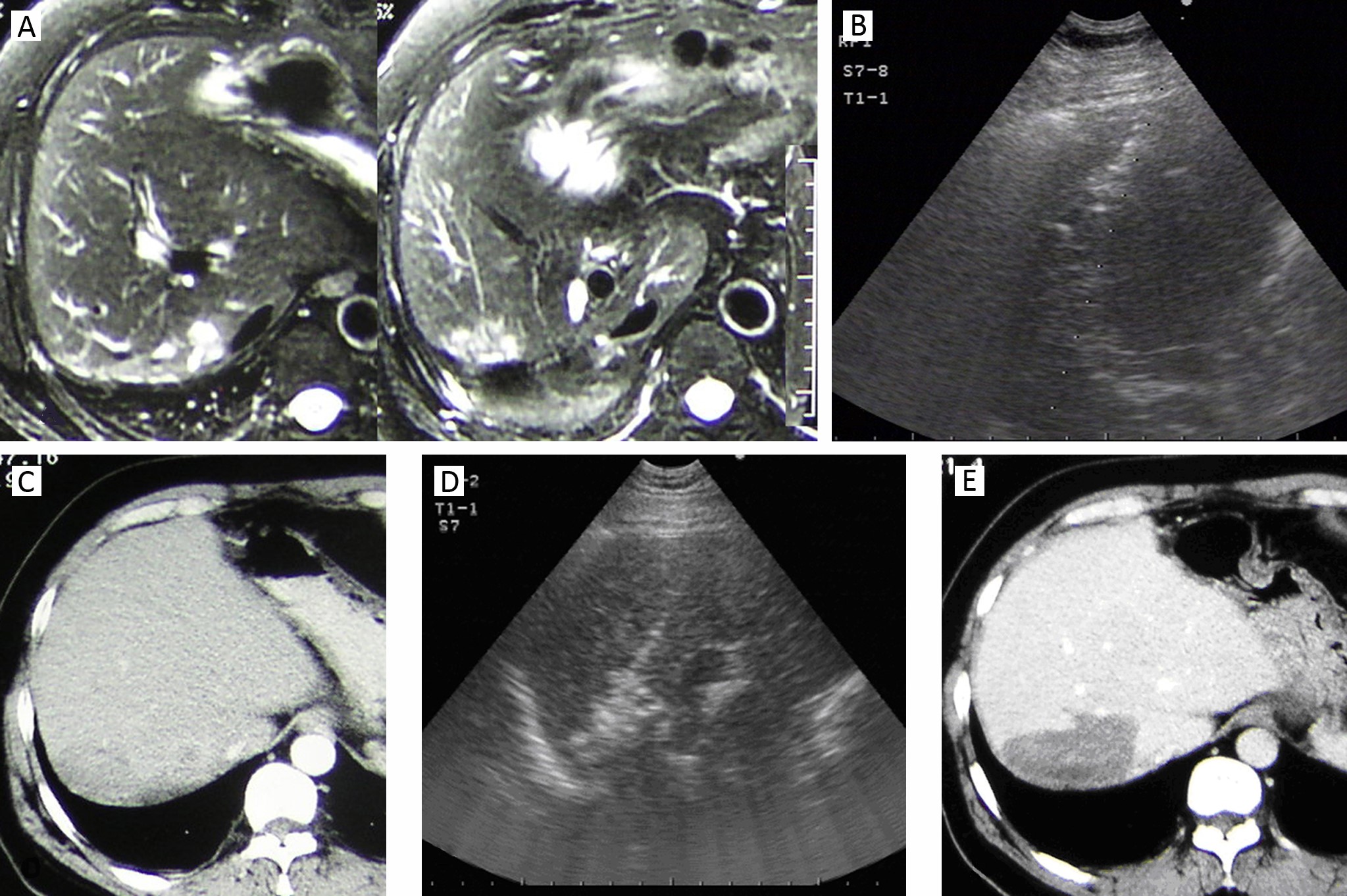
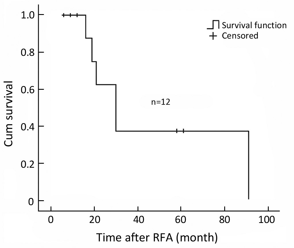
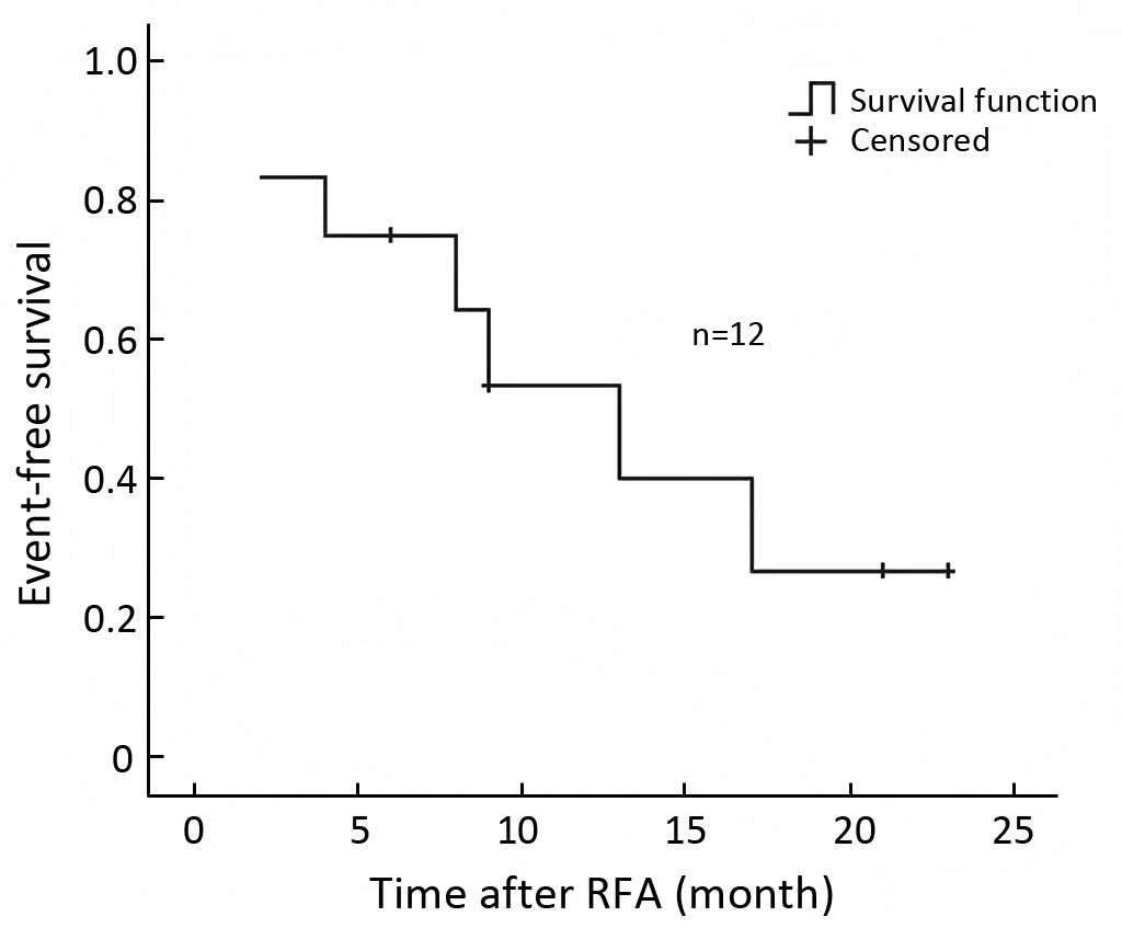
ObjectiveMost recurrent intrahepatic cholangiocarcinoma (RICC) lost the opportunity of radical resection while most nonsurgical management failed to prolong patients’ survival. The efficacy and safety of radiofrequency ablation (RFA) as a local treatment for recurrent hepatocellular carcinoma have been confirmed by many clinical studies. The purpose of this study was to evaluate the efficacy, long-term survival and complications of RFA for RICC. MethodsA total of 12 patients with 19 RICCs after radical resection were included in this study. The tumors were 1.9-6.8 cm at the maximum diameter (median, 3.2±1.6 cm). All patients were treated with ultrasound guided RFA. There were two RFA approaches including percutaneous and open. ResultsA total of 18 RFA treatment sessions were performed. Ablation was successful (evaluated by 1-month CT after the initial RFA procedure) in 18 (94.7%) of 19 tumors. By a median follow-up period of 29.9 months after RFA, 5 patients received repeated RFA because of intrahepatic lesion recurrence. The median local recurrence-free survival period and median event-free survival period after RFA were 21.0 months and 13.0 months, respectively. The median overall survival was 30 months, and the 1- and 3-year survival rates were 87.5% and 37.5%, respectively. The complication rate was 5.6% (1/18 sessions). The only one major complication was pleural effusion requiring thoracentesis. ConclusionThis study showed RFA may effectively and safely manage RICC with 3-year survival of 37.5%. It provides a treatment option for these RICC patients who lost chance for surgery.
2011, 23(4): 301-305.
doi: 10.1007/s11670-011-0301-2
Abstract:
ObjectiveBased on liver cancer model built in SD rats, the contents of trace elements (Cu, Fe, Zn, Ca and Mg), AFP, CEA, SF, TH and IGF-II in serum were measured at different stages to explore the molecular changes during the rat liver cancer development. MethodsThe SD rat liver cancer model was built by using diethylnitrosamine (DENA) as the mutagen. During 16 weeks of DENA gavage, blood samples were taken in the 14th, 28th, 56th, 77th, 105th and 112th days respectively after the first day of gavage with DENA, then the contents of five trace elements (Cu, Fe, Zn, Ca and Mg), T3, T4, IGF-II, AFP, CEA and SF in serum were determined. ResultsDuring the development of the rat liver cancer, in the test group, the Cu content significantly increased in serum, while the contents of Fe, Zn and Ca significantly decreased. The content of Mg showed no significant change. AFP and CEA of the test group showed same expression level with the control group; while the content of SF was lower than that of the control group when cancerization appeared. T3 and T4 increased at the first stage and then went down, and the content of IGF-II was always high. ConclusionCu, Fe, Zn, Ca, T3, T4, SF and IGF-II are closely related to the development of liver cancer. The changes of their contents in the development of cancer could enlighten the researches on cancer pathogenesis and prevention.
2011, 23(4): 306-311.
doi: 10.1007/s11670-011-0306-x
Abstract:
ObjectiveThe molecular mechanism of prostate cancer is poorly understood. The aim of the study was to investigate the prevalence and prognostic value of promoter hypermethylation of retinoic acid receptor beta (RARB) and p16 among benign prostatic hyperplasia (BPH) and prostate cancer patients. MethodsIn this case-control study, 63 patients were included in three groups; 21 with BPH as the control group, 21 with prostate cancer and good prognostic factors (based on prostate-specific antigen, Gleason score and stage) as good prognosis group, and 21 with prostate cancer and poor prognostic features as poor prognosis group. The prostate biopsy specimen of each individual was examined for hypermethylation of RARB and p16 promoters by methylation specific PCR (MSPCR). ResultsSeven (33.3%) patients with good prognosis and 15 (71.4%) patients with poor prognosis were positive for RARB methylation, which were significantly higher than controls (P<0.0001). p16 promoter methylation was shown in 19.0% and 47.6% patients with good and poor prognosis, respectively. The RARB and p16 promoter methylation in the poor prognosis group was significantly higher than that in the good prognosis group (P =0.02 for RARB and P<0.0001 for p16). ConclusionHypermethylation of RARB and p16 promoters may predict prognosis in prostate cancer.
Expression and Distribution Characteristics of Human Ortholog of Mammalian Enabled (hMena) in Glioma
2011, 23(4): 312-316.
doi: 10.1007/s11670-011-0312-z
Abstract:
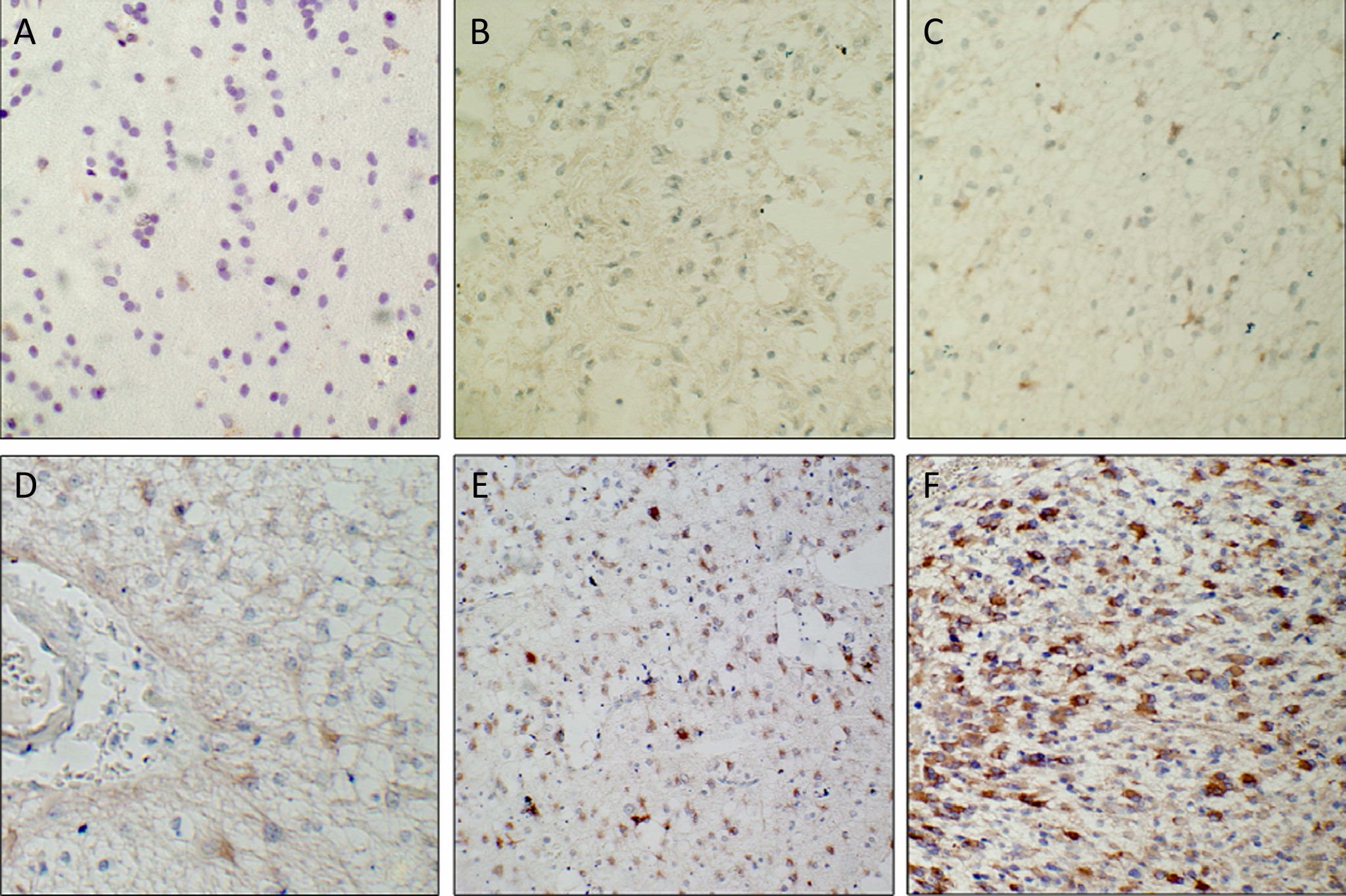
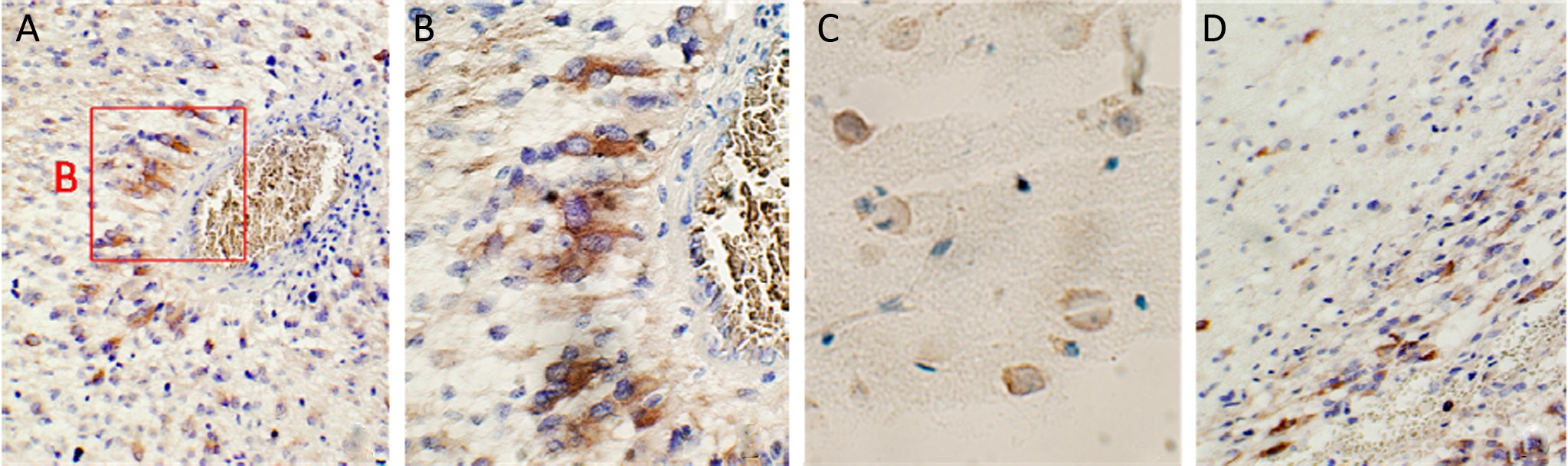
ObjectiveTo investigate the utility of hMena, a family of enabled/vasodilator-stimulated phosphoprotein (Ena/VASP), we sought to characterize the expression profile and distribution characteristics of hMena in a large panel of glioma samples and determine whether hMena expression levels might correlate with the pathological grade of glioma. MethodsSixty-five specimens of glioma with different pathological grades and five control brain tissues were collected. In 6 of the 21 glioblastoma patients, multi-specimens were obtained respectively from the main tumor mass, the junction zone between the tumor and the normal tissue, and adjacent brain tissue 1.5 cm away from the tumor boundary under assistance of neuronavigation system during the operation. Immunohistochemistry was used to detect the expression and distribution characteristics of hMena. hMena expression was analyzed by Western blot in 20 specimens. ResultsThe hMena expression was negative in control brain tissue but positive in different grades of glioma. The expression rate of hMena was positively correlated with the increasing grade of the World Health Orgnization (WHO) classification (rs=0.682, P=0.000). hMena was located in cytoplasm. Positive cells only distributed around the vessels within the tumor mass in low grade glioma, while in high grade glioma, these cells were able to be detected not only in the tumor but also in the boundary zone and adjacent brain parenchyma. In the tumor mass, hMena expressed highly and diffusedly. In the junction zone, hMena positive cells formed radiolitic pattern around the vessels. In adjacent brain parenchyma, single positive cell was scattered. hMena expression was markedly elevated in Grade III and IV glioma compared with Grade II and I. ConclusionOur data suggested that the expression of hMena is closely related to malignant grade of glioma. hMena can label the migrating cells, and indicate the migrating path of glioma cells from the tumor to adjacent tissue along with the vascular basement membranes and tracts of white matter.

2011, 23(4): 317-322.
doi: 10.1007/s11670-011-0317-7
Abstract:
ObjectiveAlthough a new matrix formulation fentanyl has been used throughout the world for cancer pain management, few data about its efficacy and clinical outcomes associated with its use in Chinese patients have been obtained. This study aimed to assess the efficacy and safety of the new system in Chinese patients with moderate to severe cancer pain. MethodsA total of 474 patients with moderate to severe cancer pain were enrolled in this study and were treated with the new transdermal fentanyl matrix patch (TDF) up to 2 weeks. All the patients were asked to record pain intensity, side effects, quality of life (QOL), adherence and global satisfaction. The initial dose of fentanyl was 25 μg/h titrated with opioid or according to National Comprehensive Cancer Network (NCCN) guidelines. Transdermal fentanyl was changed every three days. ResultsAfter 2 weeks. The mean pain intensity of the 459 evaluated patients decreased significantly from 5.63±1.26 to 2.03±1.46 (P<0.0001). The total remission rate was 91.29%, of which moderate remission rate 53.16%, obvious remission rate 25.49% and complete remission rate 12.64%. The rate of adverse events was 33.75%, 18.78% of which were moderate and 3.80% were severe. The most frequent adverse events were constipation and nausea. No fatal events were observed. The quality of life was remarkably improved after the treatment (P<0.0001). ConclusionThe new TDF is effective and safe in treating patients with moderate to severe cancer pain, and can significantly improve the quality of life.

2011, 23(4): 323-323.
doi: 10.1007/s11670-011-0323-9
Abstract:
2011, 23(4): 324-324.
doi: 10.1007/s11670-011-0324-8
Abstract: