2012 Vol.24(2)
Display Mode: |
2012, 24(2): 83-89.
doi: 10.1007/s11670-012-0083-1
 Abstract
 FullText HTML
 PDF 493KB
Abstract:
To treat many types of cancer, ionizing radiation (IR) is primarily used as external-beam radiotherapy, brachytherapy, and targeted radionuclide therapy. Exposure of tumor cells to IR can induce DNA damage as well as generation of reactive oxygen species (ROS) and reactive nitrogen species (RNS) which can cause non-DNA lesions or extracellular damage like lipid perioxidation. The initial radiation-induced cell responses to DNA damage and ROS like the proteolytic processing, as well as synthesis and releasing ligands (such as growth factors, cytokines, and hormone) can cause the delayed secondary responses in irradiated and unirradiated bystander cells through paracrine and autocrine pathways.
To treat many types of cancer, ionizing radiation (IR) is primarily used as external-beam radiotherapy, brachytherapy, and targeted radionuclide therapy. Exposure of tumor cells to IR can induce DNA damage as well as generation of reactive oxygen species (ROS) and reactive nitrogen species (RNS) which can cause non-DNA lesions or extracellular damage like lipid perioxidation. The initial radiation-induced cell responses to DNA damage and ROS like the proteolytic processing, as well as synthesis and releasing ligands (such as growth factors, cytokines, and hormone) can cause the delayed secondary responses in irradiated and unirradiated bystander cells through paracrine and autocrine pathways.
2012, 24(2): 90-96.
doi: 10.1007/s11670-012-0090-2
Abstract:
Lymph node status is a key prognostic factor in penile squamous cell carcinoma. Recently, growing evidence indicates a multimodality approach consisting of neoadjuvant chemotherapy followed by consolidation surgery improves the outcome of locally advanced penile cancer. Thus, accurate estimation of survival probability in node-positive penile cancer is critical for treatment decision making, counseling of patients and follow-up scheduling. This article reviewed evolving developments in assessing the risk for cancer progression based on lymph node related variables, such as the number of metastatic lymph nodes, bilateral lymph node metastases, the ratio of positive lymph nodes, extracapsular extension of metastatic lymph nodes, pelvic lymph node metastases, metastatic deposit in sentinel lymph nodes and N stage in TNM classification. Controversial issues surrounding the prognostic value of these nodal related predictors were also discussed.
Lymph node status is a key prognostic factor in penile squamous cell carcinoma. Recently, growing evidence indicates a multimodality approach consisting of neoadjuvant chemotherapy followed by consolidation surgery improves the outcome of locally advanced penile cancer. Thus, accurate estimation of survival probability in node-positive penile cancer is critical for treatment decision making, counseling of patients and follow-up scheduling. This article reviewed evolving developments in assessing the risk for cancer progression based on lymph node related variables, such as the number of metastatic lymph nodes, bilateral lymph node metastases, the ratio of positive lymph nodes, extracapsular extension of metastatic lymph nodes, pelvic lymph node metastases, metastatic deposit in sentinel lymph nodes and N stage in TNM classification. Controversial issues surrounding the prognostic value of these nodal related predictors were also discussed.
2012, 24(2): 97-102.
doi: 10.1007/s11670-012-0097-8
Abstract:
ObjectiveTo evaluate the efficacy and safety of nedaplatin/gemcitabine (NG) and carboplatin/gemcitabine (CG) in the management of untreated advanced non-small cell lung cancer (NSCLC). MethodsSixty-two patients with previously untreated advanced NSCLC were recruited between June 2006 and November 2007. Subjects were randomly assigned to the NG arm (n=30) and the CG arm (n=32). Only patients (24 and 25 in the NG and CG arms, respectively) who completed ≥2 chemotherapy cycles were included in the data analysis. The primary outcome measure was the objective response rate (ORR). The secondary outcome measures included progression-free survival (PFS), overall survival (OS) and adverse events. ResultsThere were no statistically significant differences in the efficacy measures (ORR, P=0.305; median PFS, P=0.198; median OS, P=0.961) or in the major adverse events (grade 3/4 neutropenia, P=0.666; grade 3/4 anemia, P=0.263; grade 3/4 thrombocytopenia, P=0.212) between the two treatment arms. However, there was a trend towards higher ORR (37.5% vs. 24.0%), longer PFS (6.0 vs. 5.0 months), and less adverse events in the NG arm. ConclusionNG regimen seems to be superior over CG regimen for advance NSCLS, but further investigation is needed to validate this superiority.


2012, 24(2): 103-108.
doi: 10.1007/s11670-012-0103-1
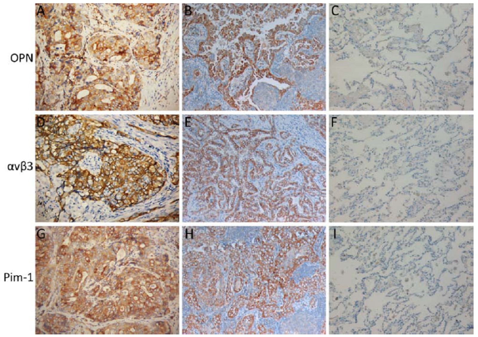
Abstract:
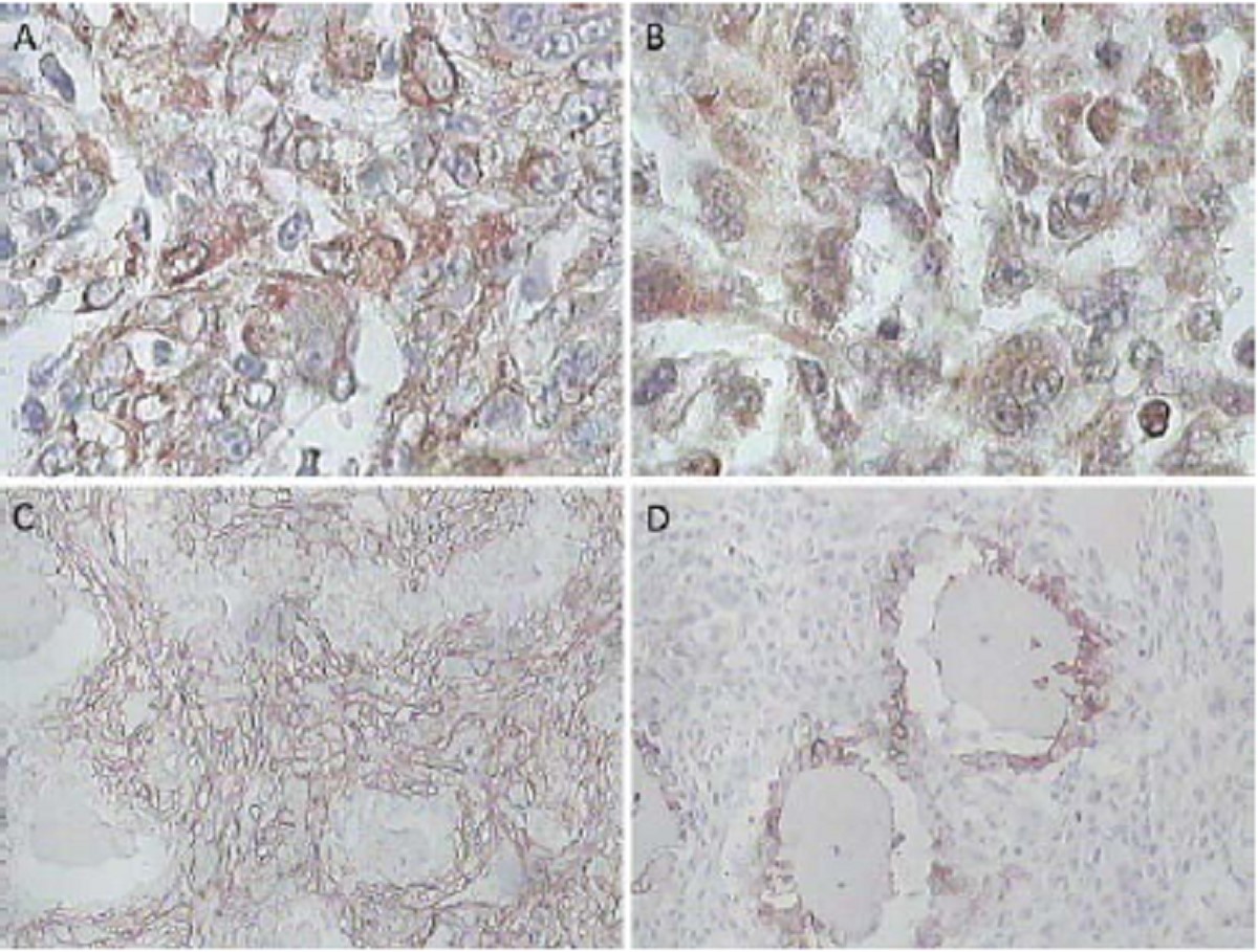
ObjectiveTo examine the expressions of osteopontin (OPN), α ν β3 and Pim-1 in non-small cell lung cancer (NSCLC), and investigate their potential pathogenic roles in the development of NSCLC. MethodsImmunohistochemistry was used to examine the expressions of OPN, α ν β3 and Pim-1 in cohort (136 cases) of NSCLC samples and their adjacent normal lung tissue specimens. Statistical analysis was performed to evaluate the relationships among expressions of OPN, α ν β3 and Pim-1 and their associations with patients clinico- pathological parameters. ResultsThe expressions of OPN and Pim-1 were predominantly observed in cytoplasm. The expression of α ν β3 was mostly detected in cytoplasm and/or membrane. In NSCLC samples, the positive rates of OPN, α ν β3 and Pim-1 expressions were 68.4% (93/136), 77.2% (105/136) and 57.4% (78/136), respectively. In normal lung tissues, in contrast, the positive rates of OPN, α ν β3 and Pim-1 were 24.0% (12/50), 26.0% (13/50) and 16.0% (8/50), respectively. There were significant differences of the positive expression rates of OPN, α ν β3 and Pim-1 between NSCLCs samples and normal lung tissues (P<0.01). In addition, the positive expression of OPN, α ν β3 and Pim-1 in NSCLCs samples was significantly associated with increased pathological grade, lymph node metastasis and advanced clinical stage (P<0.01), and they were independent of other clinicopathological parameters (P>0.05). Furthermore, a significantly positive correlation between the expression of OPN and α ν β3 (r=0.38, P<0.01), OPN and Pim-1 (r=0.37, P<0.01), or α ν β3 and Pim-1 (r=0.20, P<0.05) was evaluated in our NSCLC cohort. ConclusionOPN, α ν β3 and Pim-1 proteins are frequently overexpressed in NSCLC, and they may play important roles in the development and/or progression of NSCLC.
2012, 24(2): 109-115.
doi: 10.1007/s11670-012-0109-8
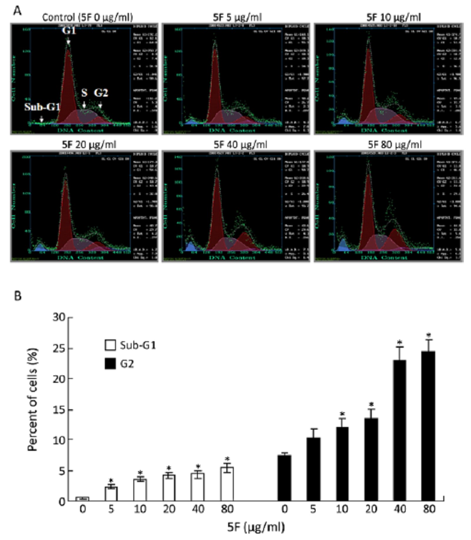
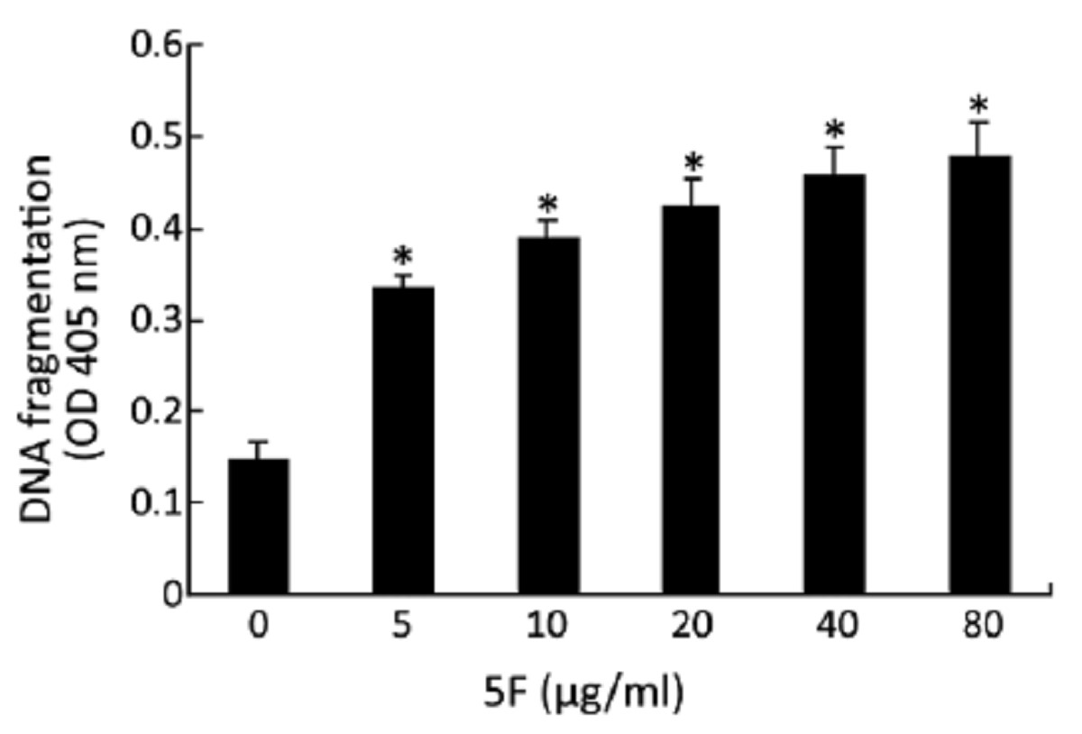
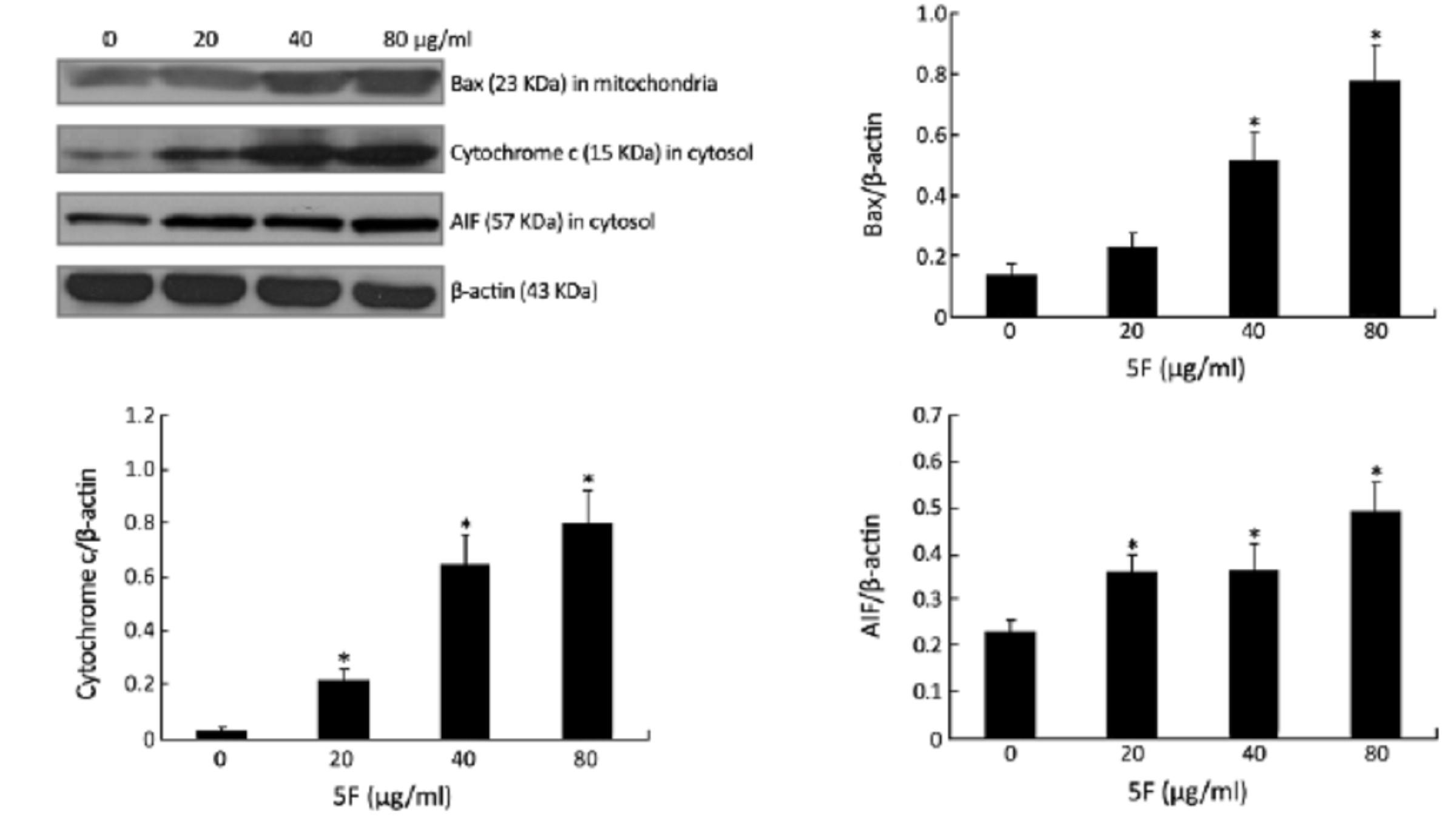
Abstract:
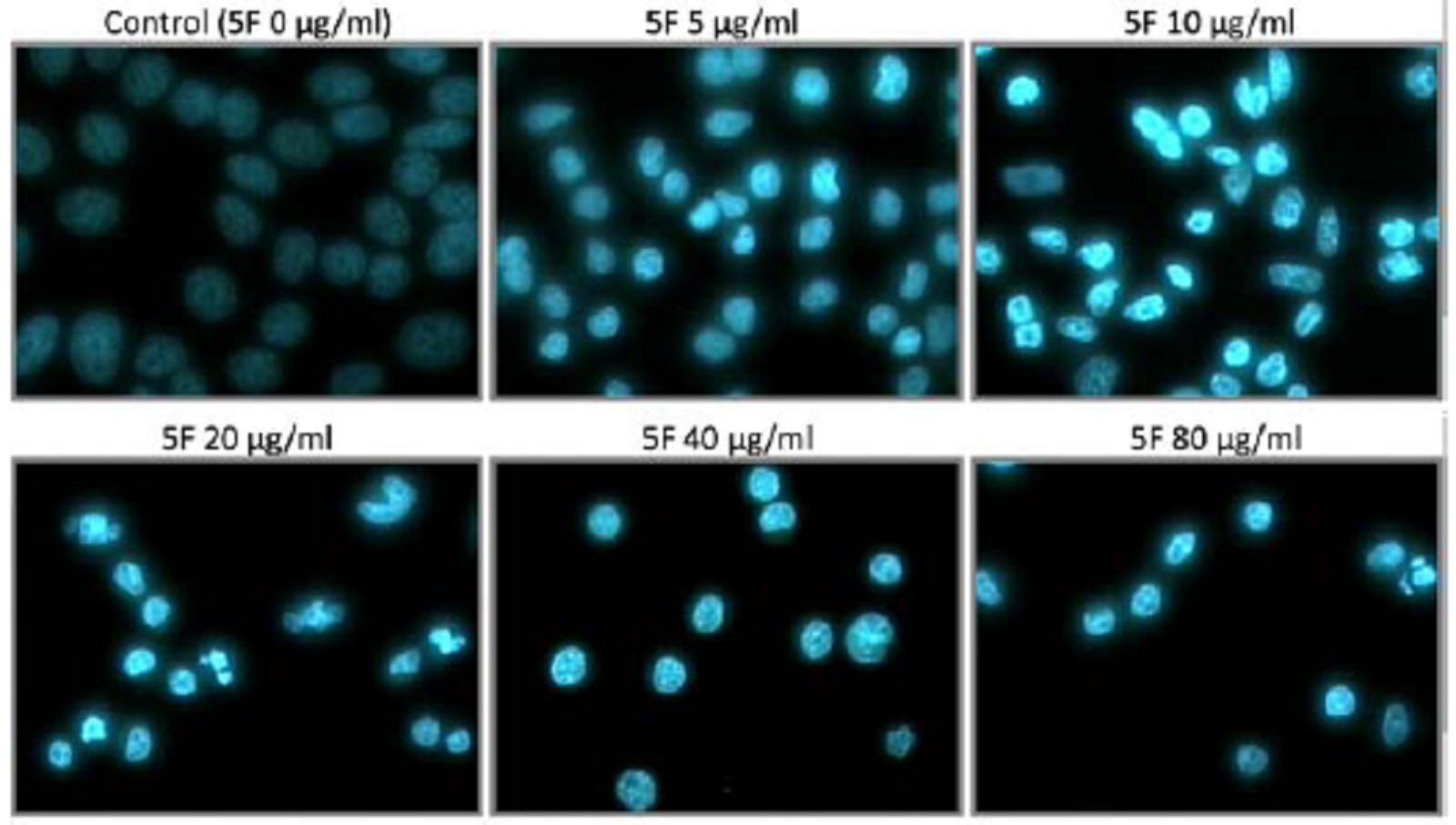
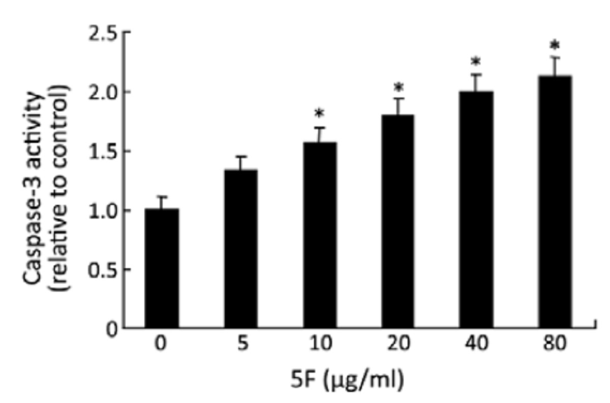
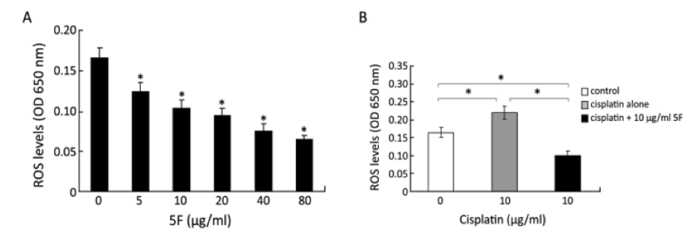
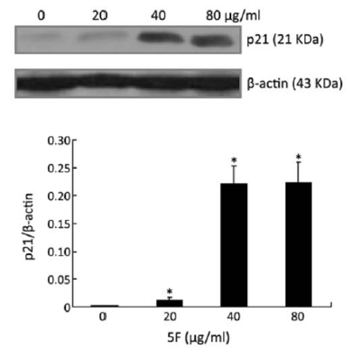
ObjectiveTo examine the apoptotic effect of ent-11α-hydroxy-15-oxo-kaur-16-en-19-oic-acid (5F), a compound isolated from Pteris semipinnata L (PsL), in human lung cancer A549 cells. MethodsA549 cells were treated with 5F (0–80 μg/ml) for different time periods. Cytotoxicity was examined using a MTT method. Cell cycle was examined using propidium iodide staining. Apoptosis was examined using Hoechst 33258 staining, enzyme-linked immunosorbent assay (ELISA) and caspase-3 activity analysis. Expression of representative apoptosis-related proteins was evaluated by Western blot analysis. Reactive oxygen species (ROS) level was measured using standard protocols. Potential interaction of 5F with cisplatin was also examined. Results5F inhibited the proliferation of A549 cells in a concentration- and time-dependent manner. 5F increased the accumulation of cells in sub-G1 phase and arrested the cells in the G2 phase. Exposure to 5F induced morphological changes and DNA fragmentation that are characteristic of apoptosis. The expression of p21 was increased. 5F exposure also increased Bax expression, release of cytochrome c and apoptosis inducing factor (AIF), and activation of caspase-3. 5F significantly sensitized the cells to cisplatin toxicity. Interestingly, treatment with 5F did not increase ROS, but reduced ROS production induced by cisplatin. Conclusion5F could inhibit the proliferation of A549 cells by arresting the cells in G2 phase and by inducing mitochondrial-mediated apoptosis.

2012, 24(2): 116-123.
doi: 10.1007/s11670-012-0116-9
Abstract:
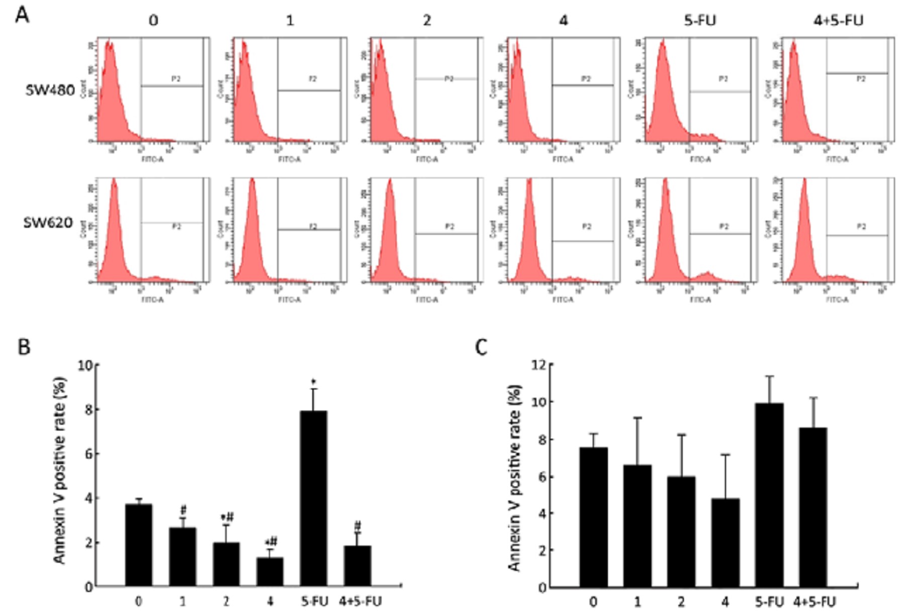
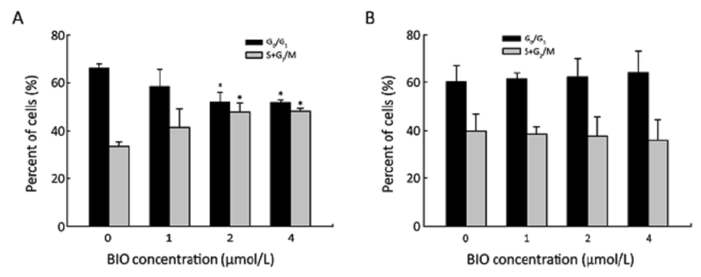
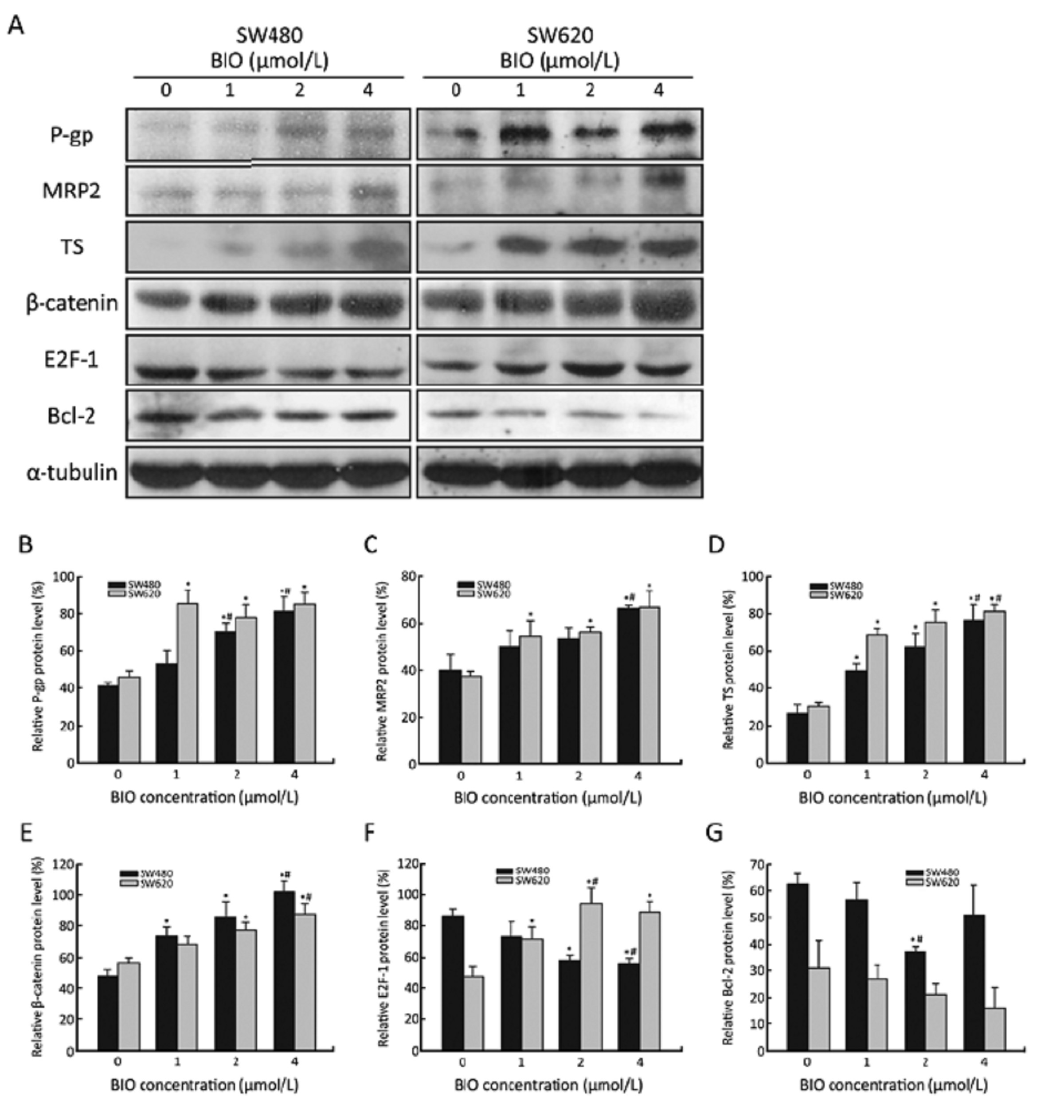
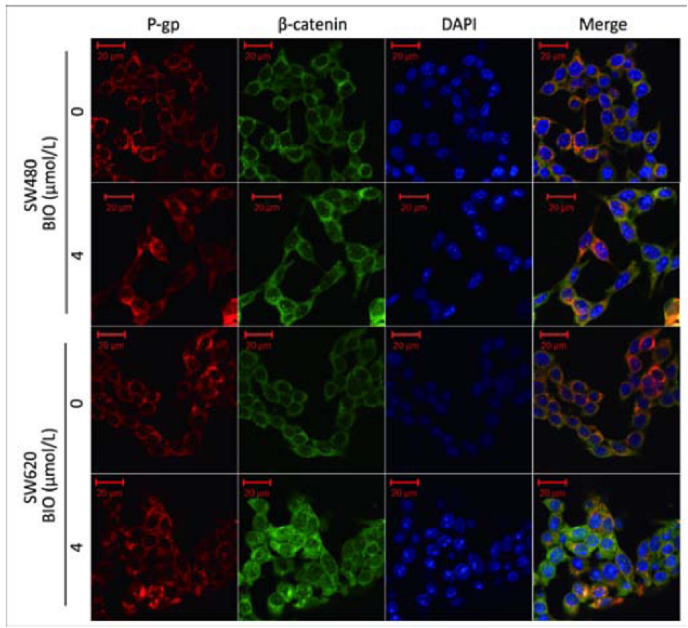
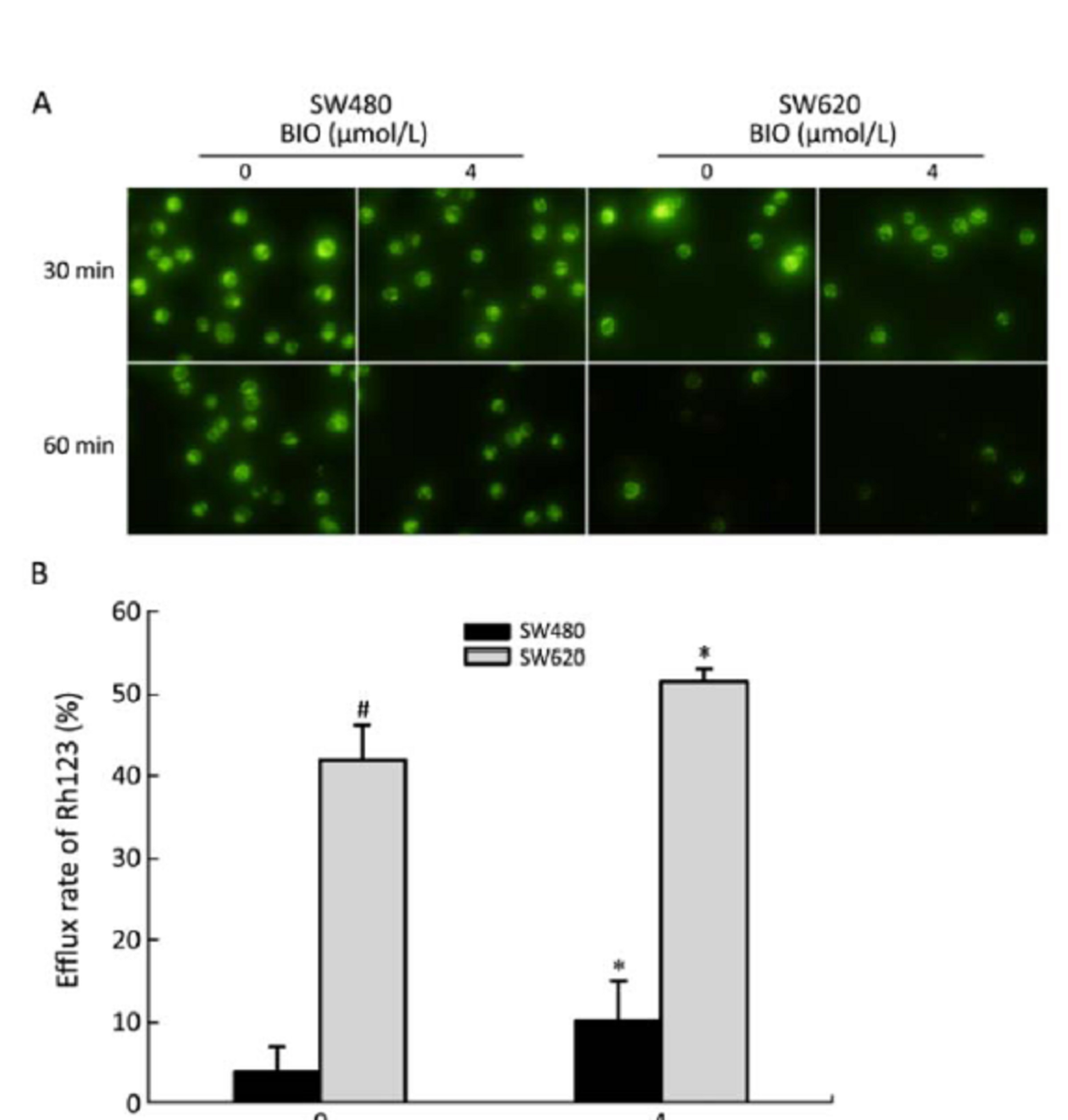
ObjectiveTo explore the effects and mechanism of glycogen synthase kinase 3β (GSK-3β) inhibitor (2'Z,3'E)-6-bromo-indirubin-3'-oxime (BIO) on drug resistance in colon cancer cells. MethodsThe colon cancer SW480 and SW620 cells were treated with BIO, 5-fluorouracil (5-FU) and BIO/5-FU, separately. Cell cycle distribution, apoptosis level and efflux ability of rhodamine 123 (Rh123) were detected by flow cytometry. The protein expressions of P-glycoprotein (P-gp), multidrug resistance protein 2 (MRP2), thymidylate synthase (TS), β-catenin, E2F-1 and Bcl-2 were detected by Western blot. β-catenin and P-gp were stained with double immunofluorescence and observed under a confocal microscope. ResultsBIO up-regulated β-catenin, P-gp, MRP2 and TS, enhanced the efflux ability of Rh123, decreased Bcl-2 protein and gave the opposite effect to E2F-1 protein in SW480 and SW620 cells. Furthermore, BIO significantly inhibited cell apoptosis, increased S and G2/M phase cells, and reduced the cell apoptosis induced by 5-FU in SW480 cells, whereas the effects were slight or not obvious in SW620 cells. ConclusionGSK-3β was involved in drug resistance regulation, and activation of β-catenin and inhibition of E2F-1 may be the most responsible for the enhancement of 5-FU chemotherapy resistance induced by GSK-3β inhibitor BIO in colon cancer.
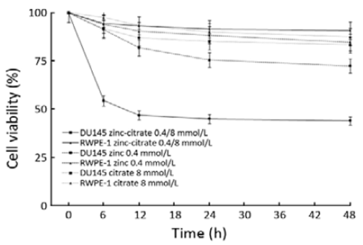
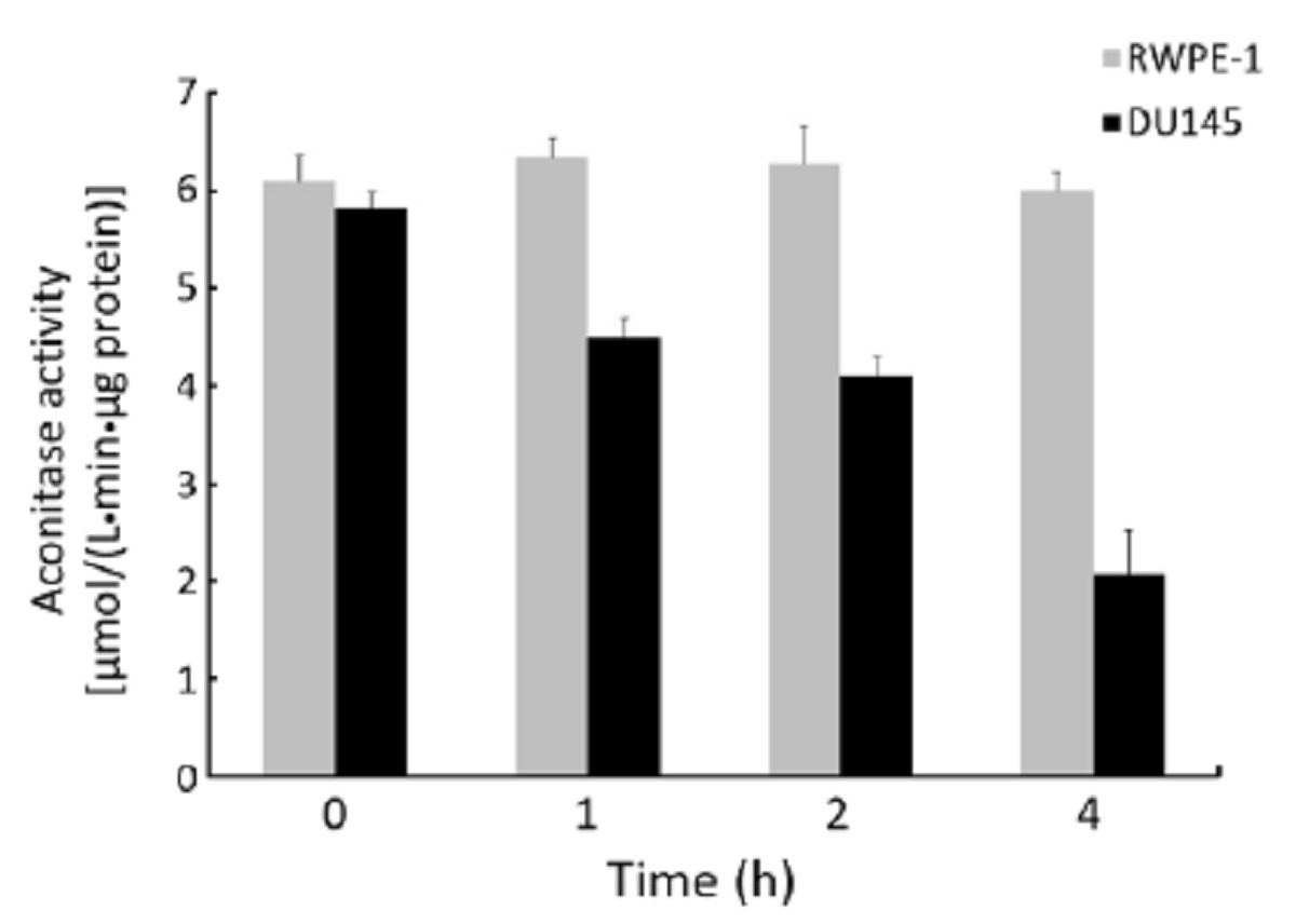
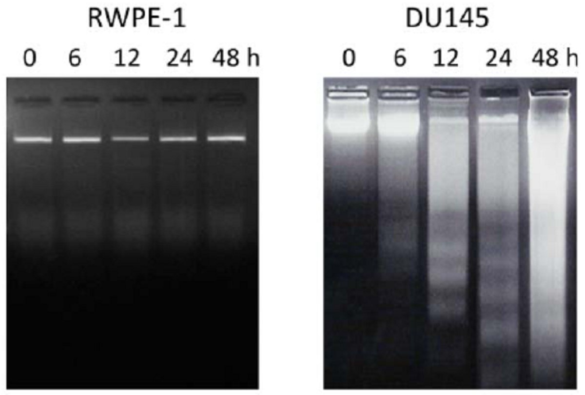
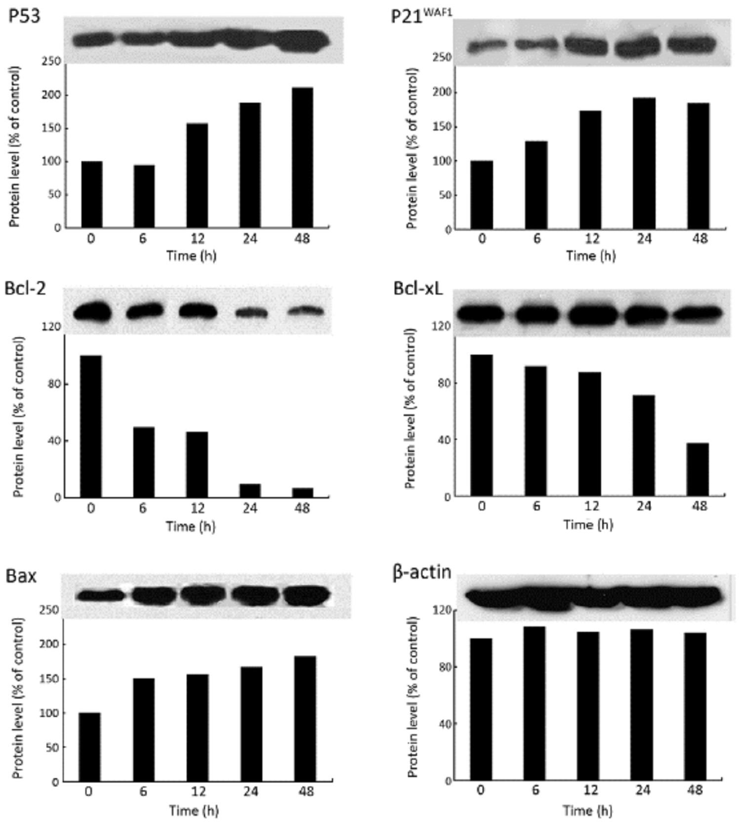
2012, 24(2): 124-129.
doi: 10.1007/s11670-012-0124-9
Abstract:
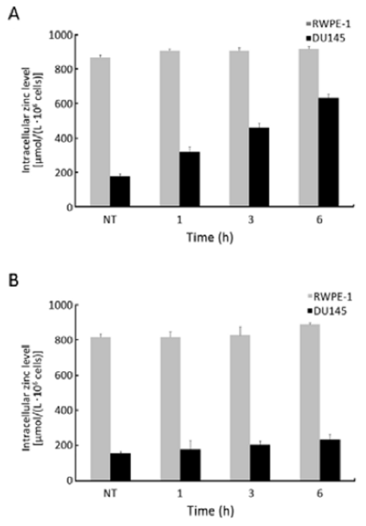
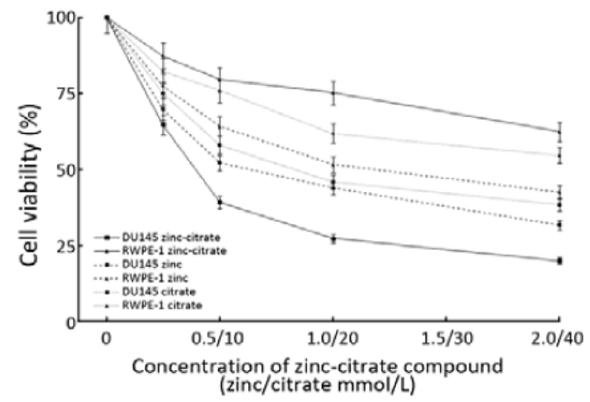
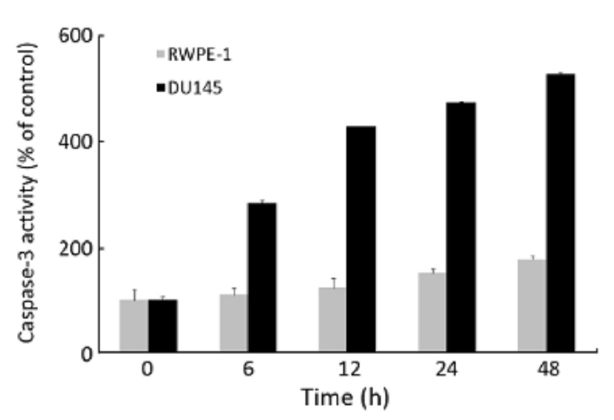
ObjectiveTo investigate the antiproliferative effects of zinc-citrate compound on hormone refractory prostate cancer (HRPC). MethodsHRPC cell line (DU145) and normal prostate cell line (RWPE-1) were treated with zinc, citrate and zinc-citrate compound at different time intervals and concentrations to investigate the effect of zinc-citrate compound. Mitochondrial (m)-aconitase activity was determined using aconitase assay. DNA laddering analysis was performed to investigate apoptosis of DU145 cells. Molecular mechanism of apoptosis was investigated by Western blot analysis of P53, P21waf1, Bcl-2, Bcl-xL and Bax, and also caspase-3 activity analysis. ResultsTreatment with zinc-citrate compound resulted in a time- and dose-dependent decrease in cell number of DU145 cells in comparison with RWPE-1. M-aconitase activity was significantly decreased. DNA laddering analysis indicated apoptosis of DU145 cells. Zinc-citrate compound increased the expression of P21waf1 and P53, and reduced the expression of Bcl-2 and Bcl-xL proteins but induced the expression of Bax protein. Zinc-citrate compound induced apoptosis of DU145 cells by activation of the caspase-3 pathway. ConclusionZinc-citrate compound can induce apoptotic cell death in DU145, by caspase-3 activation through up-regulation of apoptotic proteins and down-regulation of antiapoptotic proteins.
2012, 24(2): 130-137.
doi: 10.1007/s11670-012-0130-y
Abstract:
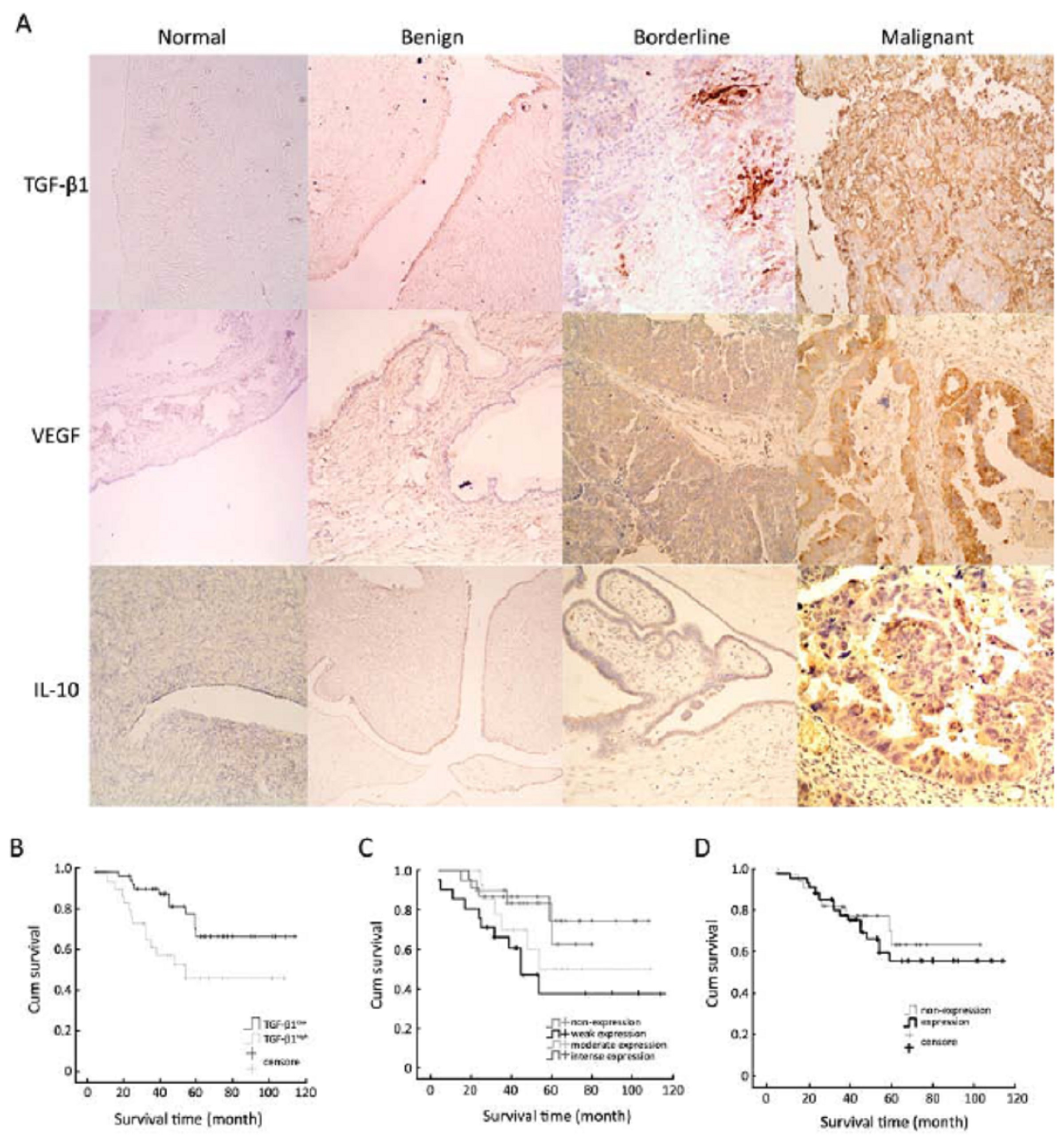
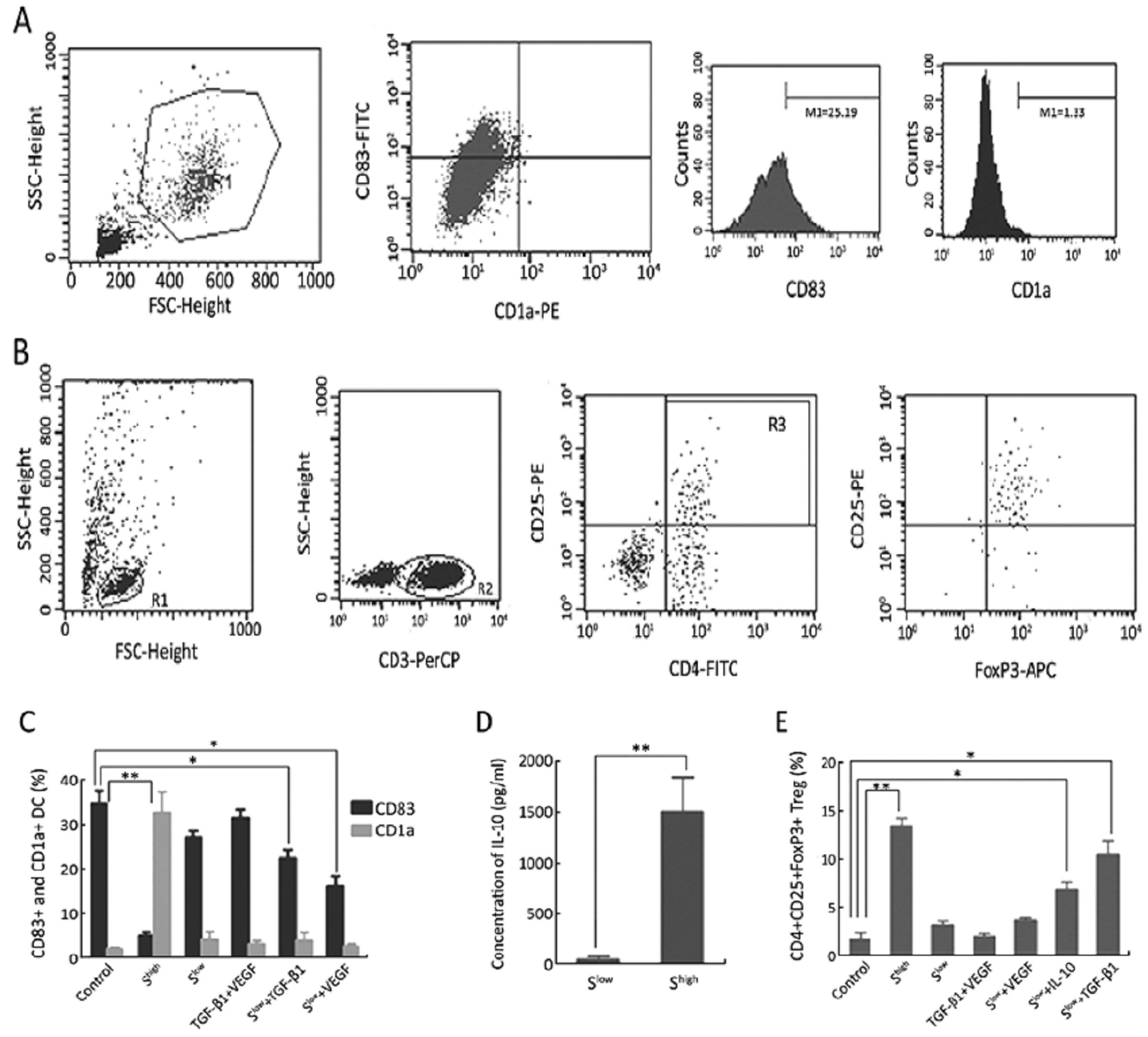
ObjectiveTransforming growth factor-1 (TGF-β1), vascular endothelial growth factor (VEGF), and interleukin-10 (IL-10) may be critical cytokines in the microenvironment of a tumor, playing roles in immune suppression. This study was conducted to elucidate the roles and immunosuppressive functions of these cytokines in epithelial ovarian cancer (EOC). MethodsThe expression levels of TGF-β1, VEGF and IL-10 in malignant tissue were evaluated by immune- histochemistry and compared with corresponding borderline, benign, and tumor-free tissues. Moreover, relationships among the levels of these cytokines and correlations between expression and the prognosis of EOC were analyzed by Pearson rank correlations and multi-factor Logistic regression. The roles of TGF-β1, VEGF, and IL-10 in the immunosuppressive microenvironment of ovarian cancer were studied through dendritic cell (DC) maturation and CD4+CD25+FoxP3+ Treg generation in vitro experiments. ResultsTGF-β1, VEGF, and IL-10 were expressed in 100%, 74.69%, and 54.96% of EOC patients, respectively. TGF-β1 was an independent prognostic factor for EOC. IL-10 was significantly co-expressed with VEGF. In vitro, VEGF and TGF-β1 strongly interfered with DC maturation and consequently led to immature DCs, which secreted high levels of IL-10 that accumulated around the tumor site. TGF-β1 and IL-10 induced Treg generation without antigen presentation in DCs. ConclusionsTGF-β1, VEGF and IL-10 play important roles in EOC and can lead to frequent immune evasion events.
2012, 24(2): 138-142.
doi: 10.1007/s11670-012-0138-3
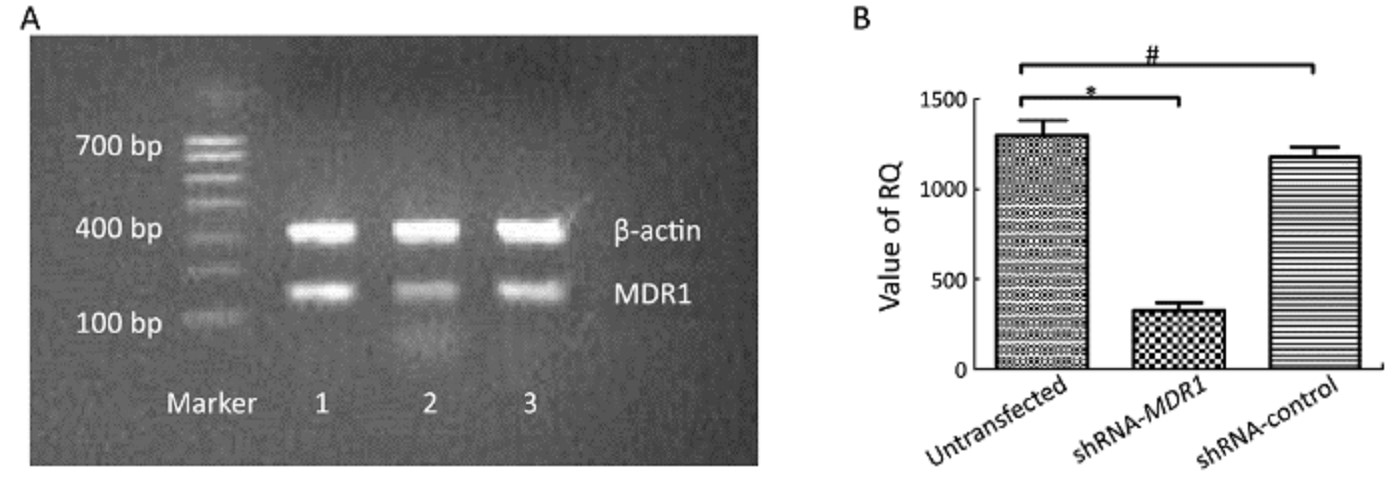
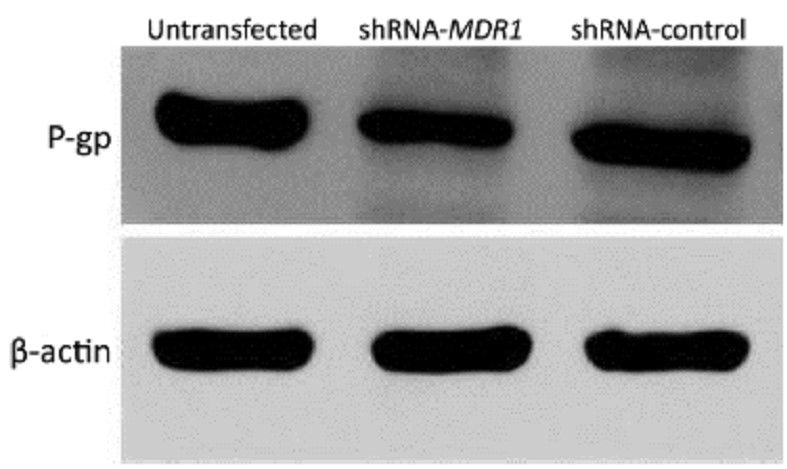
Abstract:
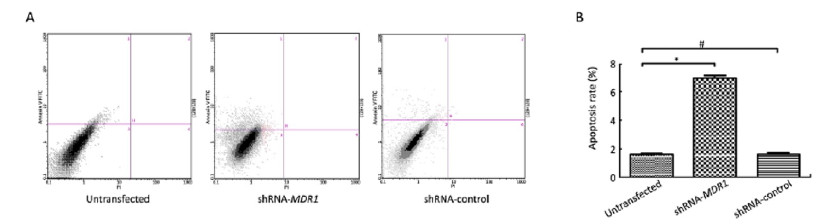
ObjectiveRecurrent ovarian cancer is often resistant to drugs such as paclitaxel. Short hairpin RNA (shRNA) targeting MDR1, a gene involved in the process of drug resistance, may be a promising strategy to overcome drug resistance. MethodsConstruction and identification of eukaryotic expression plasmid of shRNA targeting on MDR1 gene. The plasmid was transiently transfected into human ovarian cancer cell line A2780/Taxol. Apoptosis was determined by flow cytometry using annexin V-FITC/PI double labeling. Expression of MDR1 mRNA was detected by quantitative polymerase chain reaction (qPCR) and P-glycoprotein expression was detected using Western blot. ResultsThe IC50 of paclitaxel in MDR1shRNA-transfected group was significantly reduced (1.986±0.153) μmol/ml as compared with that in negative control (5.246±0.107) μmol/ml and empty vector-transfected group (5.212±0.075) μmol/ml (P<0.05). The percent of the relative reverse sensitivity to paclitaxel on A2780/Taxol cells was 67.1%, and the apoptotic rate was significantly increased [(6.977±0.333)%] compared with control [(1.637±0.111)%] and empty vector-transfected group [(1.663±0.114)%] (P<0.05). Expressions of MDR1 mRNA and P-glycoprotein were significantly reduced compared with control (P<0.05). ConclusionThe present study demonstrated that the eukaryotic expression plasmid of shRNA targeting on MDR1 inhibited the expression of MDR1 effectively, thus enhance the sensitivity of A2780/Taxol cells to paclitaxel.
2012, 24(2): 143-150.
doi: 10.1007/s11670-012-0143-6
Abstract:
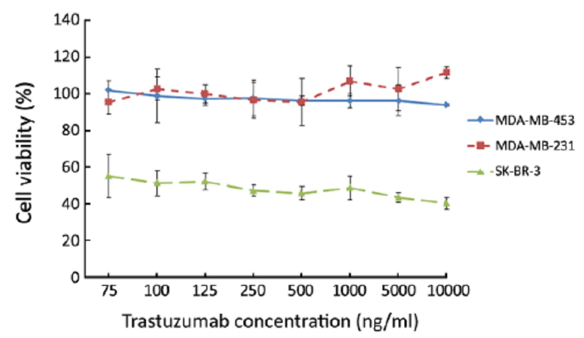
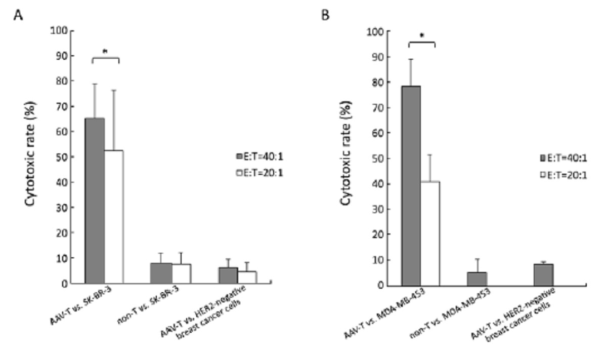
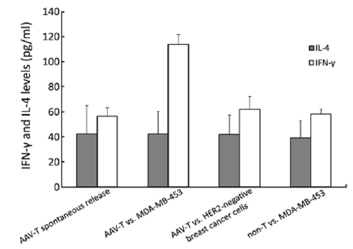
ObjectiveAlthough the development of trastuzumab has improved the outlook for women with human epidermal growth factor receptor 2 (HER2)-positive breast cancer, the resistance to anti-HER2 therapy is a growing clinical dilemma. We aim to determine whether HER2-specific T cells generated from dendritic cells (DCs) modified with HER2 gene could effectively kill the HER2-positive breast cancer cells, especially the trastuzumab-resistant cells. MethodsThe peripheral blood mononuclear cells (PBMCs) from healthy donors, whose HLA haplotypes were compatible with the tumor cell lines, were transfected with reconstructive human adeno-association virus (rhAAV/HER2) to obtain the specific killing activities of T cells, and were evaluated by lactate dehydrogenase (LDH) releasing assay. ResultsTrastuzumab produced a significant inhibiting effect on SK-BR-3, the IC50 was 100ng/ml. MDA-MB-453 was resistant to trastuzumab even at a concentration of 10,000 ng/ml in vitro. HER2-specific T lymphocytes killed effectively SK-BR-3 [(69.86±13.41)%] and MDA-MB-453 [(78.36±10.68)%] at 40:1 (effector:target ratio, E:T), but had no significant cytotoxicity against HER2-negative breast cancer cell lines MDA-MB-231 or MCF-7 (less than 10%). ConclusionThe study showed that HER2-specific T lymphocytes generated from DCs modified by rhAAV/HER2 could kill HER2-positive breast cancer cell lines in a HER2-dependent manner, and result in significantly high inhibition rates on the intrinsic trastuzumab-resistant cell line MDA-MB-453 and the tastuzumab-sensitive cell line SK-BR-3. These results imply that this immunotherapy might be a potential treatment to HER2-positive breast cancer.
2012, 24(2): 151-156.
doi: 10.1007/s11670-012-0151-6
Abstract:
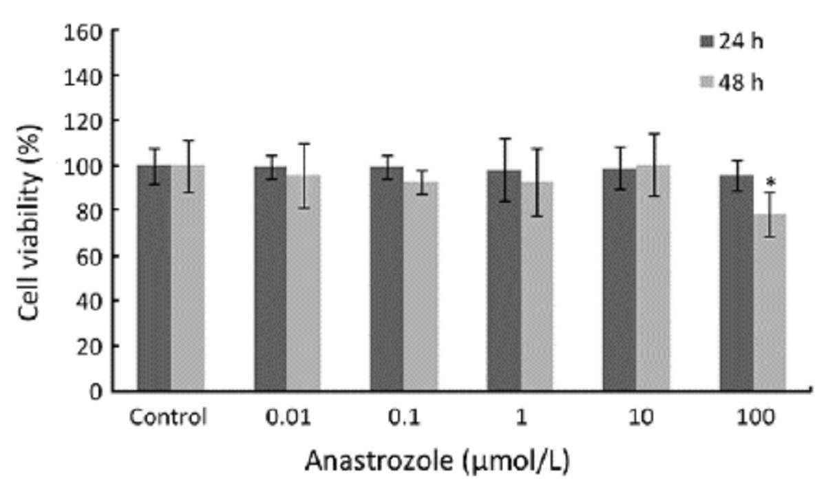
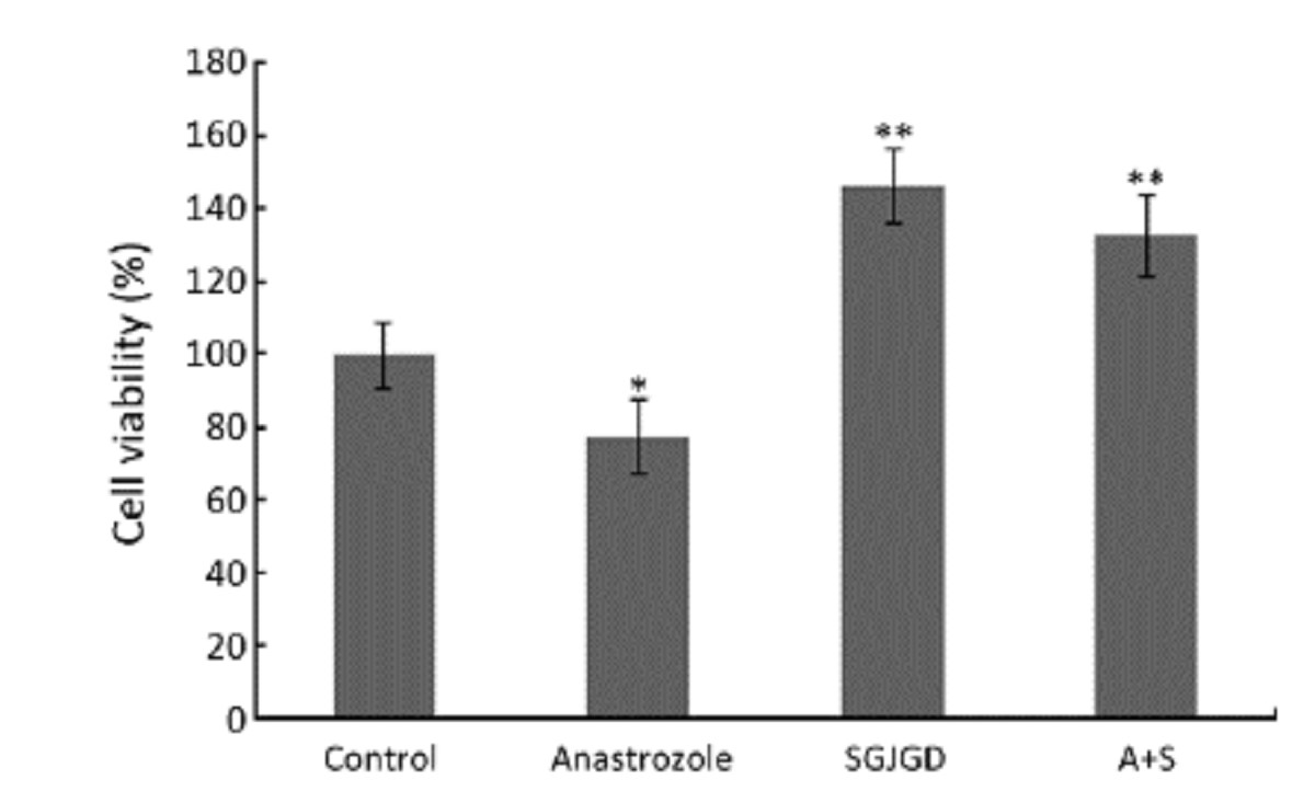
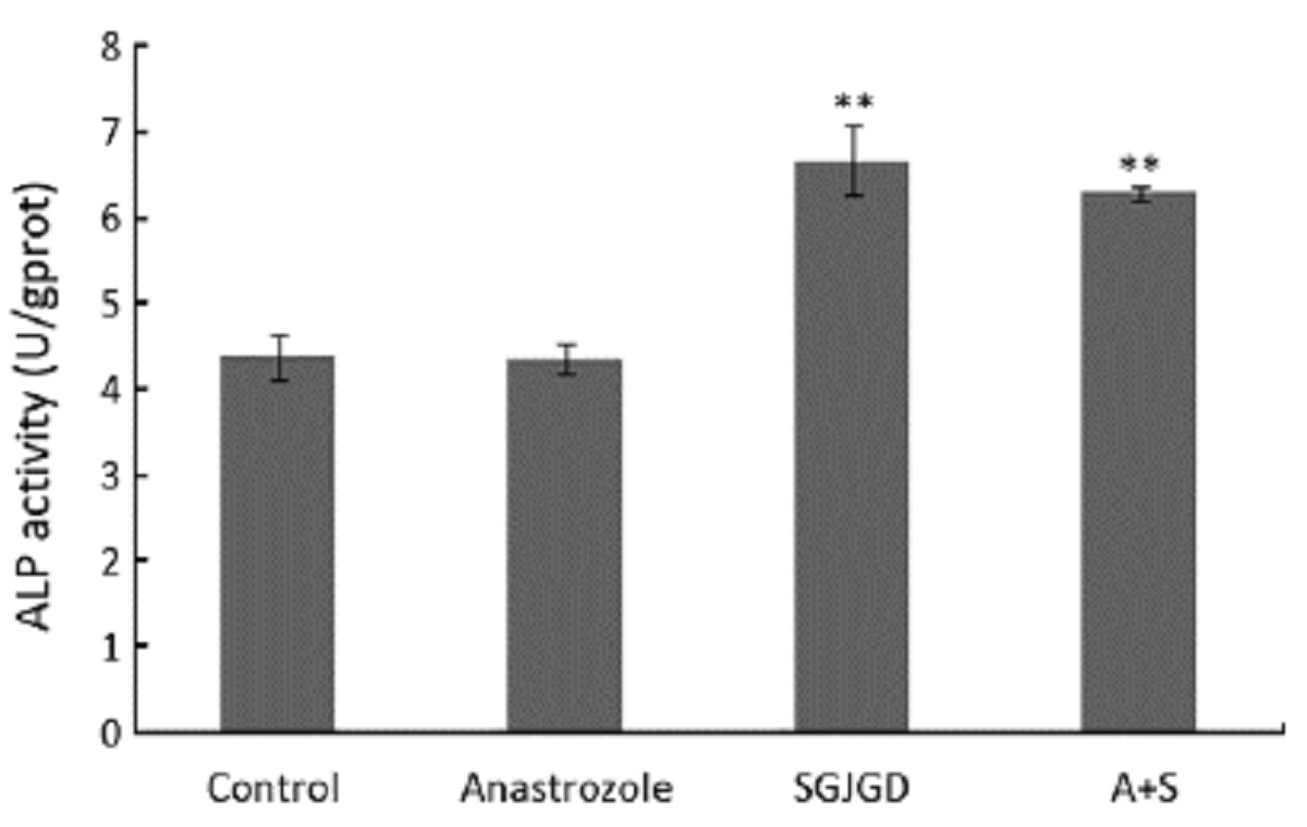
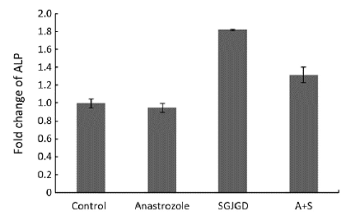
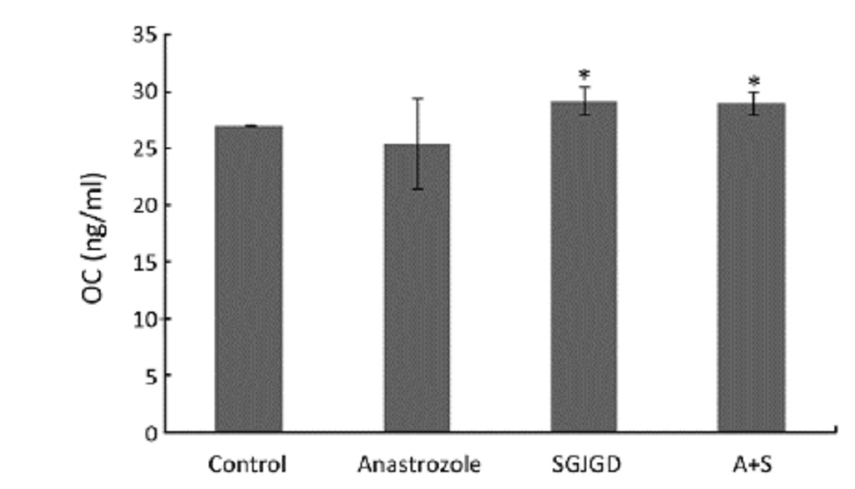
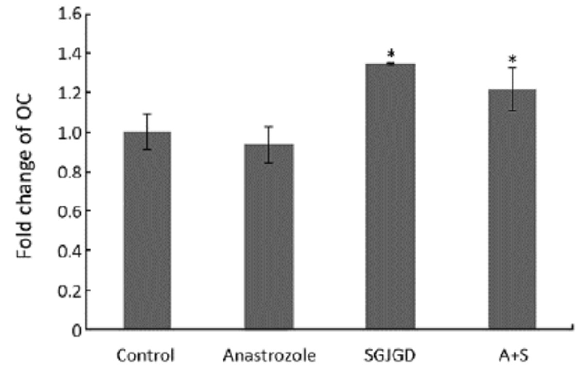
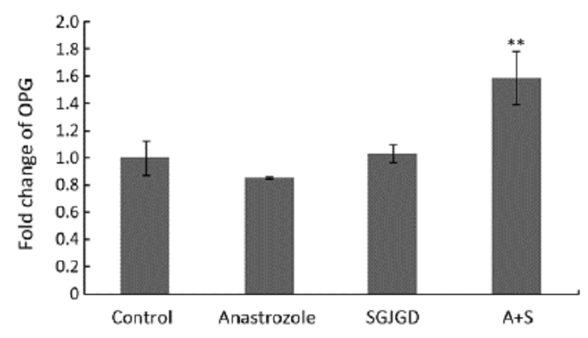
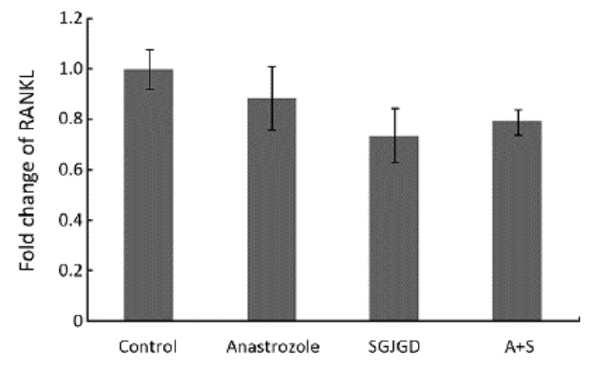
ObjectiveTo investigate the effects of anastrozole combined with Shuganjiangu decoction on osteoblast-like cells. MethodsHuman osteoblast-like cells MG-63 were cultured and divided into four groups: control, anastrozole, Shuganjiangu decoction (SGJGD), and anastrozole combined with SGJGD. Cell proliferation was investigated by MTT assay. Alkaline phosphatase (ALP) and osteocalcin, the indicators of cell differentiation, were evaluated by p-nitrophenyl- phosphate method and radioimmunoassay, respectively. Gene expressions of ALP, osteocalcin, osteoprotegerin (OPG) and receptor activator of nuclear factor kappa B ligand (RANKL) were examined by real-time PCR. ResultsAs evidenced by MTT assay, cell proliferation of MG-63 was inhibited by anastrozole, but stimulated with treatment of SGJGD alone and combined with anastrozole (P<0.01). Compared with control group, ALP activity was increased by the treatment of SGJGD alone and combined with anastrozole (P<0.01). Also, osteocalcin secretion was enhanced with the treatment of SGJGD single and combination with anastrozole (P<0.05). In the real-time PCR assay, gene expressions of ALP and osteocalcinwere significantly increased (P<0.01 for ALP, P<0.05 for osteocalcin) by the treatment of SGJGD and anastrozole combined with SGJGD, but the expression of RANKL was decreased (P<0.05). Moreover, anastrozole combined with SGJGD upregulated gene expression of OPG (P<0.01). ConclusionSGJGD may alleviate the injury effects of anastrozole on MG-63 cells through adjusting bone formation and resorption indicators.

2012, 24(2): 157-160.
doi: 10.1007/s11670-012-0157-0
Abstract:
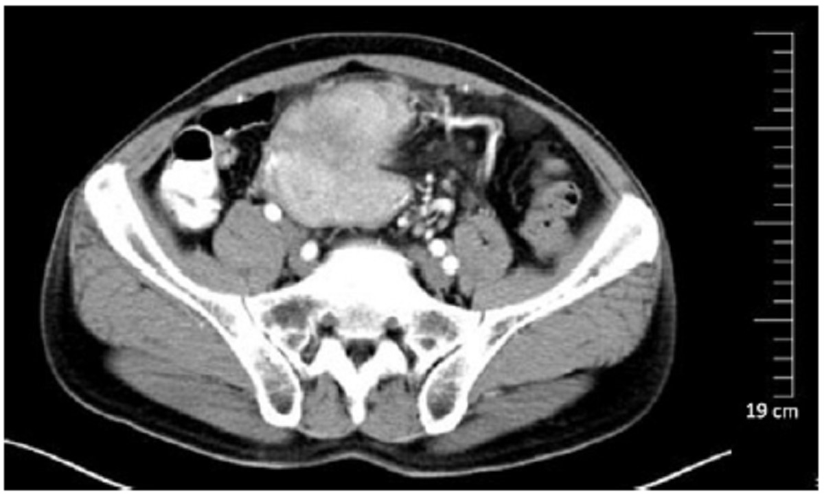
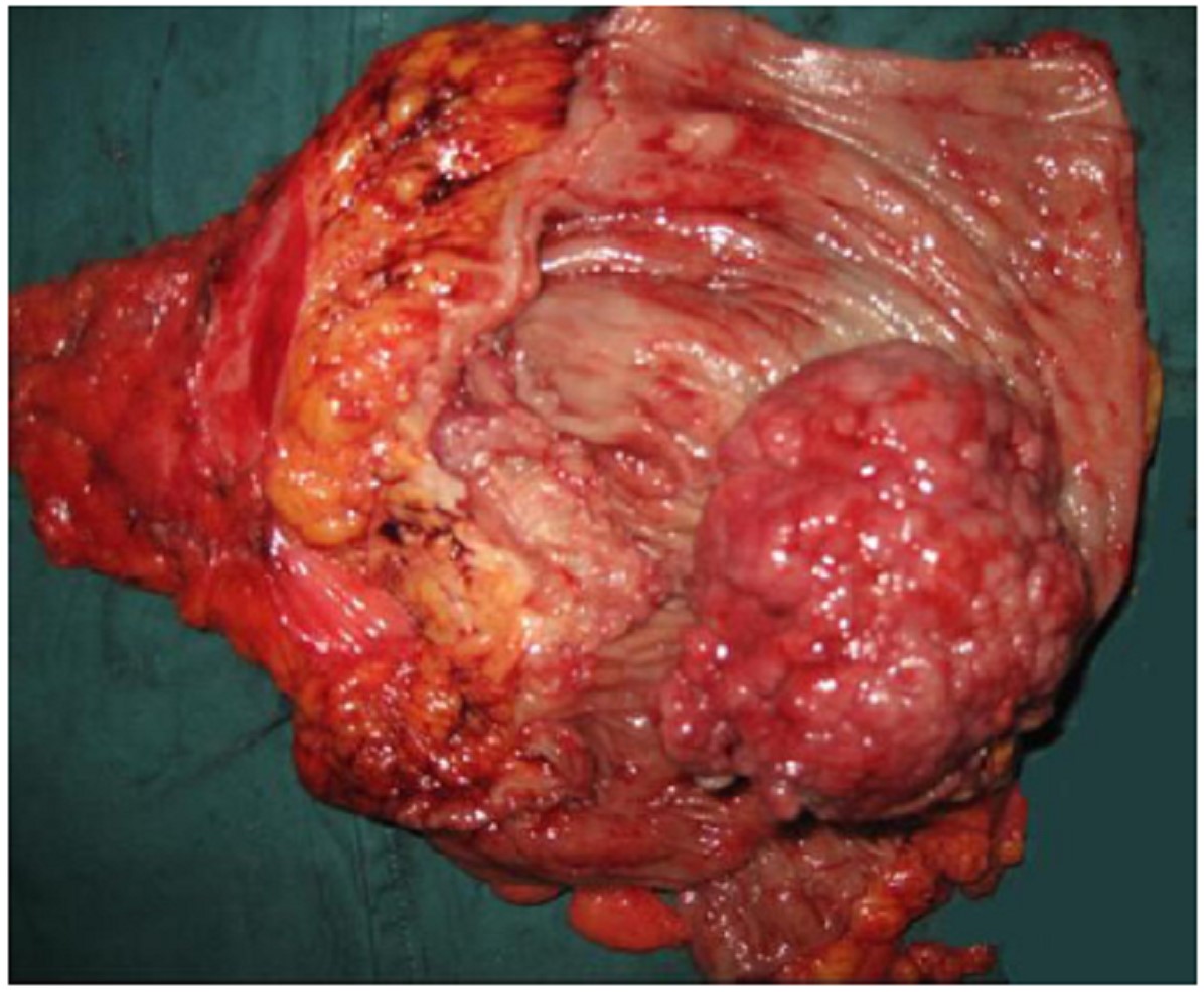
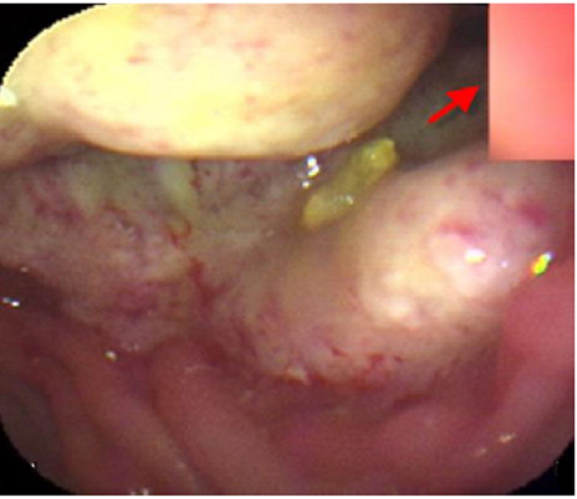
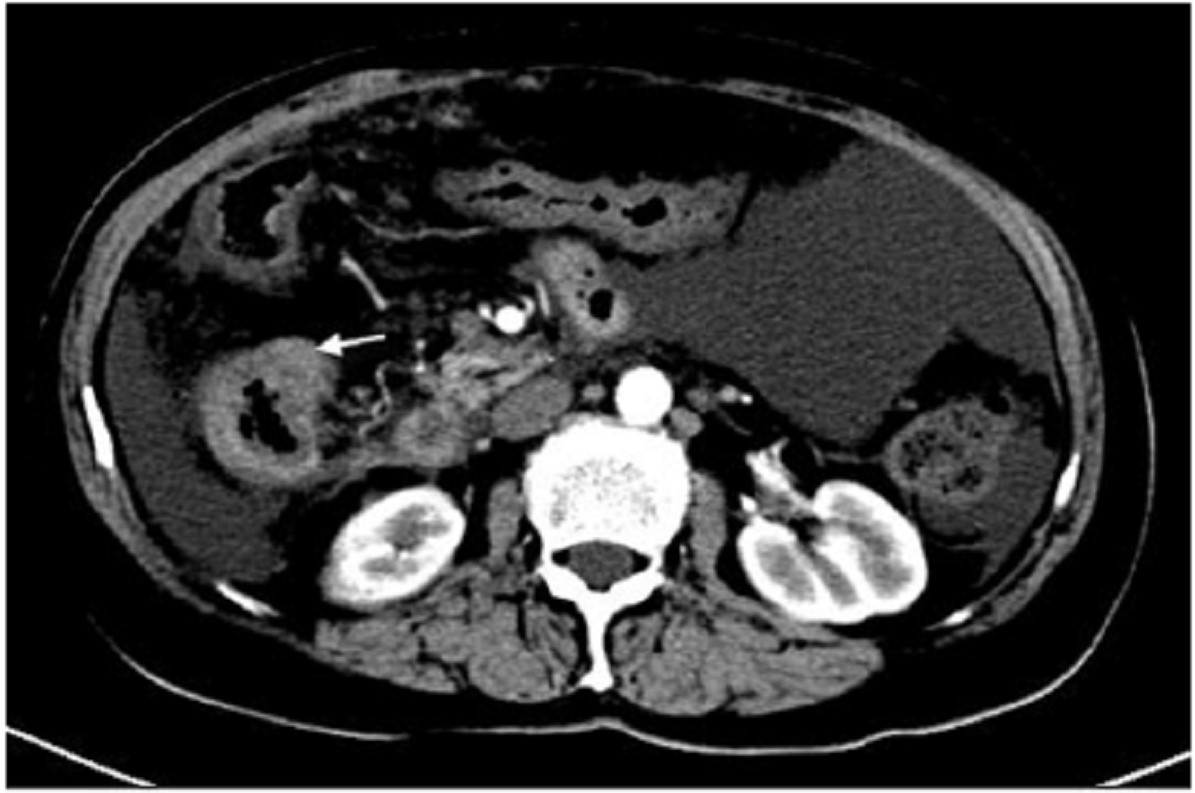
Fever of unknown origin (FUO) was originally defined as a body temperature greater than 38.3°C on several occasions longer than 3 weeks, with a diagnosis that remains unclear after 7 days of obligatory investigation. Only a few types of solid tumors have been associated with FUO. We described 2 patients who had recurrent fever but no other specific gastrointestinal symptoms where carcinoma of the colon was the only identifiable cause. In the first case, a mass arising from the sigmoid colon was found without any nodal metastasis, and the fever was resolved after three days of the surgical resection. In the second case, advanced adenocarcinoma was found in the ascending colon together with liver cirrhosis. Although it was not possible to surgically remove this tumor, prolonged fever in the patient was most likely due to the carcinoma. These cases indicate that clinicians should consider carcinoma of the colon in the differential diagnosis of patients with FUO.
Fever of unknown origin (FUO) was originally defined as a body temperature greater than 38.3°C on several occasions longer than 3 weeks, with a diagnosis that remains unclear after 7 days of obligatory investigation. Only a few types of solid tumors have been associated with FUO. We described 2 patients who had recurrent fever but no other specific gastrointestinal symptoms where carcinoma of the colon was the only identifiable cause. In the first case, a mass arising from the sigmoid colon was found without any nodal metastasis, and the fever was resolved after three days of the surgical resection. In the second case, advanced adenocarcinoma was found in the ascending colon together with liver cirrhosis. Although it was not possible to surgically remove this tumor, prolonged fever in the patient was most likely due to the carcinoma. These cases indicate that clinicians should consider carcinoma of the colon in the differential diagnosis of patients with FUO.
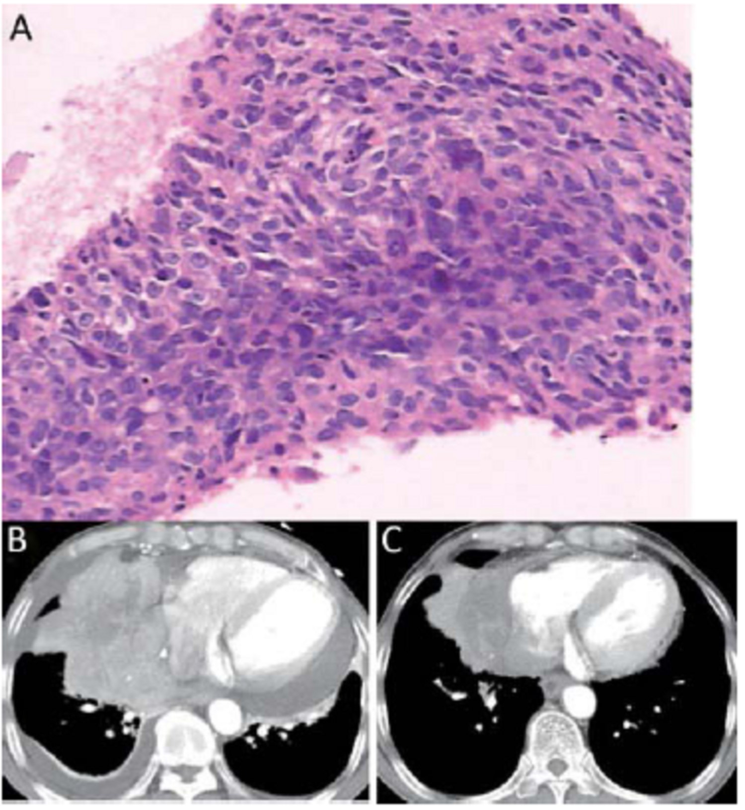
2012, 24(2): 161-163.
doi: 10.1007/s11670-012-0161-4
Abstract:
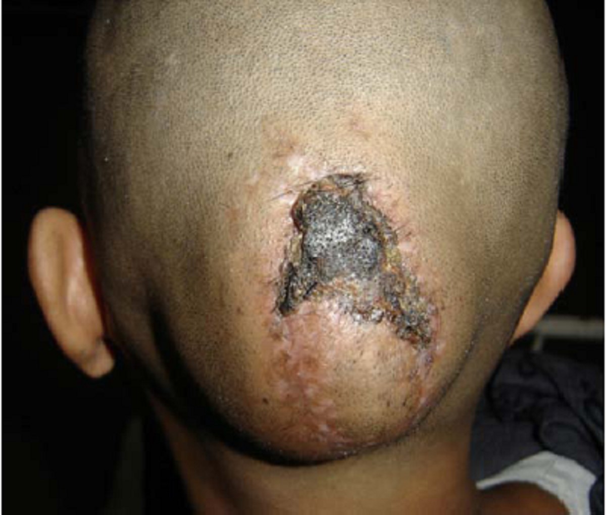
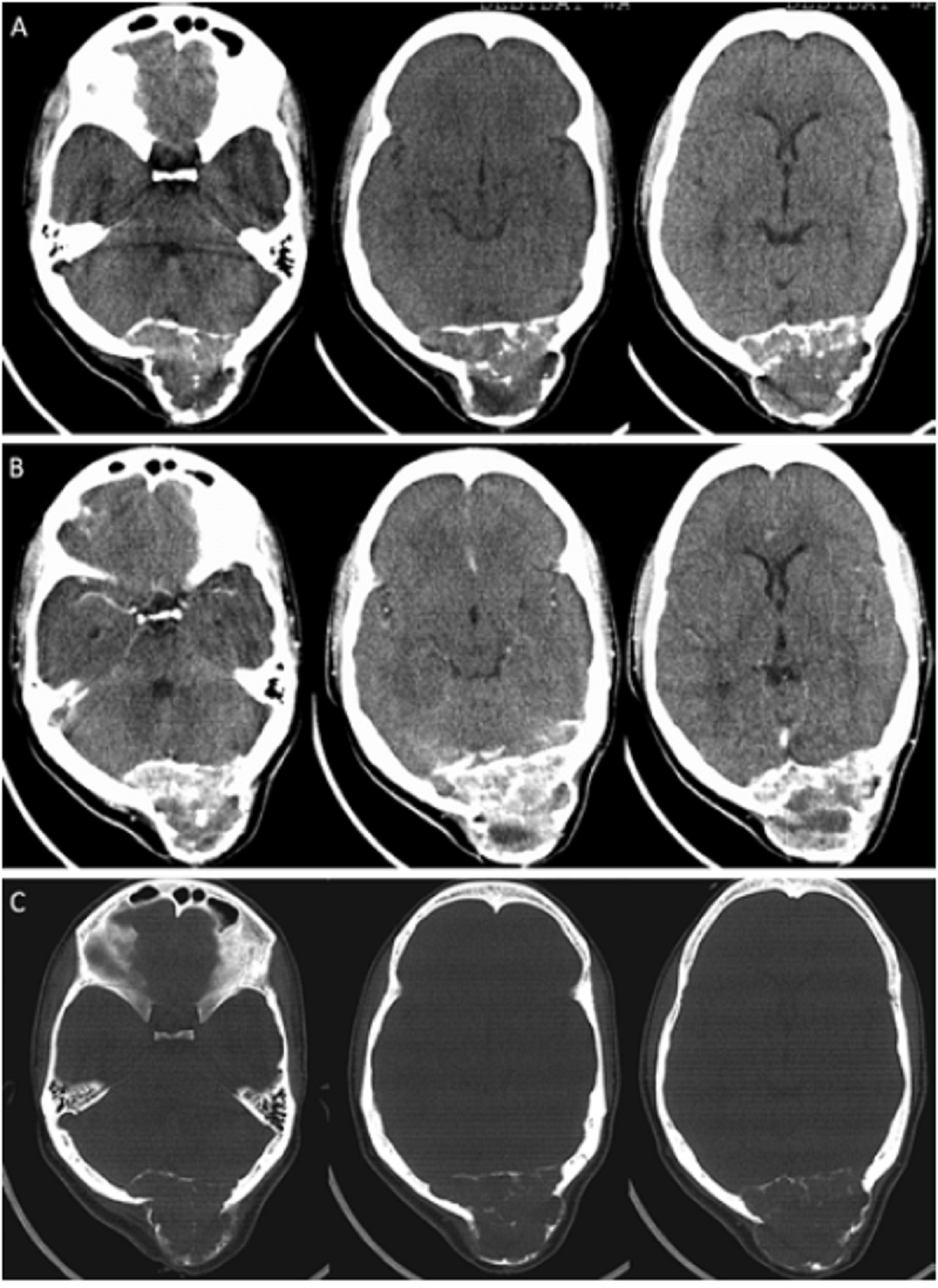
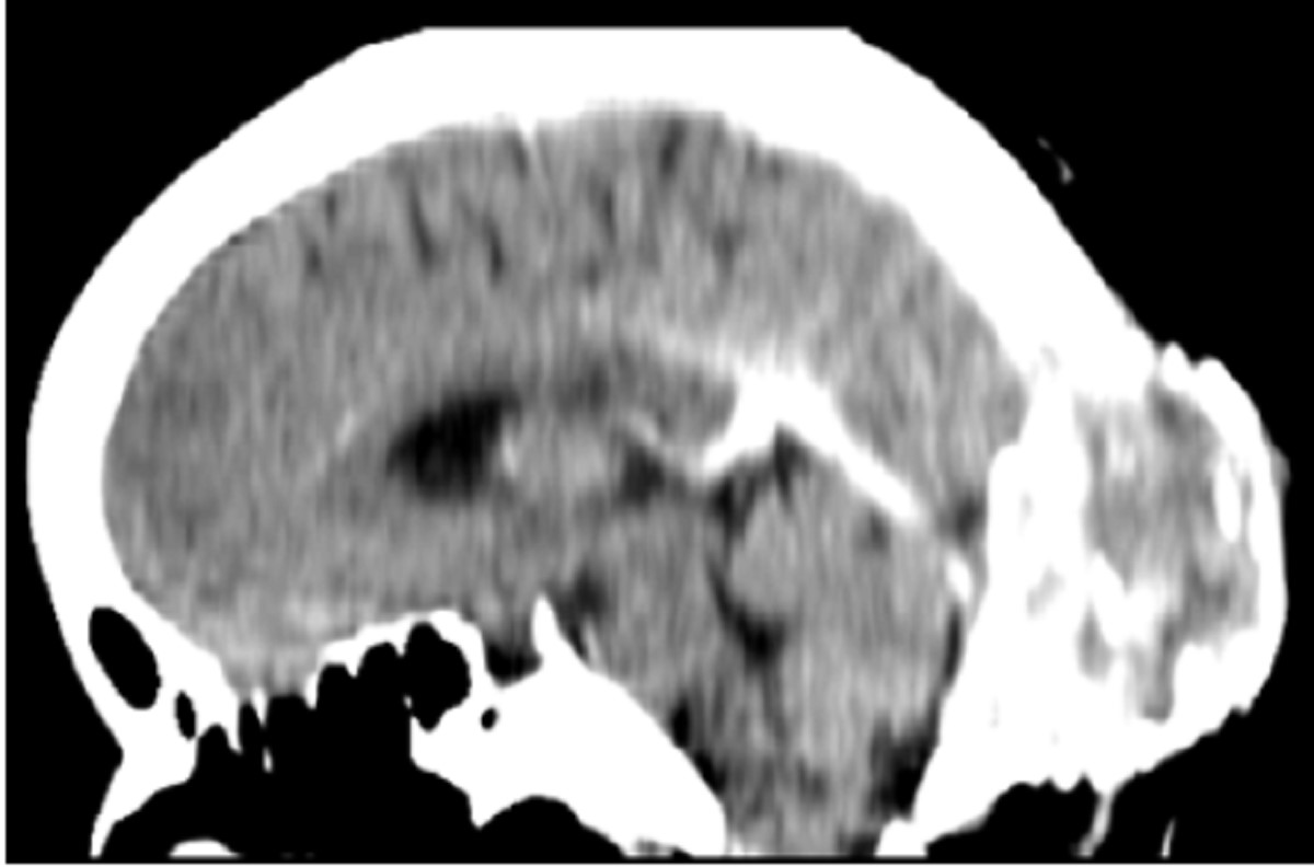
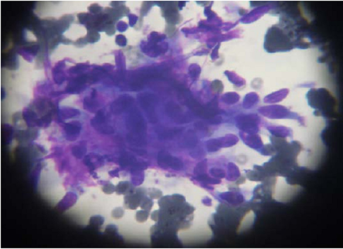
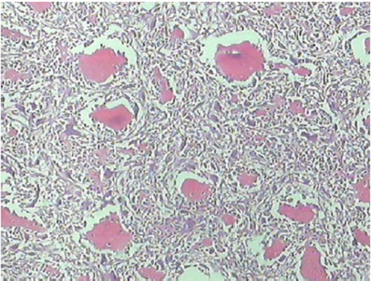
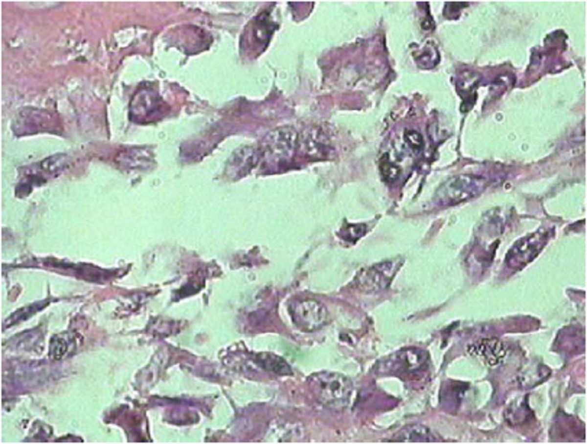
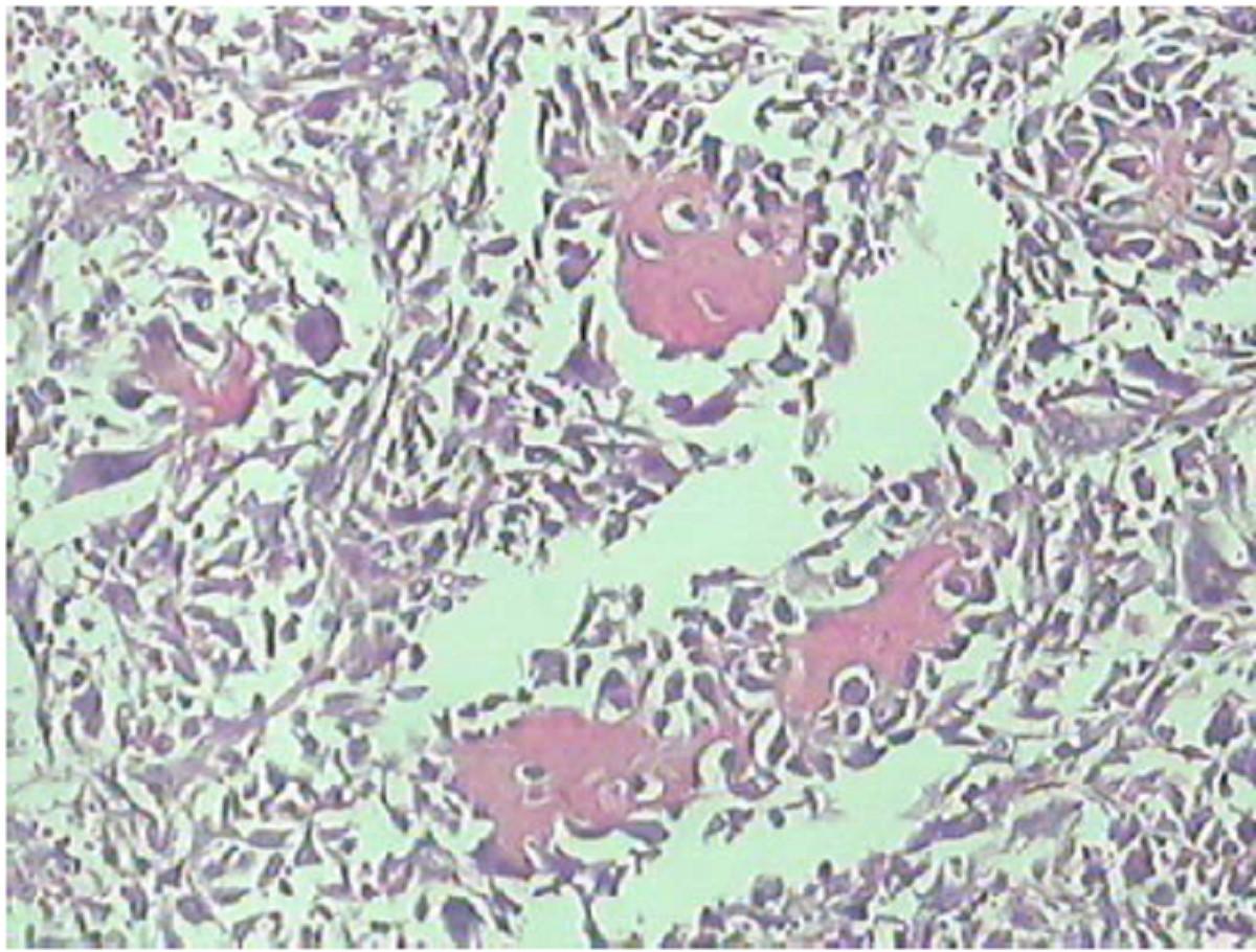
Epithelioidhemangioendothelioma is a rare vascular tumor of bone, and rarely these lesions can present as unique and extremely aggressive tumor. We report a case of highly aggressive epithelioidhemangioendothelioma and discuss the imaging findings. CT brain plain study revealed a poorly-defined, mixed density expansile and lytic lesion involving the occipital bone with extension to the left side with poorly defined trabecula formation. There was significant but irregular enhancement after intravenous administration of contrast material and also marked bone destruction. Microscopic examination of the fine needle aspiration cytology showed a tumor composed of vascular channels lined by plump endothelial cells, which had enlarged hyperchromatic nuclei. In view of the extensive infiltration the patient was submitted for the radiotherapy.
Epithelioidhemangioendothelioma is a rare vascular tumor of bone, and rarely these lesions can present as unique and extremely aggressive tumor. We report a case of highly aggressive epithelioidhemangioendothelioma and discuss the imaging findings. CT brain plain study revealed a poorly-defined, mixed density expansile and lytic lesion involving the occipital bone with extension to the left side with poorly defined trabecula formation. There was significant but irregular enhancement after intravenous administration of contrast material and also marked bone destruction. Microscopic examination of the fine needle aspiration cytology showed a tumor composed of vascular channels lined by plump endothelial cells, which had enlarged hyperchromatic nuclei. In view of the extensive infiltration the patient was submitted for the radiotherapy.
2012, 24(2): 164-166.
doi: 10.1007/s11670-012-0164-1
Abstract:
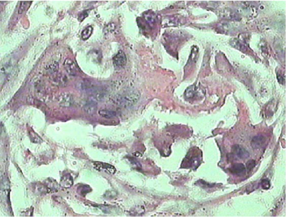
Extraskeletal osteosarcoma (EOS) is rare and commonly arises in the retroperitoneum, limbs, head and neck. There is no significant difference between EOS and other malignant tumors in soft tissue. Localized pain and swelling are the common presenting symptoms. Clinical diagnosis of EOS is difficult, imaging techniques may be helpful and careful, and the histopathological analysis is necessary. The common histological variants of EOS include: osteoblastoma, chondroblastoma, and fibroblastoma, and other unusual subtypes were reported occasionally. It should be distinguished with myositis ossificans, malignant mesenchymoma, giant cell tumor and parosteal osteosarcoma. We present an EOS arising in the penis. The primary site and histological category of the tumor were extremely rare. We hope the case will be helpful to the recognition of clinical signs, iconography and histopathology of EOS.
Extraskeletal osteosarcoma (EOS) is rare and commonly arises in the retroperitoneum, limbs, head and neck. There is no significant difference between EOS and other malignant tumors in soft tissue. Localized pain and swelling are the common presenting symptoms. Clinical diagnosis of EOS is difficult, imaging techniques may be helpful and careful, and the histopathological analysis is necessary. The common histological variants of EOS include: osteoblastoma, chondroblastoma, and fibroblastoma, and other unusual subtypes were reported occasionally. It should be distinguished with myositis ossificans, malignant mesenchymoma, giant cell tumor and parosteal osteosarcoma. We present an EOS arising in the penis. The primary site and histological category of the tumor were extremely rare. We hope the case will be helpful to the recognition of clinical signs, iconography and histopathology of EOS.
2012, 24(2): 167-170.
doi: 10.1007/s11670-012-0167-y
Abstract:
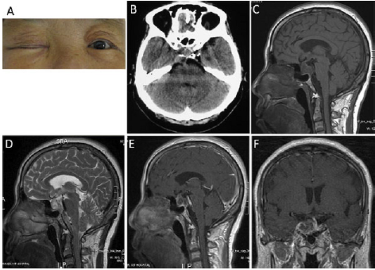
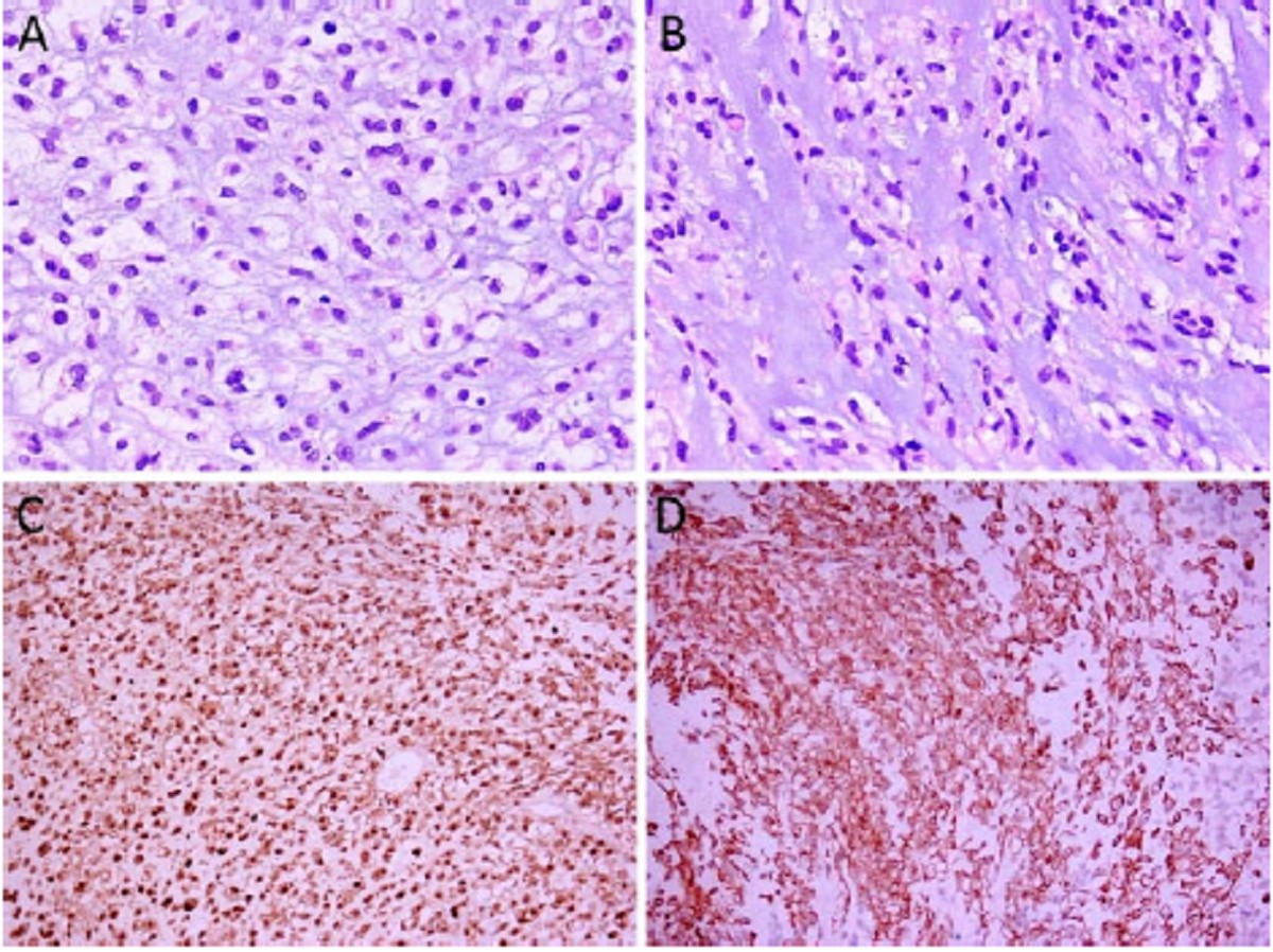
We described a 61-year-old female with a sellarchordoma, which presented as pseudo-macroprolactinoma with unilateral third cranial nerve palsy. Physical examination revealed that her right upper lid could not be raised by itself, right eyeball movement limited to the abduction direction, right pupil dilated to 4.5 mm with negative reaction to light, and hemianopsia in bitemporal sides. CT scanning showed a hyperdense lesion at sellar region without bone destruction. Magnetic resonance imaging (MRI) revealed the tumor was 2.3 cm×1.8 cm×2.6 cm, with iso-intensity on T1WI, hyper-intensity on T2WI and heterogeneous enhancement on contrast imaging. Endocrine examination showed her serum prolactin level increased to 1,031.49 mIU/ml. The tumor was sub-totally resected via pterional craniotomy under microscope and was histologically proven to be a chordoma. Postoperatively, she recovered uneventfully but ptosis and hemianopsia remained at the 6th month.
We described a 61-year-old female with a sellarchordoma, which presented as pseudo-macroprolactinoma with unilateral third cranial nerve palsy. Physical examination revealed that her right upper lid could not be raised by itself, right eyeball movement limited to the abduction direction, right pupil dilated to 4.5 mm with negative reaction to light, and hemianopsia in bitemporal sides. CT scanning showed a hyperdense lesion at sellar region without bone destruction. Magnetic resonance imaging (MRI) revealed the tumor was 2.3 cm×1.8 cm×2.6 cm, with iso-intensity on T1WI, hyper-intensity on T2WI and heterogeneous enhancement on contrast imaging. Endocrine examination showed her serum prolactin level increased to 1,031.49 mIU/ml. The tumor was sub-totally resected via pterional craniotomy under microscope and was histologically proven to be a chordoma. Postoperatively, she recovered uneventfully but ptosis and hemianopsia remained at the 6th month.