2012 Vol.24(3)
Display Mode: |
2012, 24(3): 171-180.
doi: 10.1007/s11670-012-0171-2
 Abstract
 FullText HTML
 PDF 470KB
Abstract:
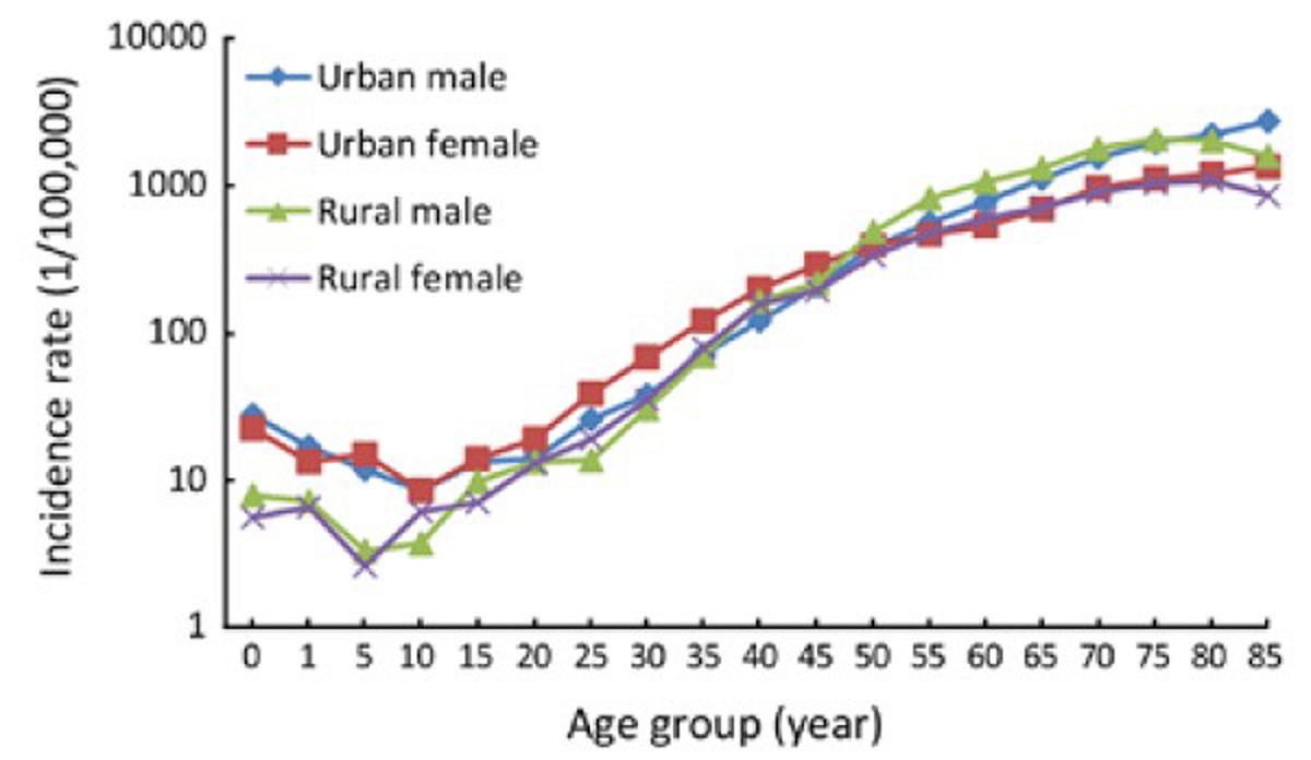
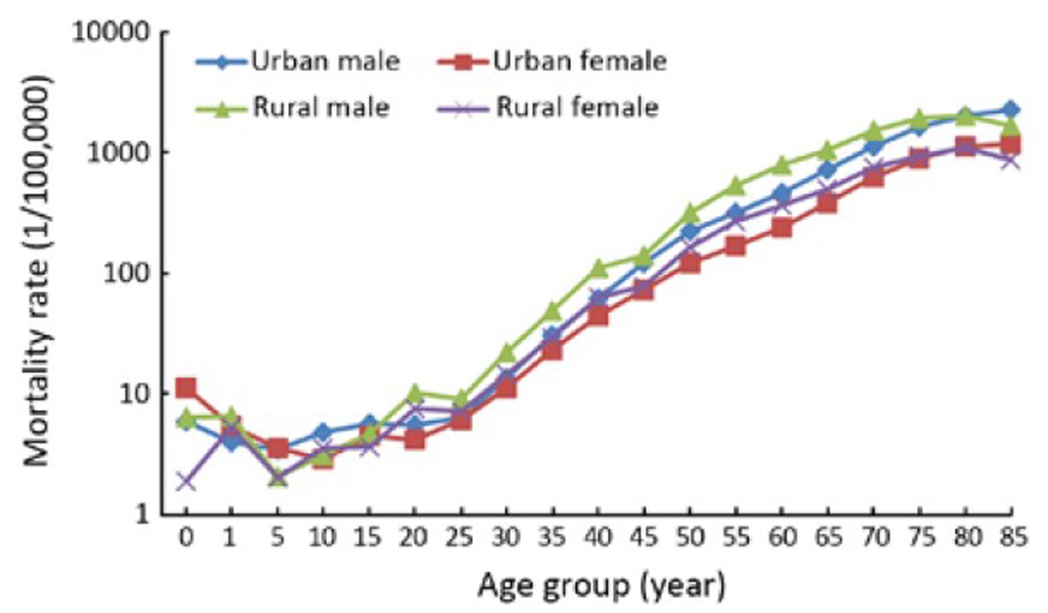
ObjectiveAnnual cancer incidence and mortality in 2008 were provided by National Central Cancer Registry in China, which data were collected from population-based cancer registries in 2011. MethodsThere were 56 registries submitted their data in 2008. After checking and evaluating the data quality, total 41 registries’ data were accepted and pooled for analysis. Incidence and mortality rates by area (urban or rural areas) were assessed, as well as the age- and sex-specific rates, age-standardized rates, proportions and cumulative rate. ResultsThe coverage population of the 41 registries was 66,138,784 with 52,158,495 in urban areas and 13,980,289 in rural areas. There were 197,833 new cancer cases and 122,136 deaths in cancer with mortality to incidence ratio of 0.62. The morphological verified rate was 69.33%, and 2.23% of cases were identified by death certificate only. The crude cancer incidence rate in all areas was 299.12/100,000 (330.16/100,000 in male and 267.56/100,000 in female) and the age-standardized incidence rates by Chinese standard population (ASIRC) and world standard population (ASIRW) were 148.75/100,000 and 194.99/100,000, respectively. The cumulative incidence rate (0–74 years old) was of 22.27%. The crude incidence rate in urban areas was higher than that in rural areas. However, after adjusted by age, the incidence rate in urban was lower than that in rural. The crude cancer mortality was 184.67/100,000 (228.14/100,000 in male and 140.48/100,000 in female), and the age-standardized mortality rates by Chinese standard population (ASMRC) and by world population were 84.36/100,000 and 114.32/100,000, respectively. The cumulative mortality rate (0–74 years old) was of 12.89%. Age-adjusted mortality rates in urban areas were lower than that in rural areas. The most common cancer sites were lung, stomach, colon-rectum, liver, esophagus, pancreas, brain, lymphoma, breast and cervix which accounted for 75% of all cancer incidence. Lung cancer was the leading cause of cancer death, followed by gastric cancer, liver cancer, esophageal cancer, colorectal cancer and pancreas cancer, which accounted for 80% of all cancer deaths. The cancer spectrum varied by areas and sex in rural areas, cancers from digestive system were more common, such as esophageal cancer, gastric cancer and liver cancer, while incidence rates of lung cancer and colorectal cancer were much higher in urban areas. In addition, breast cancer was the most common cancer in urban women followed by liver cancer, gastric cancer and colorectal cancer. ConclusionLung cancer, gastric cancer, colorectal cancer, liver cancer, esophageal cancer and female breast cancer contributed to the increased incidence of cancer, which should be paid more attention to in further national cancer prevention and control program. Different cancer control strategies should be carried out due to the varied cancer spectrum in different groups.
2012, 24(3): 181-189.
doi: 10.1007/s11670-012-0181-0
Abstract:
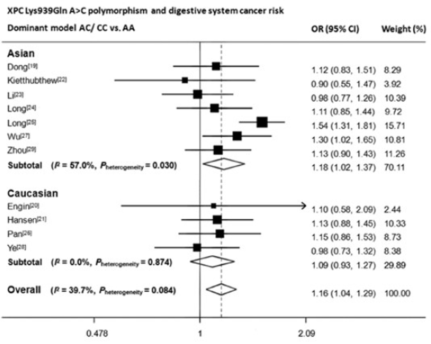
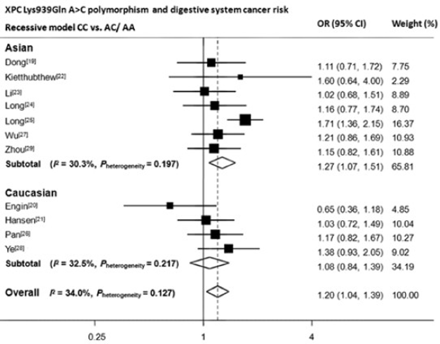
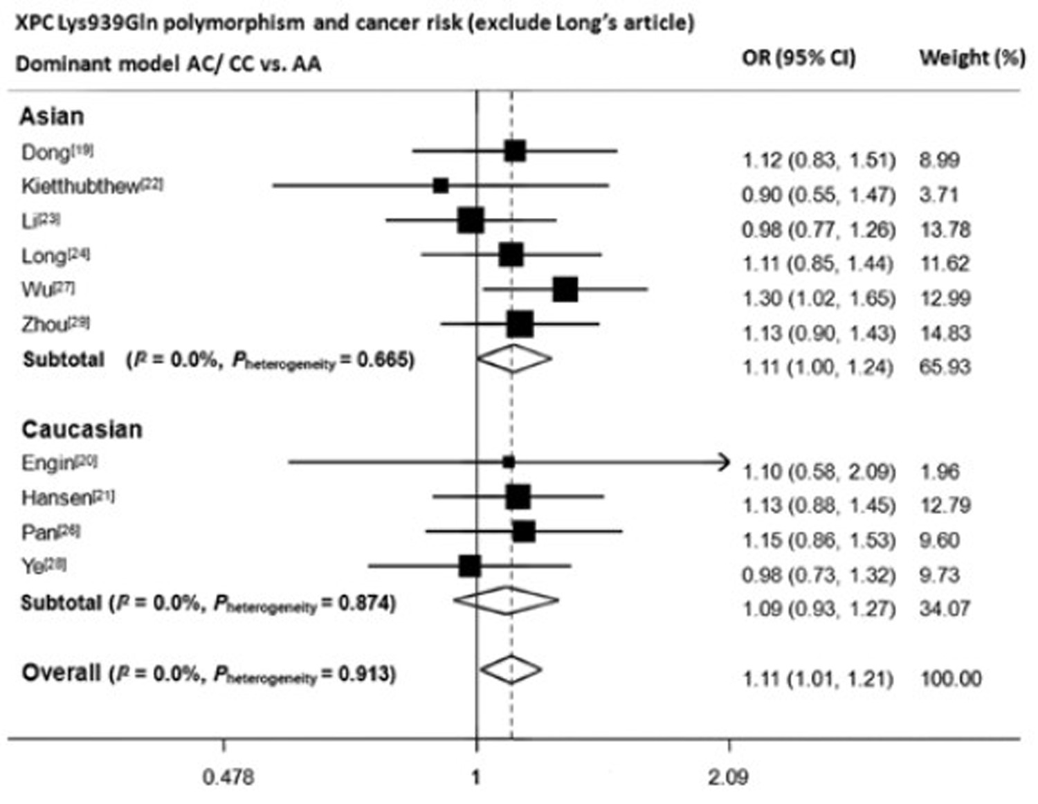
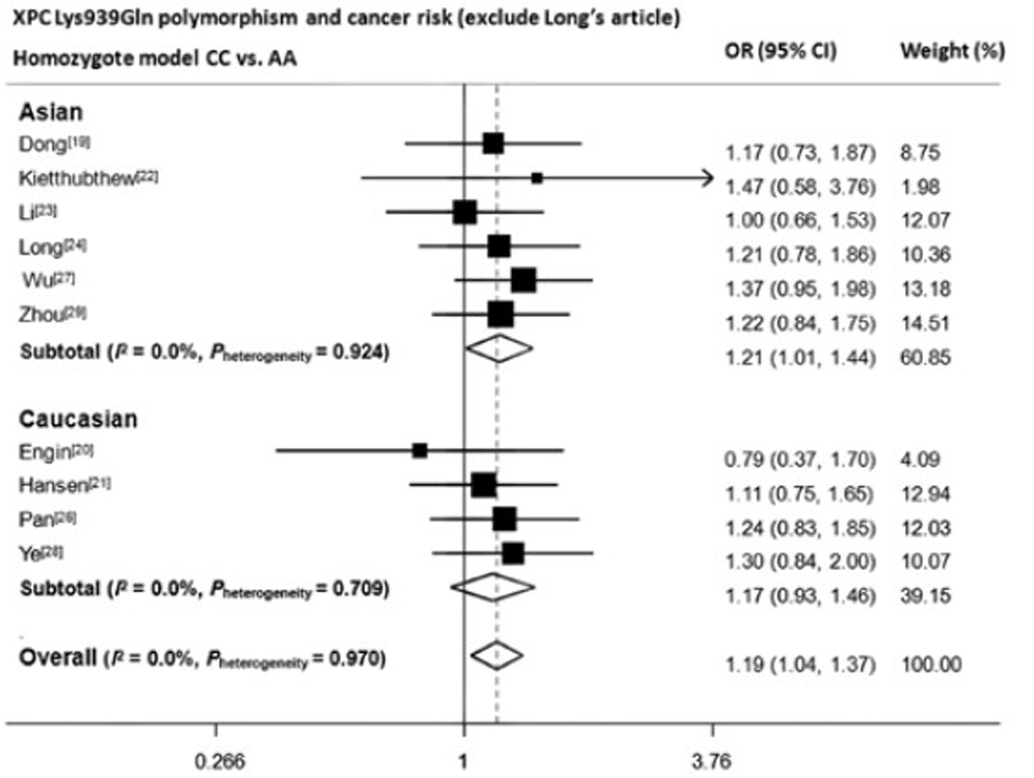
ObjectiveXeroderma pigmentosum complementation group C (XPC) participates in the initial recognition of DNA damage during nucleotide excision repair process in global genomic repair. Our meta-analysis was performed to evaluate the association between three polymorphisms (Lys939Gln, PAT+/– and Ala499Val) of XPC gene and risk of digestive system cancers. MethodsAll the relevant case-control studies published to April 2011 were identified through searching PubMed. Digestive system cancer risk with the three polymorphisms was estimated for each study by odds ratio (OR) with its 95% confidence interval (95% CI). ResultsWe found an increased overall risk for digestive system cancers in all three models of Lys939Gln A>C (AC/CC vs. AA: OR, 1.20; 95% CI, 1.11–1.30; CC vs. AC/AA: OR, 1.24; 95% CI, 1.11–1.39; CC vs. AA: OR, 1.36; 95% CI, 1.21–1.53). When stratified by ethnicity, results remained significant in Asian population (AC/CC vs. AA: OR, 1.18; 95% CI, 1.02–1.37; CC vs. AC/AA: OR, 1.32; 95% CI, 1.1–1.51; CC vs. AA: OR, 1.35; 95% CI, 1.08–1.70), but not for Caucasians. However for Ala499Val C>T, a significant protective effect of T allele was only observed in the dominant model. Otherwise, no significant results were observed for PAT+/–. ConclusionXPC Lys939Gln A>C polymorphism may play an important role in digestive system cancer susceptibility.


2012, 24(3): 190-195.
doi: 10.1007/s11670-012-0190-z
Abstract:
ObjectiveTo evaluate the association between mucin 2 (MUC2) expression and clinicopathological characters of colorectal cancer. MethodsA literature search was performed on December 31, 2010 according to defined selection criteria. We evaluated the correlation between MUC2 (detected by immunohistochemistry) and clinicopathological characters of colorectal cancer. According to the tumor histological type, differentiation, location and TNM staging of colorectal carcinoma, we divided the clinicopathological characteristics into different subgroups. Fixed and random effects models were applied for estimation of the summarized risk ratios (RRs) and 95% confidence intervals (CIs) in different subgroups. Finally, forest plots and funnel plots were created to allow for visual comparison of the results or the effect of publication bias. ResultsAccording with the inclusive criteria, fourteen studies (n=1,558) were eligible for the meta-analysis. We observed a trend towards a correlation of MUC2 higher positivity in mucinous than non-mucinous carcinoma (RR, 2.10; 95% CI, 1.30–3.40; P=0.002) and less positivity in distal than proximal colon (RR, 0.74; 95% CI, 0.64–0.85; P=0.000). There was no statistically significance for the association between MUC2 expression and differentiation or TNM staging of colorectal cancer, but MUC2 overexpression tended to be associated with the presence of T stage tumor (RR, 1.17; P=0.052). ConclusionMUC2 overexpression was associated with the mucinous and proximal colorectal cancer.

2012, 24(3): 196-200.
doi: 10.1007/s11670-012-0196-6
Abstract:
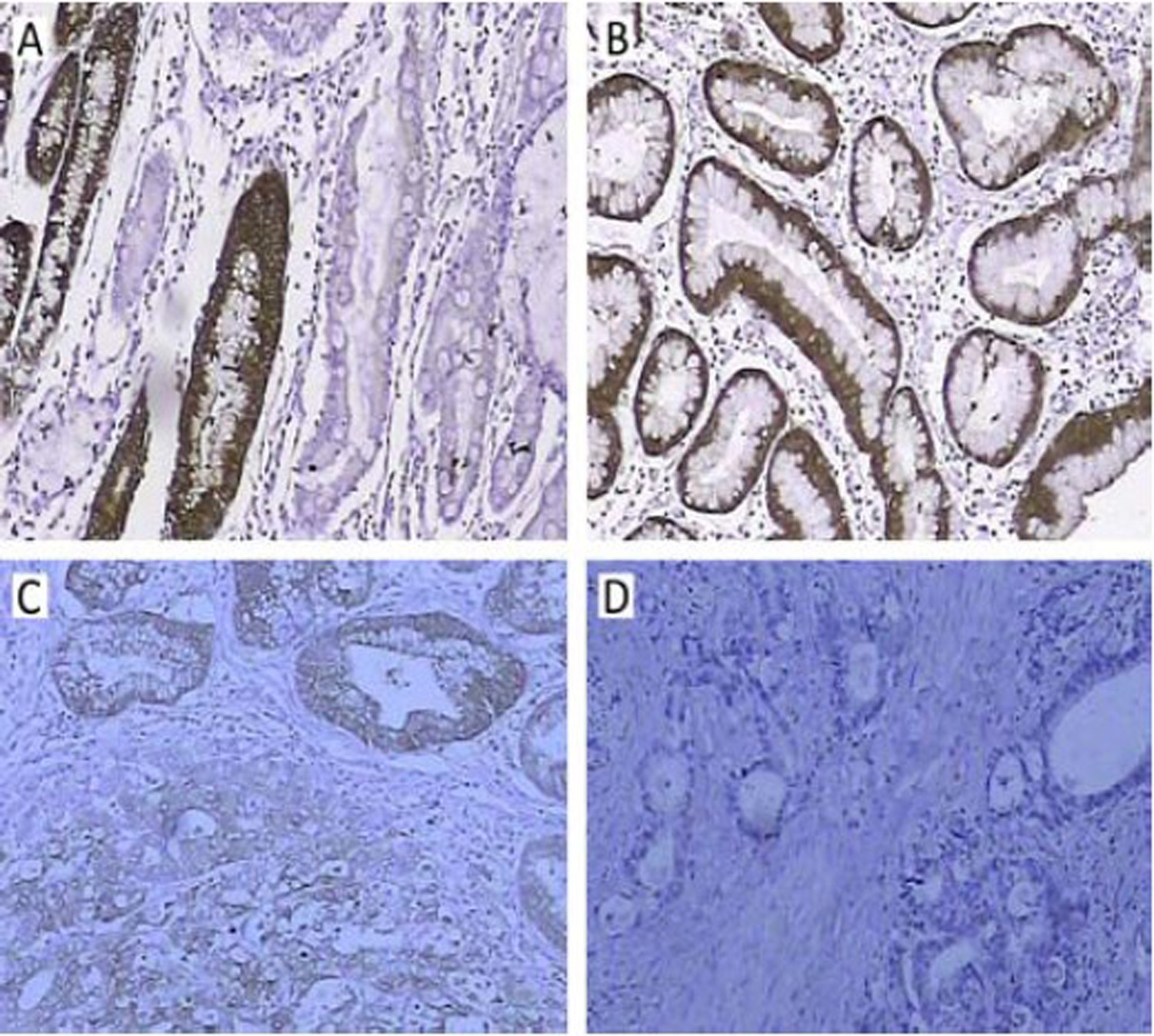
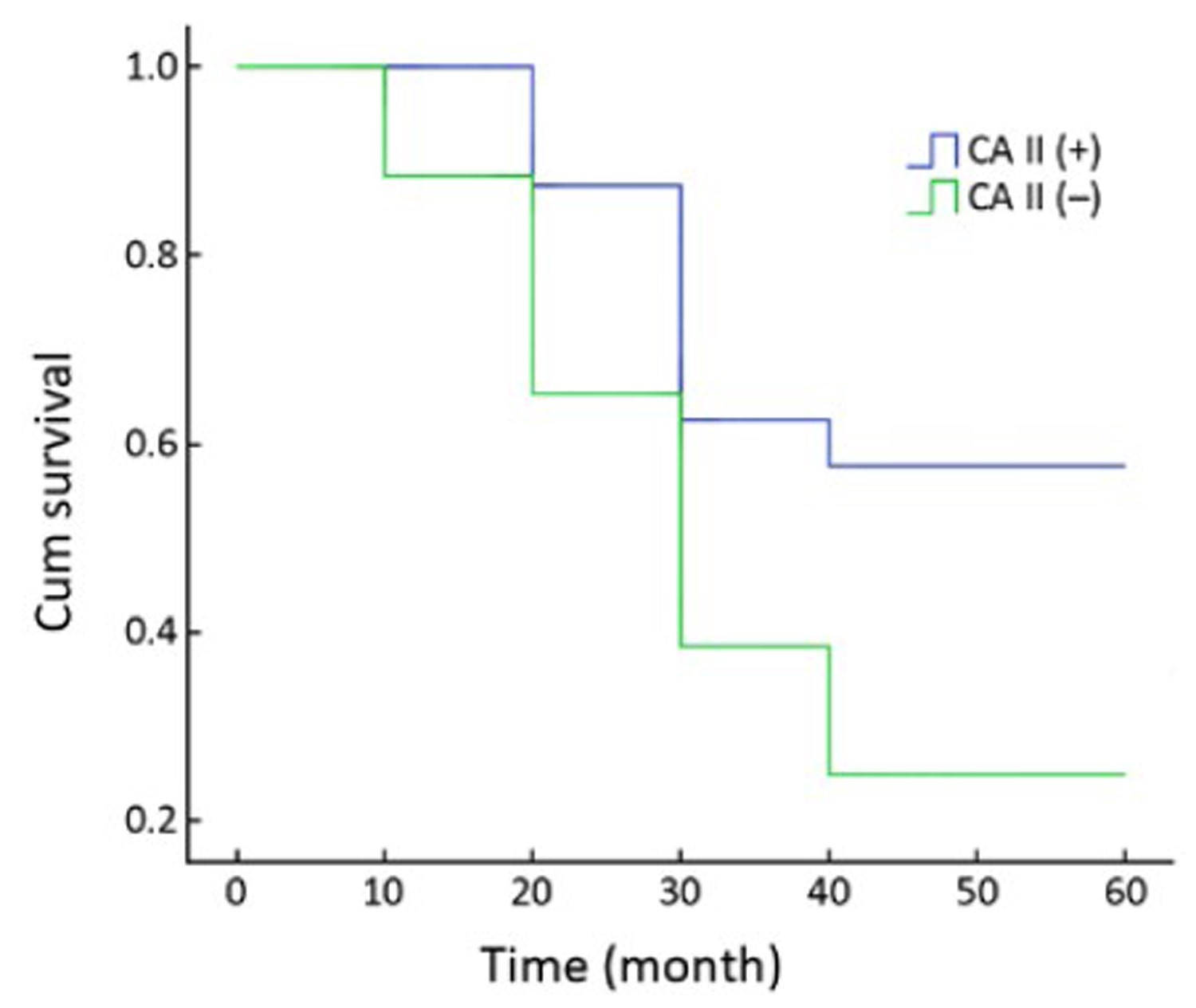
ObjectiveHuman carbonic anhydrases II (CAII) gene plays an important role in different cancer. However, its relevance to gastric cancer (GC) remains unclear. In the present study, we aimed to investigate the expression of CAII in GC and explore its correlation with some clinicopathologic characteristics of GC. MethodsThe expression of CAII in 20 specimens of normal gastric mucosa, 38 specimens of intraepithelial neoplasia and 112 specimens of gastric carcinoma were detected by immunohistochemical techniques. Survival in GC with CAII expression was studied. ResultsThe positive rate of CAII protein in normal gastric mucosa was significantly higher than that in intraepithelial neoplasia and gastric carcinoma (100% vs. 63.16% and 28.57%, P<0.001). The positive rate of CAII protein was significantly higher in gastric carcinoma at early stages than that at advanced stages (70.0% vs. 19.57%, P<0.001). The positive rate of CAII protein was significantly lower in gastric carcinoma with lymph node metastases than that without lymph node metastases (10.81% vs. 37.33%, P<0.05). Furthermore, the positive rate of CAII protein was significantly lower in poorly-differentiated gastric carcinoma than in moderately- or well-differentiated gastric carcinoma (15.94% vs. 31.03% or 60.00%, P<0.05). Moreover, CAII expression was not related with sex, age and tumor size. The patients with CAII-positive tumors showed a better survival rate than those with CAII-negative tumors (P=0.024, log-rank test). ConclusionCAII expression was related with stages and lymph node metastases in gastric carcinoma. The reduction of CAII expression in GC might promote tumor cell motility and contribute to tumor growth and metastasis.
2012, 24(3): 201-206.
doi: 10.1007/s11670-012-0201-0
Abstract:
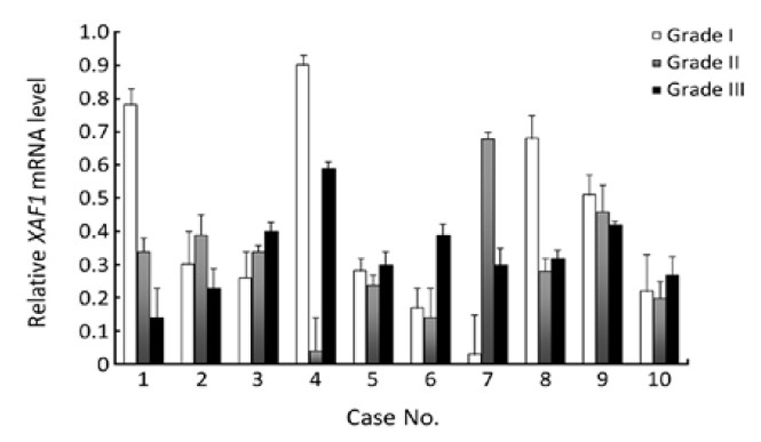
ObjectiveXIAP-associated factor 1 (XAF1) expression has been shown to be related with apoptosis in hepatocellular carcinoma (HCC). However, the correlation of XAF1 expression with HCC tumor grade has not been intensively assessed. XIAP-associated factor-1 (XAF1) is an important apoptosis inducer in human HCC. The aim of this study is to determine the correlation between XAF1 expression and HCC histopathological grades. MethodsThe mRNA levels of XAF1 in 24 paired HCC-nonneoplastic specimens were quantified by real-time reverse transcription PCR (RT-PCR). Protein levels of XAF1 in 110 paired HCC-noncancer tissues were investigated by immunostaining specimens on a tissue microarray (TMA). Correlations between XAF1 mRNA levels or protein expression and clinicopathological features were assessed by statistical analysis. ResultsBoth XAF1 mRNA and protein were significantly under-expressed in HCC tissues compared to their non-neoplastic counterparts. No significant relationship was found between XAF1 mRNA or protein expression and histological tumor grade. ConclusionAll these data suggest that XAF1 is a potential biomarker for differentiating HCC with noncancerous tissues.

2012, 24(3): 207-212.
doi: 10.1007/s11670-012-0207-7
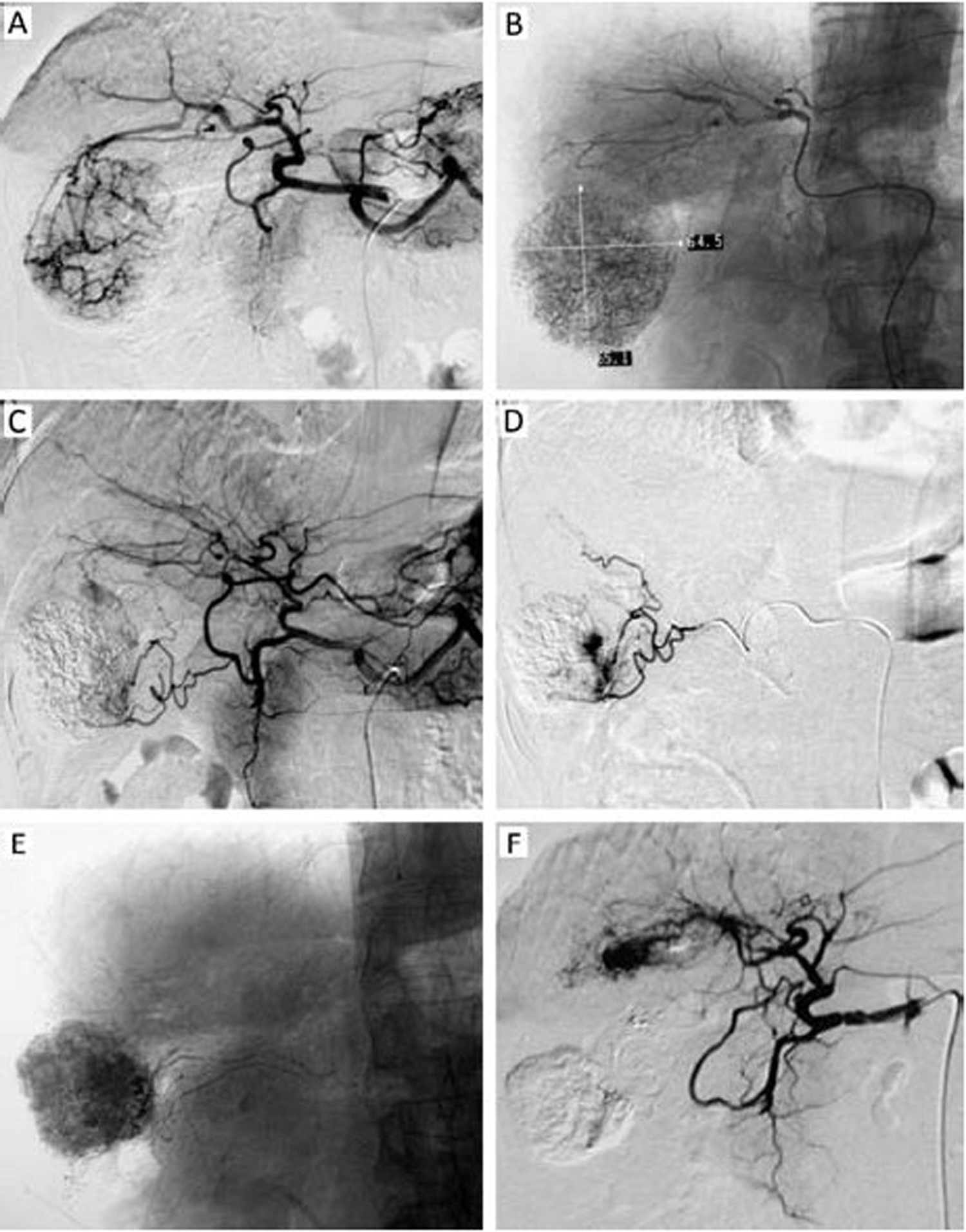
Abstract:
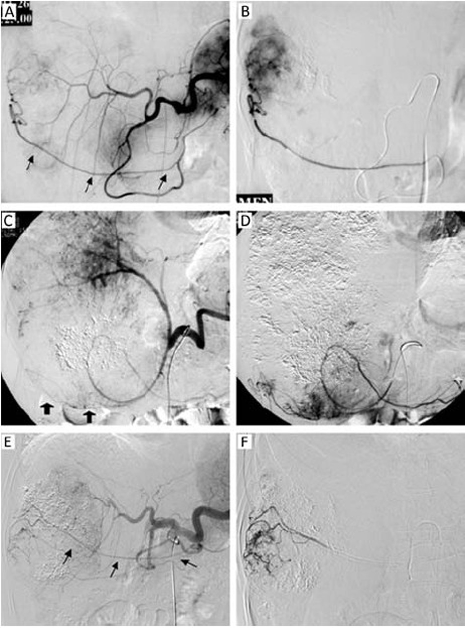
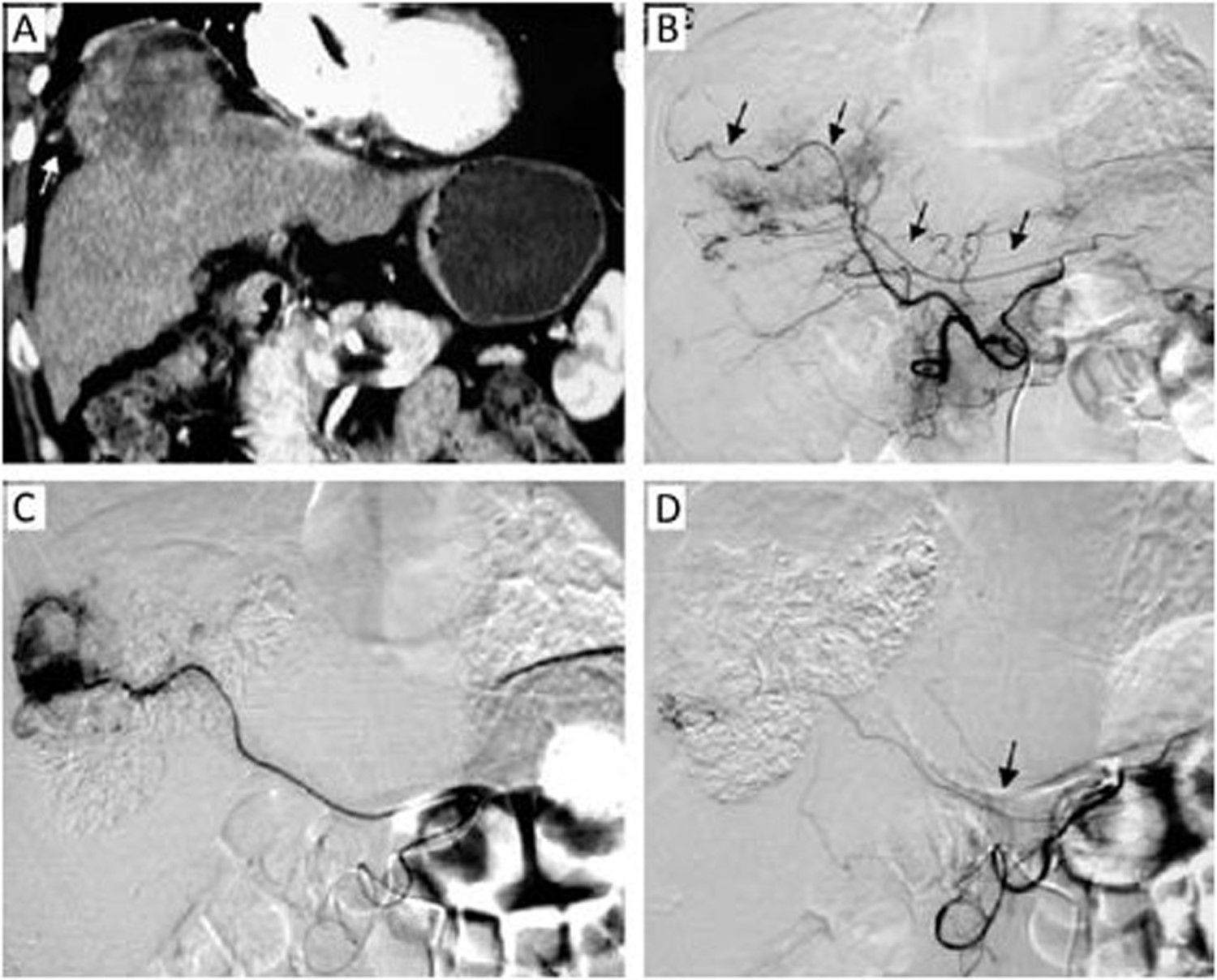
ObjectiveTo analyze angiographic appearance of hepatocellular carcinoma (HCC) with blood supply from parasitized omental artery (POA), and evaluate the technical feasibility, safety and therapeutic efficacy of chemo-embolization via the POAs. MethodsA total of 1,221 HCC patients who had undergone chemoembolization procedures were evaluated retrospectively. The evaluated indexes included the incidence rate of POAs, success rate of superselective catheterization, post-reaction after chemoembolization, and the cumulative survival rates. ResultsTotally 1,221 HCC patients had undergone 3,639 chemoembolization procedures, and 32 patients with POAs were enrolled, with 97 POAs found in 76 angiography procedures, giving an incidence rate of 2.09%. POA was observed mostly at the right lobe and left medial lobe except the segment II, and 62 POAs underwent superselective catheterization with microcatheter, giving a success rate of 63.9%. The angiographic appearance was: (1) hypertrophic POAs participating in tumor staining (n=28); (2) stiff and distorted POA (n=11), displaced due to tumor’s oppression (n=8); and (3) defective tumor staining close to either gastrocolic omentum distribution or liver capsule (n=7). In 19 patients, chemoembolization via POAs was performed successfully (A group), while the remaining 13 patients failed (B group). Except 1 acute edema pancreatitis case, no serious complication was recorded. The cumulative survival rates of 6-, 12-, 18- and 24-month were 78.9%, 47.4%, 31.6% and 21.1% respectively for A group; correspondingly, 61.5%, 30.8%, 15.4% and 7.7%% for B group, in which 2 patients died of ruptured HCC. ConclusionChemoembolization with microcatheter via POAs is a relatively safe, feasible and valuable method.

2012, 24(3): 213-219.
doi: 10.1007/s11670-012-0213-9
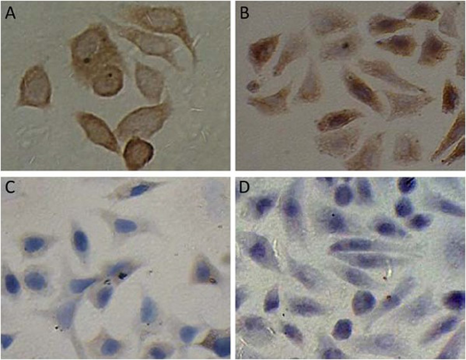
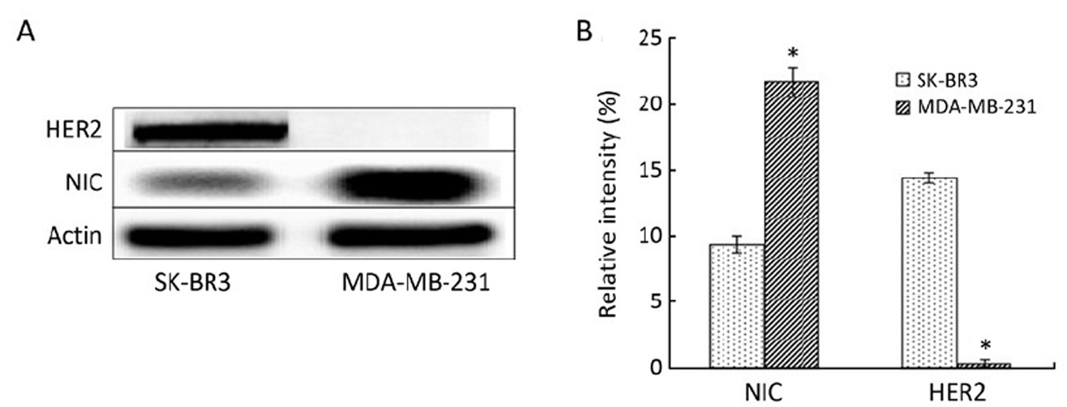
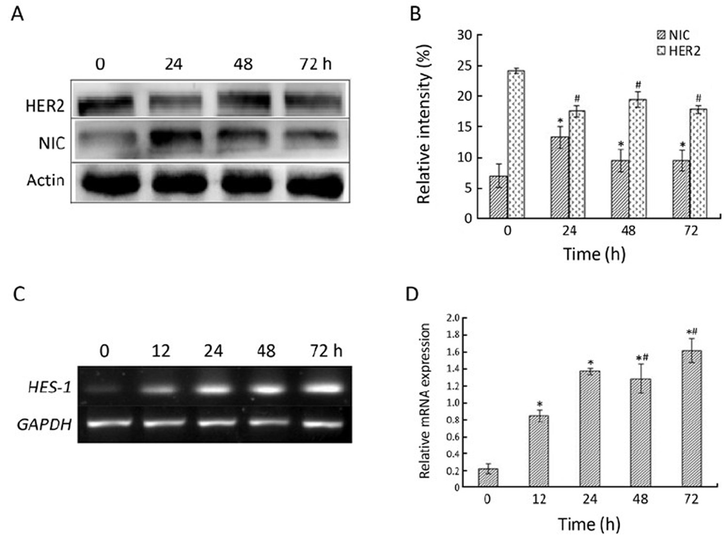
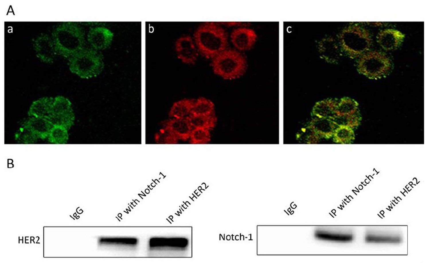
Abstract:
ObjectiveTo investigate the effects and mechanisms of trastuzumab on Notch-1 pathway in breast cancer cells, recognizing the significance of Notch-1 signaling pathway in trastuzumab resistance. MethodsImmunocytochemistry staining and Western blotting were employed to justify the expression of Notch-1 protein in HER2-overexpressing SK-BR3 cells and HER2-non-overexpressing breast cancer MDA-MB-231 cells. Western blotting and reverse transcription PCR (RT-PCR) were used to detect the activated Notch-1 and Notch-1 target gene HES-1 mRNA expression after SK-BR3 cells were treated with trastuzumab. Double immunofluorescence staining and co-immunoprecipitation were used to analyze the relationship of Notch-1 and HER2 proteins. ResultsThe level of Notch-1 nuclear localization and activated Notch-1 proteins in HER2-overexpressing cells were significantly lower than in HER2-non-overexpressing cells (P<0.01), and the expressions of activated Notch-1 and HES-1 mRNA were obviously increased after trastuzumab treatment (P<0.05), but HER2 expression did not change significantly for trastuzumab treating (P>0.05). Moreover, Notch-1 was discovered to co-localize and interact with HER2 in SK-BR3 cells. ConclusionOverexpression of HER2 decreased Notch-1 activity by the formation of a HER2-Notch1 complex, and trastuzumab can restore the activity of Notch-1 signaling pathway, which could be associated with cell resistance to trastuzumab.
2012, 24(3): 220-225.
doi: 10.1007/s11670-012-0220-x
Abstract:
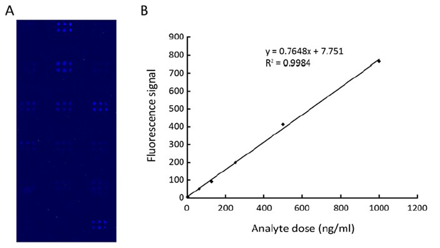
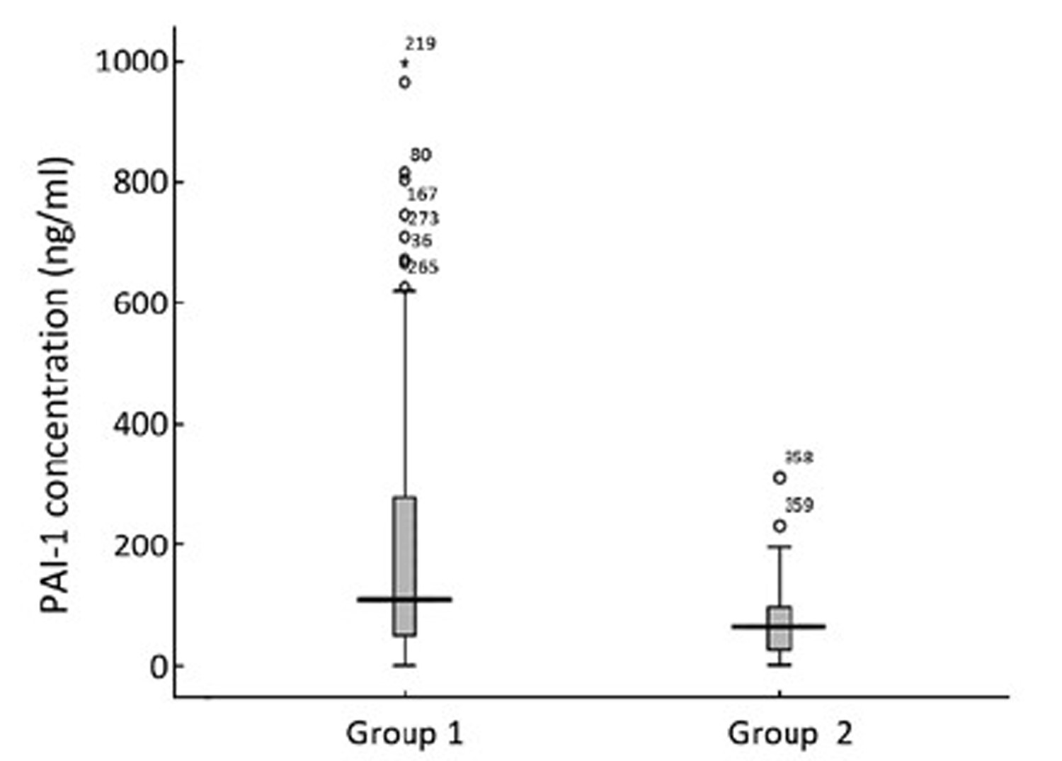
ObjectivePlasminogen activator inhibitor-1 (PAI-1), one crucial component of the plasminogen activator system, is a major player in the pathogenesis of many vascular diseases as well as in cancer. High levels of PAI-1 in breast cancer tissue are associated with poor prognosis. The aim of this study is to evaluate rigorously the potential of serum PAI-1 concentration functioning as a general screening test in diagnostic or prognostic assays. MethodsA protein-microarray-based sandwich fluorescence immunoassay (FIA) was developed to detect PAI-1 in serum. Several conditions of this microarray-based FIA were optimized to establish an efficacious method. Serum specimens of 84 healthy women and 285 women with breast cancer were analyzed using the optimized FIA microarray. ResultsThe median serum PAI-1 level of breast cancer patients was higher than that of healthy women (109.7 ng/ml vs. 63.4 ng/ml). Analysis of covariance revealed that PAI-1 levels of the two groups were significantly different (P<0.001) when controlling for an age effect on PAI-1 levels. However, PAI-1 values in TNM stage I−IV patients respectively were not significantly different from each other. ConclusionThis microarray-based sandwich FIA holds potential for quantitative analysis of tumor markers such as PAI-1.
2012, 24(3): 226-231.
doi: 10.1007/s11670-012-0226-4
Abstract:
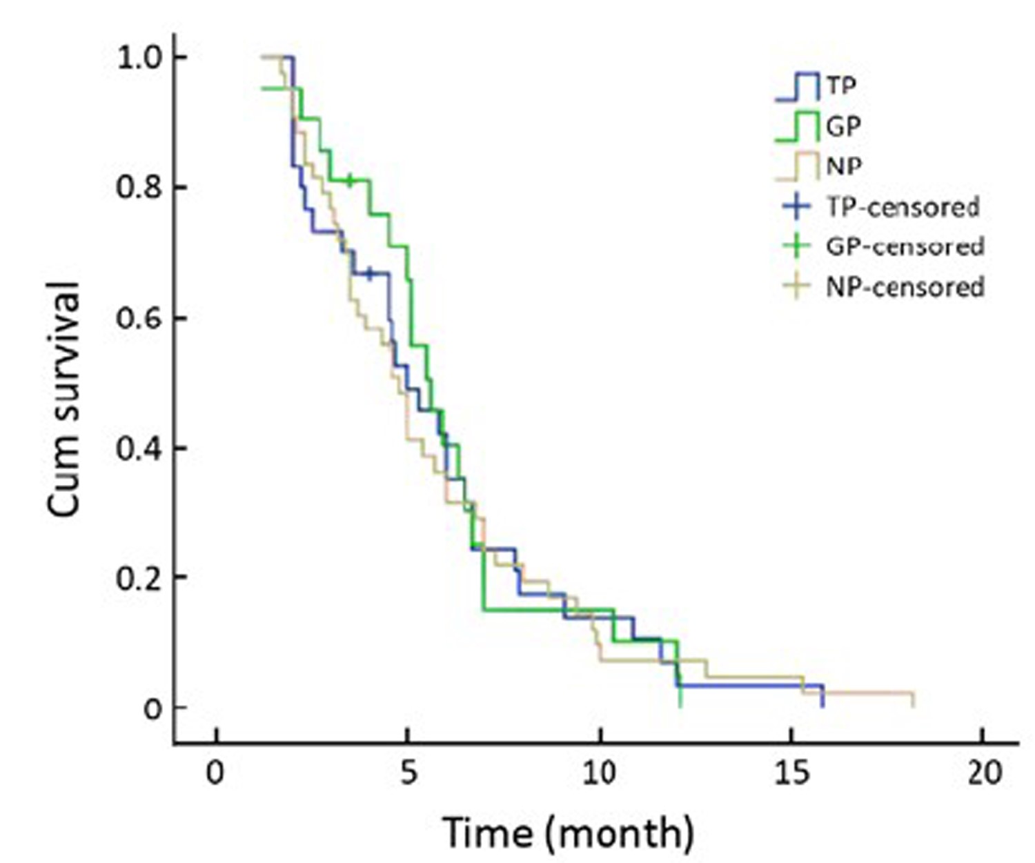
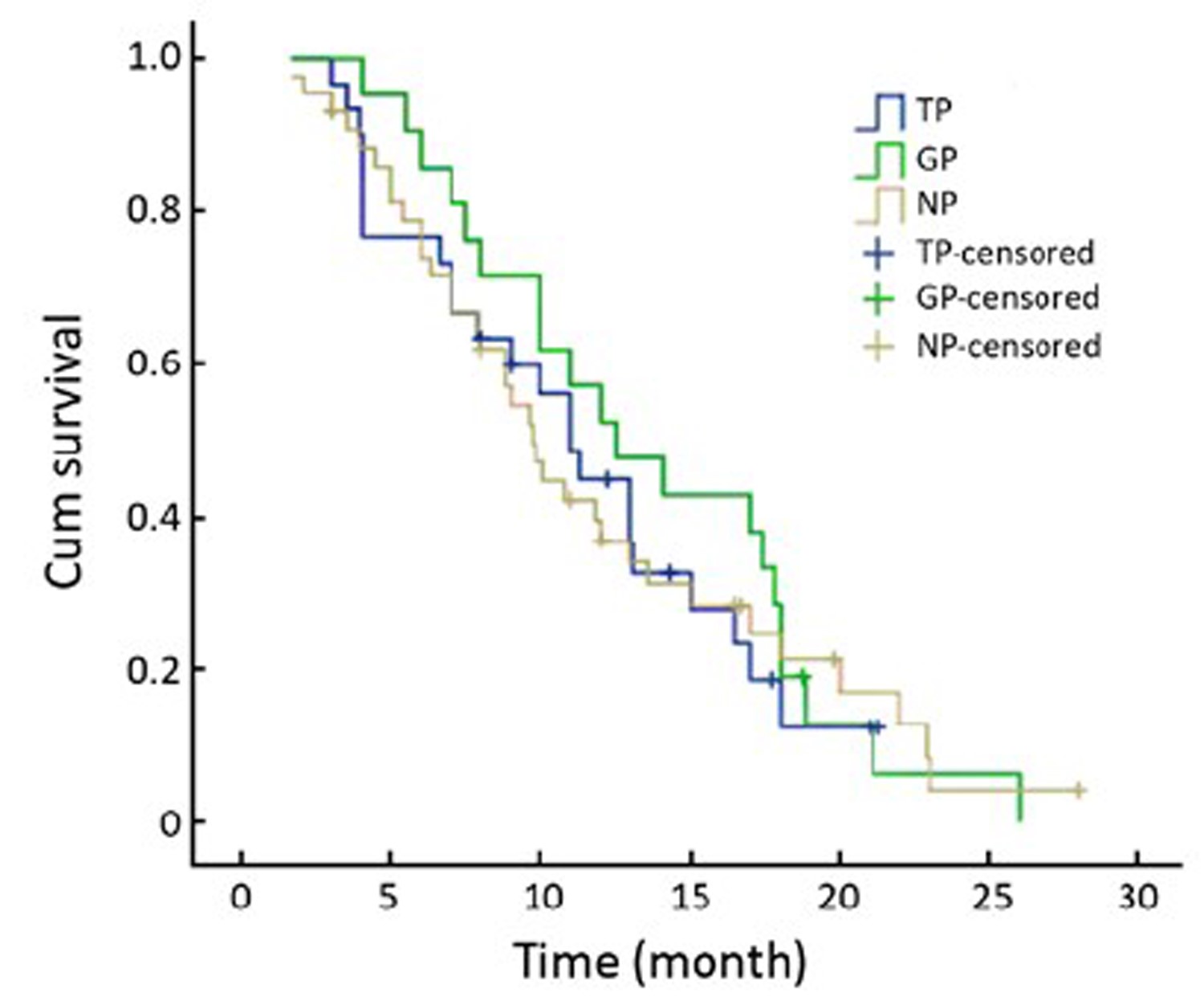
ObjectiveExperimental evidence suggests that the overexpression of breast cancer-specific tumor suppressor protein 1 (BRCA1) gene enhances sensitivity to docetaxel and resistance to cisplatin and ribonucleotide reductase M1 (RRM1) gene overexpression enhances resistance to gemcitabine. To further examine the effect of BRCA1 and RRM1 mRNA levels on outcome in advanced non-small cell lung cancer (NSCLC), we performed this non-randomized phase II clinical trial which tested the hypothesis that customized therapy would confer improved outcome over non-customized therapy. MethodsRNA was isolated from fresh tumor tissue. Patients received chemotherapy regimen based on their BRCA1 and RRM1 mRNA levels: both low–cisplatin plus gemcitabine (GP); both high–vinorelbine plus cisplatin (NP); BRCA1 low and RRM1 high–cisplatin plus docetaxel (TP); BRCA1 high and RRM1 low–vinorelbine plus gemcitabine (GN). ResultsFrom Dec 2005 to Nov 2008, 94 metastatic and locally advanced NSCLC patients from our institute were enrolled in this study. The median age was 58 years old. Among them, 21 patients received GP, 30 patients received TP and 43 patients received NP chemotherapy. GP group had a higher response rate, and longer median time to progression (TTP) and median overall survival (OS) time than the other 2 groups. The response rates in the GP, TP and NP groups were 42.9%, 36.7% and 27.9%, respectively (P=0.568). The median TTP was 5.6, 5.0, 4.8 months (P=0.975), respectively, and the median OS time was 12.5, 11.0, 9.7 months (P=0.808), respectively. ConclusionChemotherapy customized according to BRCA1 and RRM1 expression levels is associated with higher response rate and longer TTP and OS time in the GP group. This suggests that BRCA1 and RRM1 mRNA levels could be used as biomarkers in individual therapy in NSCLC.
2012, 24(3): 232-237.
doi: 10.1007/s11670-012-0232-6
Abstract:
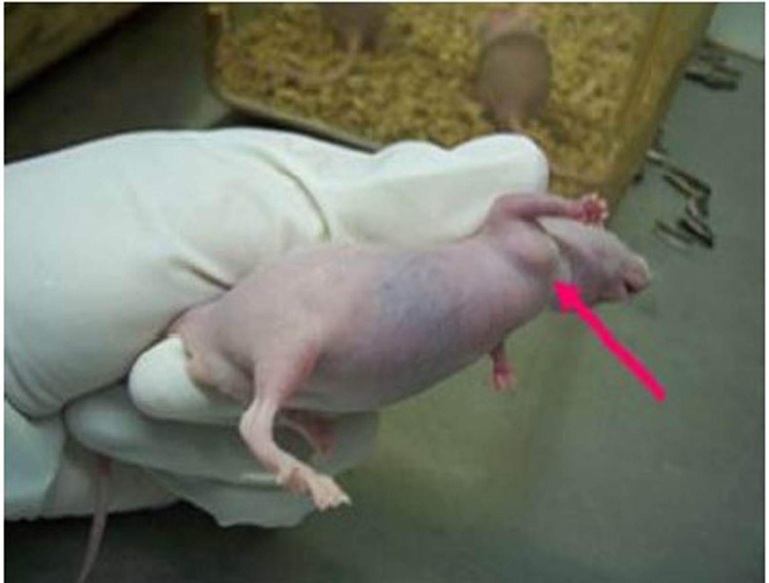
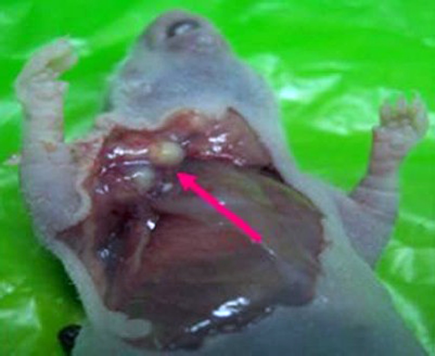
ObjectiveTo investigate the effects of 5-Aza-2'-deoxycytidine (5-Aza-Cdr) and trichostatin A (TSA) combined with p53-expressing adenovirus (Ad-p53) on Hep-2 cell line in vivo and in vitro, in order to explore its possibility in biological treatment of laryngocarcinoma. MethodsEffects of 5-Aza-Cdr and TSA in combination with Ad-p53 on Hep-2 cell line in vivo were determined by Cell Counting Kit-8 (CCK-8) assay. The effect of drug combination was calculated by Jin's formula. Effects on the cell line in vitro were investigated by establishing the nude mice model. Results5-Aza-Cdr and TSA showed inhibitory effects on the proliferation of Hep-2 cells in dose- and time-dependent manner. Ad-p53 can inhibit the growth of Hep-2 cells in vivo and in vitro. However, the combination of epigenetic reagents (5-Aza-Cdr/TSA) and Ad-p53 was less effective than individual use of Ad-p53. 5-Aza-Cdr and Ad-p53 inhibited the growth of transplanted tumors and reduced the volume of tumors, and the tumor volume of Ad-p53 group was significantly smaller than that of the control group (P<0.05). ConclusionBoth epigenetic reagents (5-Aza-Cdr/TSA) and Ad-p53 can suppress cell proliferation on Hep-2 in vivo and in vitro and there may be some antagonistic mechanism between Ad-p53 and epigenetic reagents (5-Aza-Cdr/ TSA).
2012, 24(3): 238-244.
doi: 10.1007/s11670-012-0238-0
Abstract:
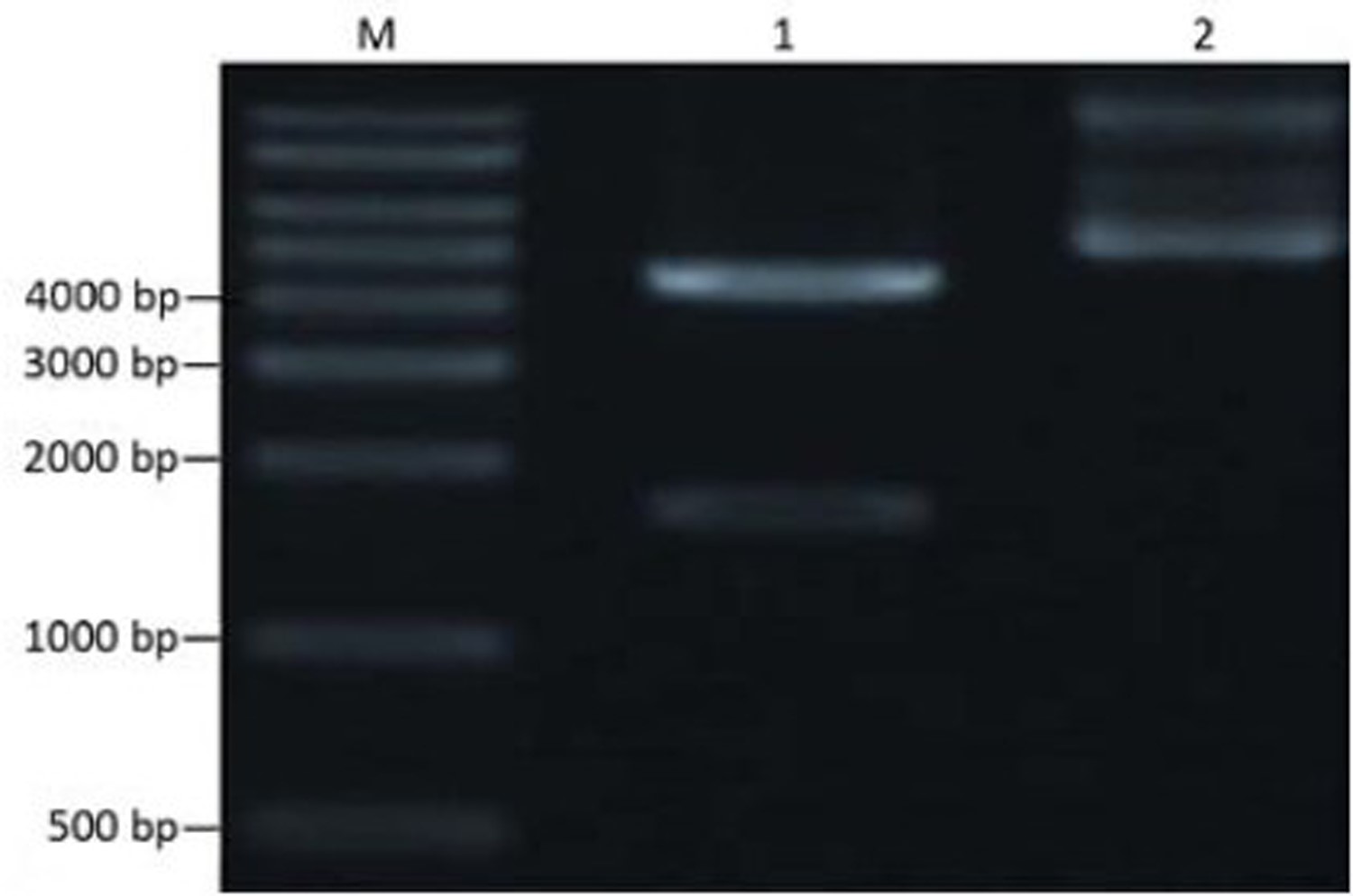
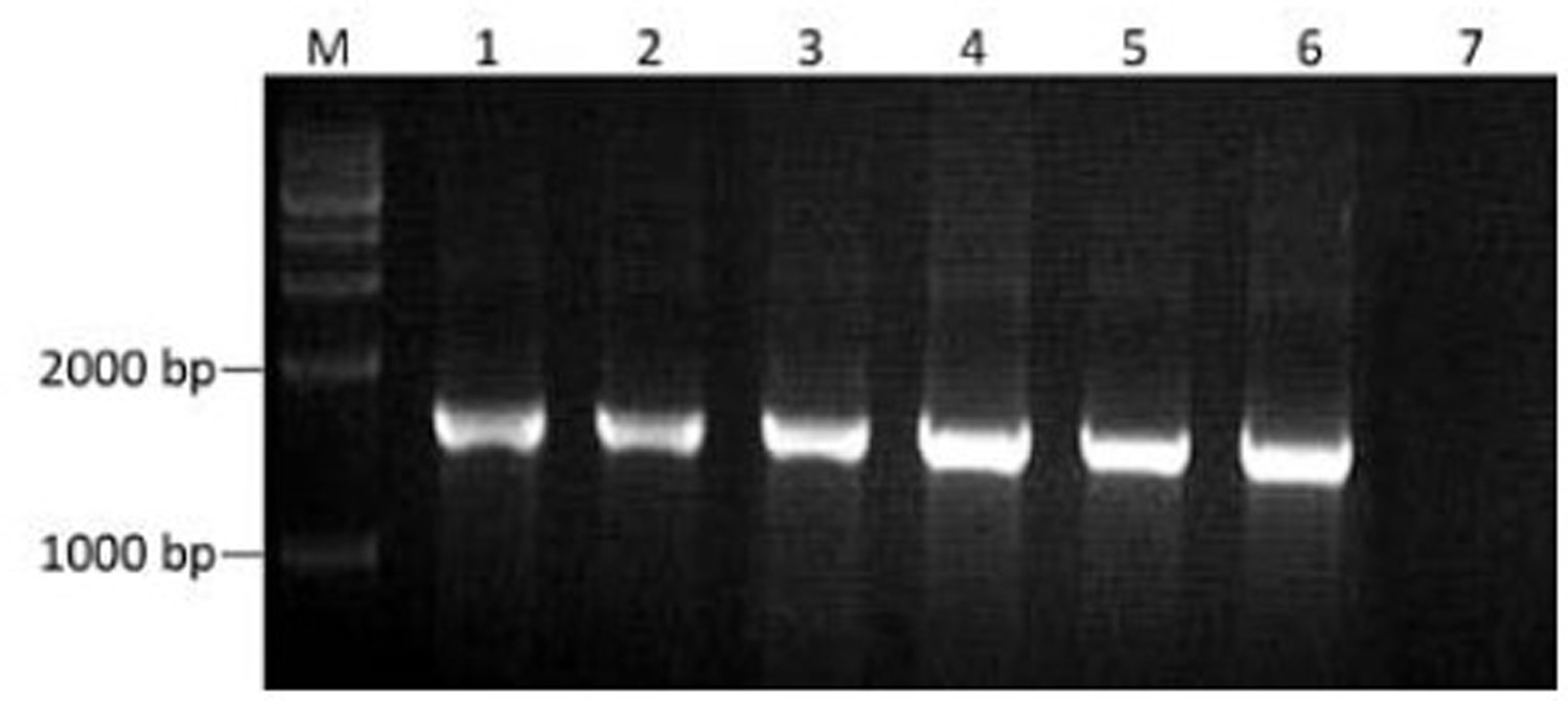
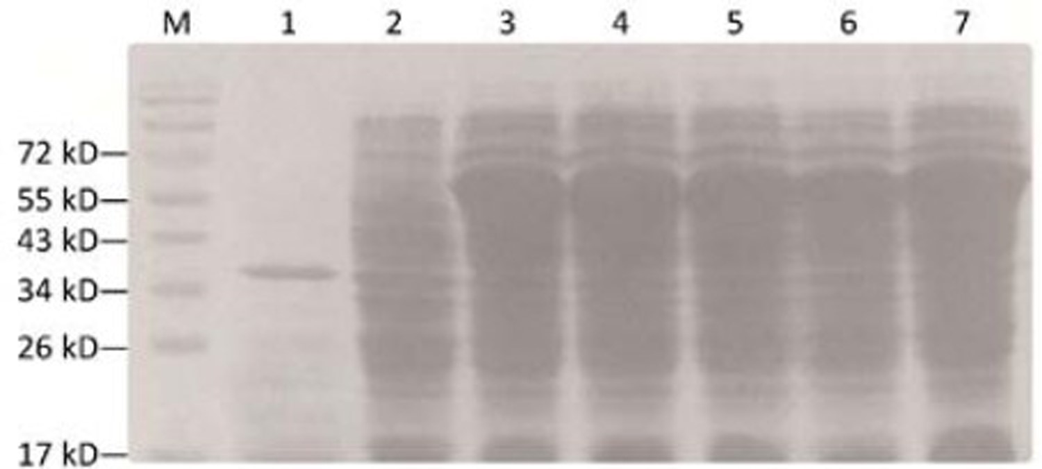
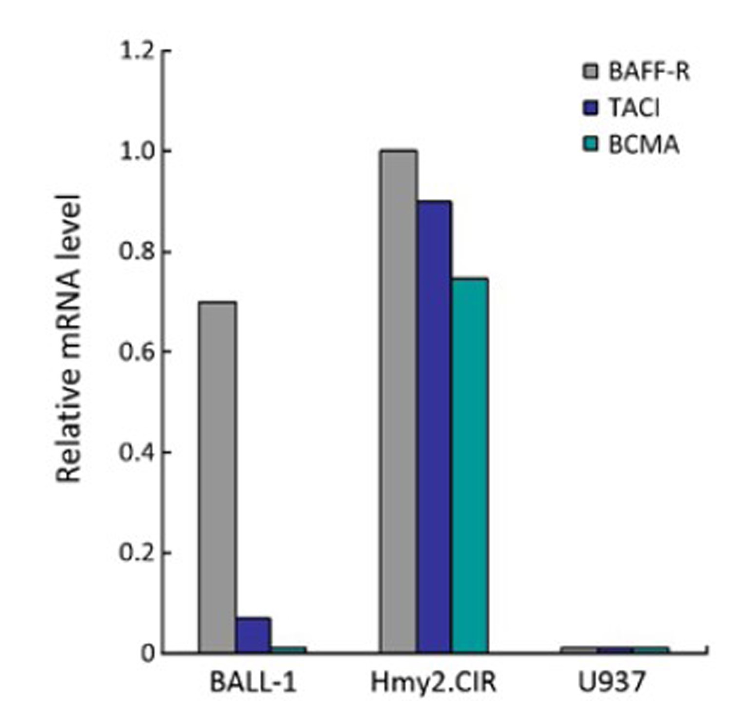
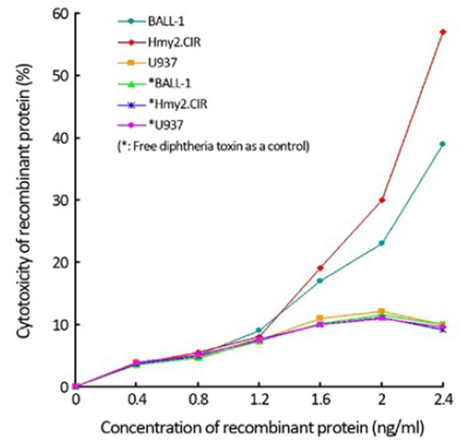
ObjectiveThis study aimed to express a fusion protein of diphtheria toxin and human B cell-activating factor (DT388sBAFF) in Escherichia coli (E. coli) and investigate its activity in human B-lineage acute lymphoblastic leukemia 1 cells (BALL-1). MethodsA fragment of DT388sBAFF fusion gene was separated from plasmid pUC57-DT388sBAFF digested with Nde I and Xho I, and inserted into the expression vector pcold II digested with the same enzymes. Recombinants were screened by the colony polymerase chain reaction (PCR) and restriction map. The recombinant expression vector was transformed into BL21 and its expression was induced by isopropyl β-D-1-thiogalactopyranoside (IPTG). The recombinant protein was identified by sodium dodecyl sulfate-polyacrylamide gel electrophoresis (SDS-PAGE) and Western blot, and then purified by Ni2+-NTA affinity chromatography. The expression level of B cell-activating factor receptor (BAFF-R) on BALL-1 cells was assessed by real-time PCR. The receptor binding capacity of recombinant protein was determined by cell fluorescent assay. The specific cytotoxicity of recombinant protein on BALL-1 cells was detected by 3-(4,5-dimethylthiazolyl-2)-2,5-diphenyltetrazolium bromide (MTT) assay. ResultsThe expression level of recombinant protein was 50% of total bacterial proteins in E. coli, and the recombinant protein could bind to BAFF-R-positive BALL-1 cells and thereby produce a cytotoxic effect on the cells. ConclusionThe fusion protein expression vector DT388sBAFF was successfully constructed and the recombinant protein with selective cytotoxicity against BALL-1 cells was obtained, providing foundation for further study of the therapy of human B-lineage acute lymphoblastic leukemia.


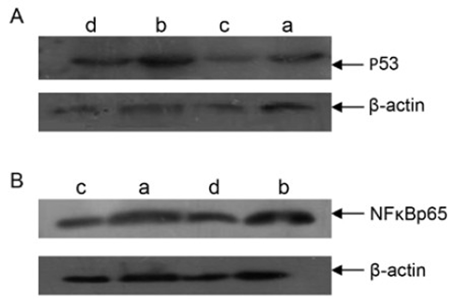
Dys-psychological Stress Effect on Expressions of P53 and NFκBp65 in Human Ovarian Carcinoma In Vivo
2012, 24(3): 245-248.
doi: 10.1007/s11670-012-0245-1
Abstract:
ObjectiveTo investigate the dys-psychological stress effect on the growth of subcutaneous xenotransplanted tumor in nude mice bearing human epithelium ovarian carcinoma, and the influence on P53 and NFκBp65 expressions. MethodsThe subcutaneous tumor xenografts were established by implanting human epithelium ovarian carcinoma tissues into nude mice and the dys-psychological stress model was established with restraint. The mice were randomized into the following four treatment groups with each group six mice respectively: tumor group (group A), normal saline intraperitoneal injection; tumor with stress group (group B), normal saline intraperitoneal injection; tumor therapy group (group C), cisplatin intraperitoneal injection; and tumor therapy with stress group (group D), cisplatin intraperitoneal injection. The expressions of P53 and NFκBp65 in tumor tissues were determined by Western blotting. ResultsThe expressions of P53 and NFκBp65 in each restraint group were enhanced compared with the control groups (P<0.05). ConclusionThe dys-psychological stress may induce the high expressions of P53 and NFκBp65 proteins and further promote tumor growth.
2012, 24(3): 249-252.
doi: 10.1007/s11670-012-0249-x
Abstract:
Although non-small cell lung cancer (NSCLC) can metastasize to almost any organ, metastasis to the gallbladder with significant clinical manifestation is relatively rare. Here, we report a case of gallbladder metastasis of NSCLC presenting as acute cholecystitis. A 79-year-old man presented with pain in the right upper quadrant and fever. A computed tomography (CT) scan of the chest and abdomen showed a cavitary mass in the right lower lobe of the lung and irregular wall thickening of the gallbladder. Open cholecystectomy and needle biopsy of the lung mass were performed. Histological examination of the gallbladder revealed a moderately-differentiated squamous cell carcinoma displaying the same morphology as the lung mass assessed by needle biopsy. Subsequent immunohistochemical examination of the gallbladder and lung tissue showed that the tumor cells were positive for P63 but negative for cytokeratin 7, cytokeratin 20 and thyroid transcription factor-1. A second primary tumor of the gallbladder was excluded by immunohistochemical methods, and the final pathological diagnosis was gallbladder metastasis of NSCLC. Although the incidence is extremely rare, acute cholecystitis can occur in association with lung cancer metastasis to the gallbladder.
Although non-small cell lung cancer (NSCLC) can metastasize to almost any organ, metastasis to the gallbladder with significant clinical manifestation is relatively rare. Here, we report a case of gallbladder metastasis of NSCLC presenting as acute cholecystitis. A 79-year-old man presented with pain in the right upper quadrant and fever. A computed tomography (CT) scan of the chest and abdomen showed a cavitary mass in the right lower lobe of the lung and irregular wall thickening of the gallbladder. Open cholecystectomy and needle biopsy of the lung mass were performed. Histological examination of the gallbladder revealed a moderately-differentiated squamous cell carcinoma displaying the same morphology as the lung mass assessed by needle biopsy. Subsequent immunohistochemical examination of the gallbladder and lung tissue showed that the tumor cells were positive for P63 but negative for cytokeratin 7, cytokeratin 20 and thyroid transcription factor-1. A second primary tumor of the gallbladder was excluded by immunohistochemical methods, and the final pathological diagnosis was gallbladder metastasis of NSCLC. Although the incidence is extremely rare, acute cholecystitis can occur in association with lung cancer metastasis to the gallbladder.
2012, 24(3): 253-256.
doi: 10.1007/s11670-012-0253-1
Abstract:
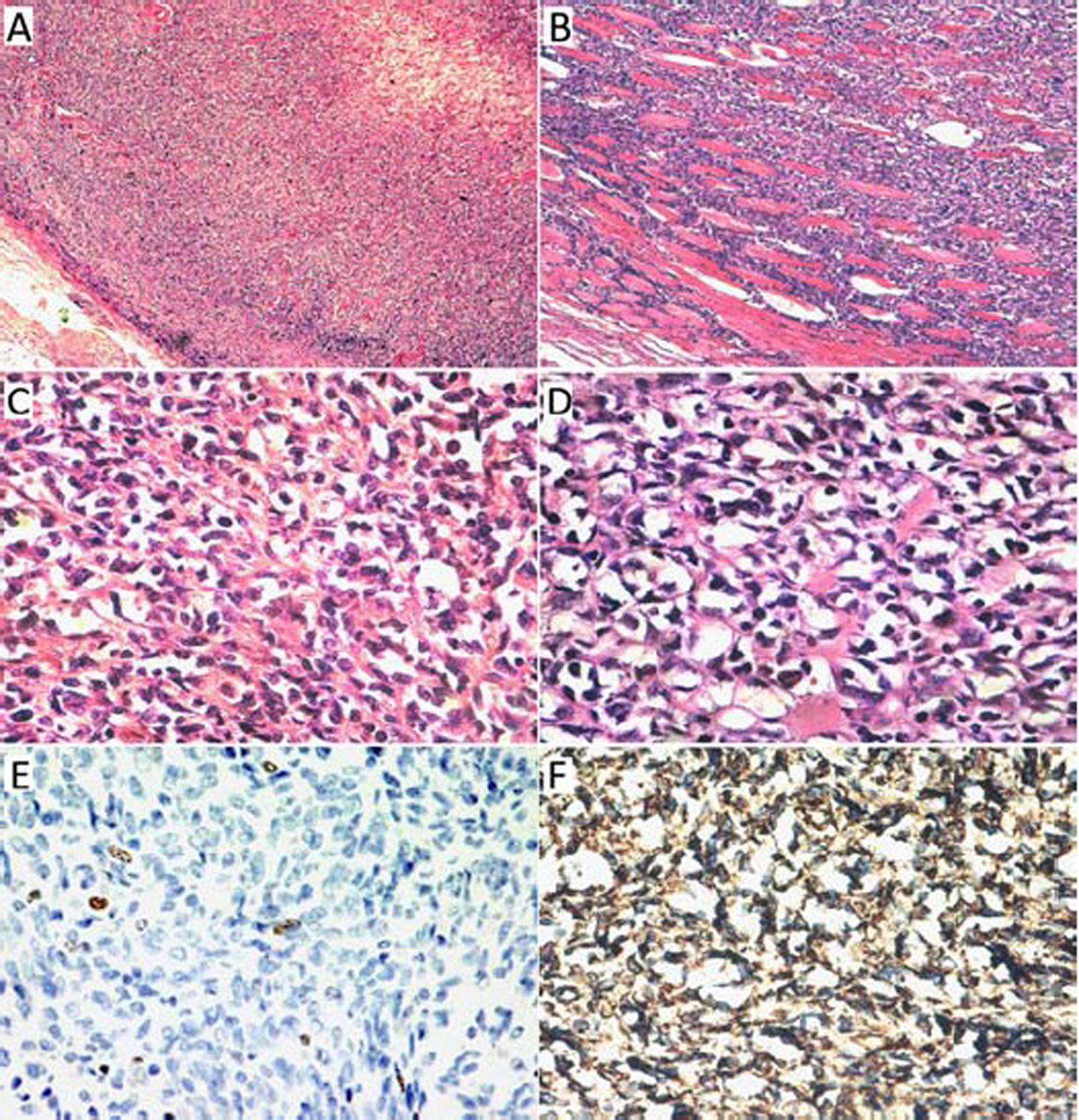
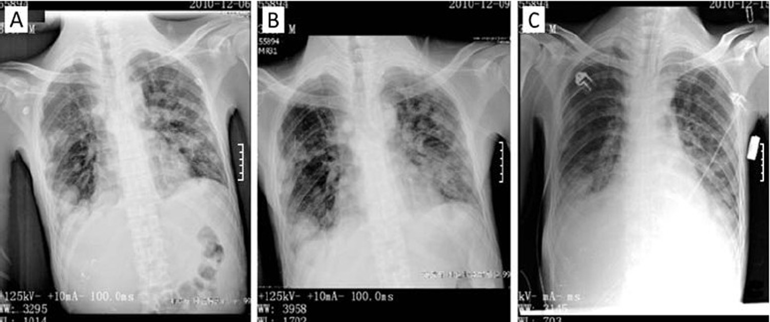
Sclerosing epithelioid fibrosarcoma (SEF) is a rare and poorly defined variant of fibrosarcoma, but generally insensitive to chemotherapy and progresses with poor prognosis. We report the marvelous effect of irinotecan hydrochloride (CPT-11) chemotherapy in rescuing a patient with atypical SEF from emergent condition, who underwent recurrences after several treatment methods. Small dose of CPT-11 was administered to the patient, with which, the size of superficial mass (cervical lymph node) gradually decreased observed by the naked eyes in 5 days. X-ray and CT image proved a marked reduction in the size of the tumor. CPT-11 is valuable for the treatment of this aggressive sarcoma. In condition of emergency caused by sarcoma oppression, administering a tolerable small dose of topoisomerase I-inhibiting drug could be a beneficial choice.
Sclerosing epithelioid fibrosarcoma (SEF) is a rare and poorly defined variant of fibrosarcoma, but generally insensitive to chemotherapy and progresses with poor prognosis. We report the marvelous effect of irinotecan hydrochloride (CPT-11) chemotherapy in rescuing a patient with atypical SEF from emergent condition, who underwent recurrences after several treatment methods. Small dose of CPT-11 was administered to the patient, with which, the size of superficial mass (cervical lymph node) gradually decreased observed by the naked eyes in 5 days. X-ray and CT image proved a marked reduction in the size of the tumor. CPT-11 is valuable for the treatment of this aggressive sarcoma. In condition of emergency caused by sarcoma oppression, administering a tolerable small dose of topoisomerase I-inhibiting drug could be a beneficial choice.