2013 Vol.25(2)
Display Mode: |
2013, 25(2): 128-129.
doi: 10.3978/j.issn.1000-9604.2013.02.05
 Abstract
 FullText HTML
 PDF 85KB
Abstract:
2013, 25(2): 130-131.
doi: 10.3978/j.issn.1000-9604.2013.02.04
Abstract:
2013, 25(2): 132-133.
doi: 10.3978/j.issn.1000-9604.2013.01.11
Abstract:
2013, 25(2): 134-142.
doi: 10.3978/j.issn.1000-9604.2013.03.02
Abstract:
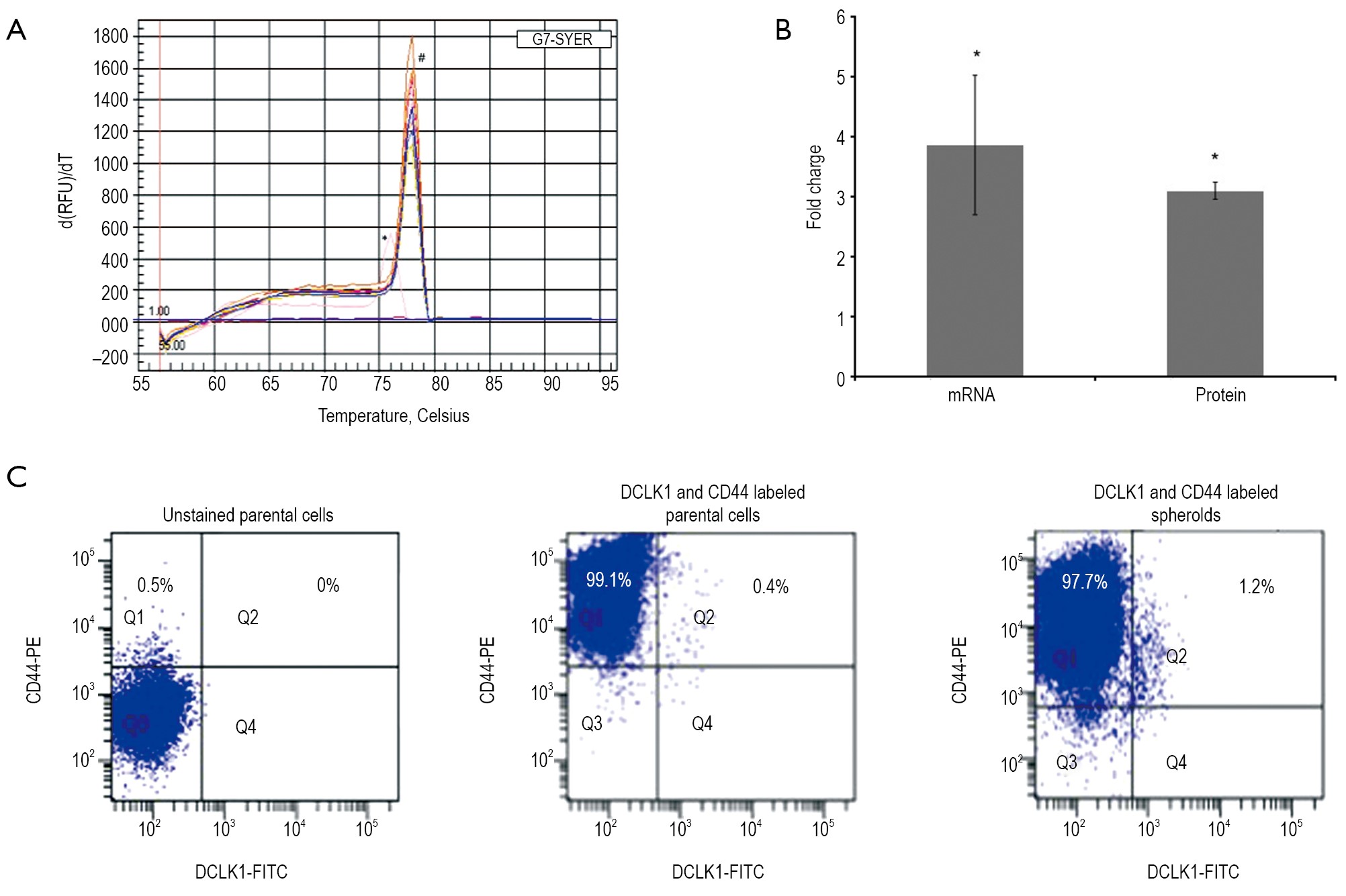
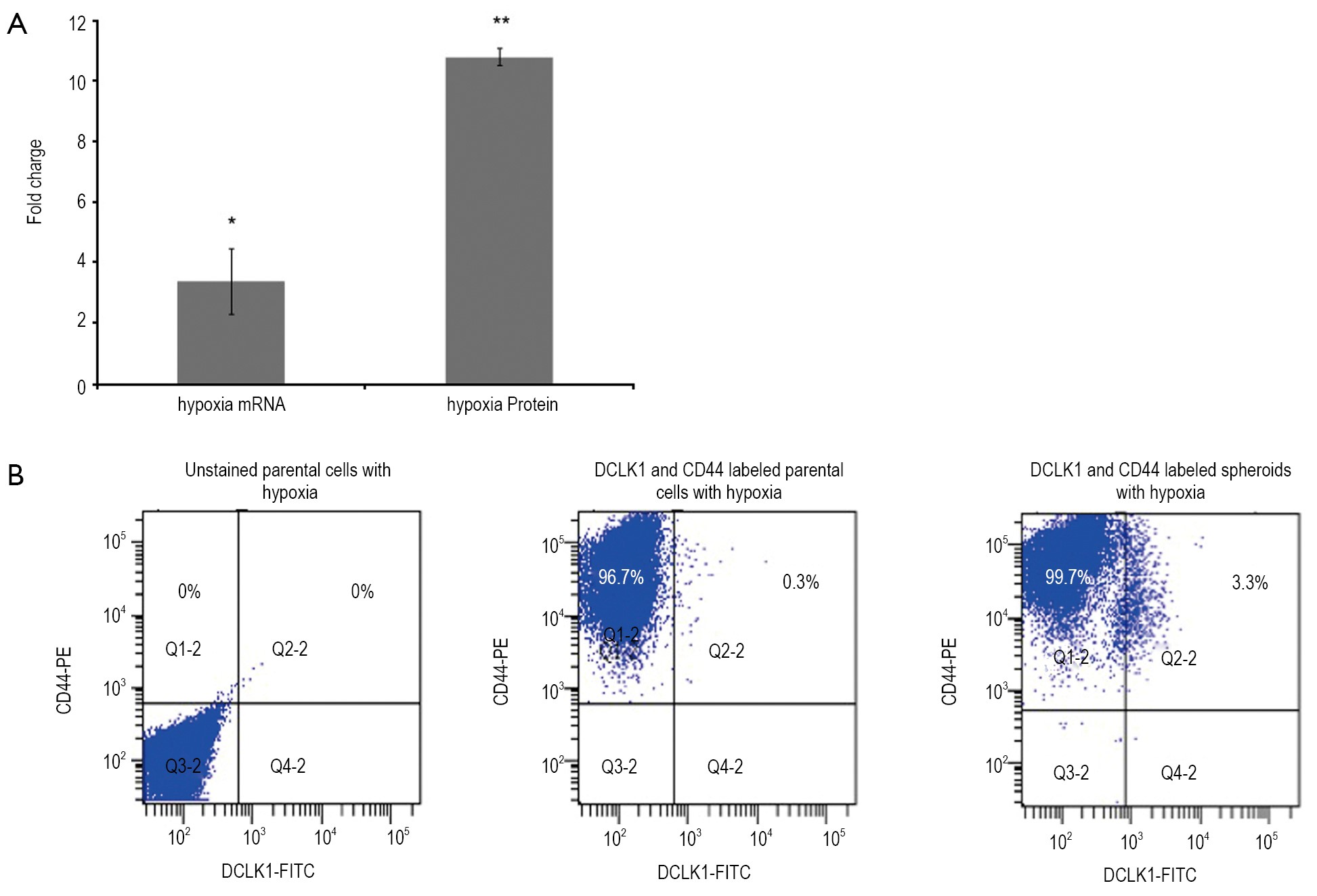
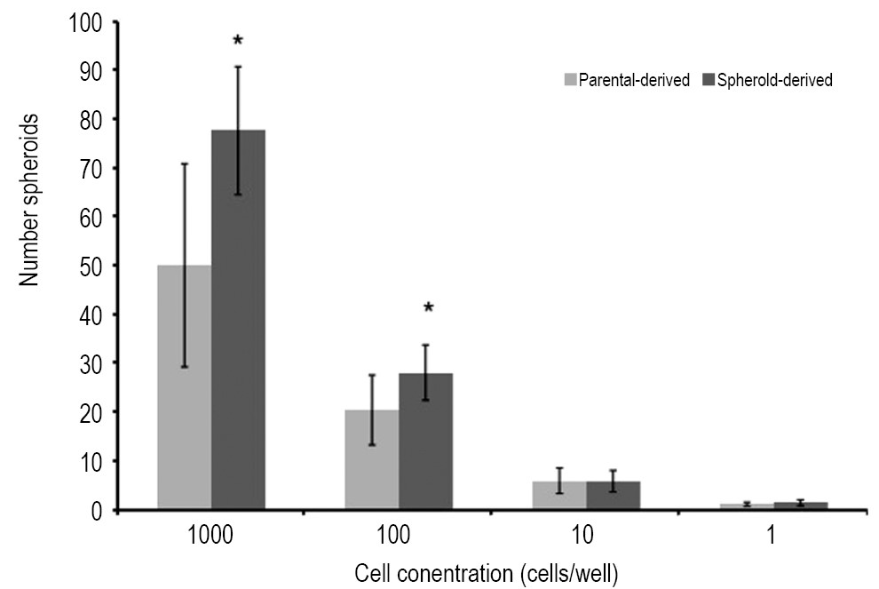
ObjectiveColon cancer stem cells (CSCs) are implicated in colorectal cancer carcinogenesis, metastasis, and therapeutic resistance. The identification of these cells could help to develop novel therapeutic strategies. Doublecortin-like kinase 1 (DCLK1) has been viewed as a marker for gastrointestinal stem cells that fuel the self-renewal process, however others view them as a marker of Tuft cells or as an enteroendocrine subtype. The purpose of this study was to use a colon cancer cell line to identify and characterize the stem-like characteristics of the DCLK1+ cell population. MethodsTo enrich stem-like cells, HCT116 cells (derived from colon adenocarcinomas) were cultured using serum-free media to form spheres under both normal oxygen and hypoxia condition. DCLK1 transcript expression in the adherent parental cells and spheroids was quantified using quantitative real time reverse transcription- polymerase chain reaction [(q)RT-PCR]. DCLK1 protein expression was determined using flow cytometry. Self-renewal capability from adherent parental cells and spheroids was determined using extreme limiting dilution analysis (ELDA). ResultsUnder both normal oxygen and hypoxia condition, the adherent parental cells were composed of cells that express low levels of DCLK1. However, spheroids exhibited an increased frequency of cells expressing DCLK1 on both mRNA and protein levels. Cells derived from spheroids also possess stronger self-renewal capability. ConclusionsThe higher fraction of DCLK1+ cells exhibited by spheroids and hypoxia reflects the stem-like characteristics of these cells. DCLK1 may represent an ideal marker to study and develop effective strategies to overcome chemo-resistance and relapse of colon cancer.
2013, 25(2): 143-154.
doi: 10.3978/j.issn.1000-9604.2013.01.02
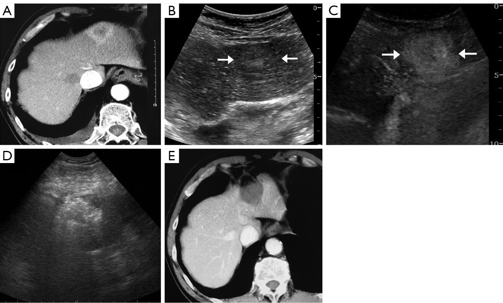
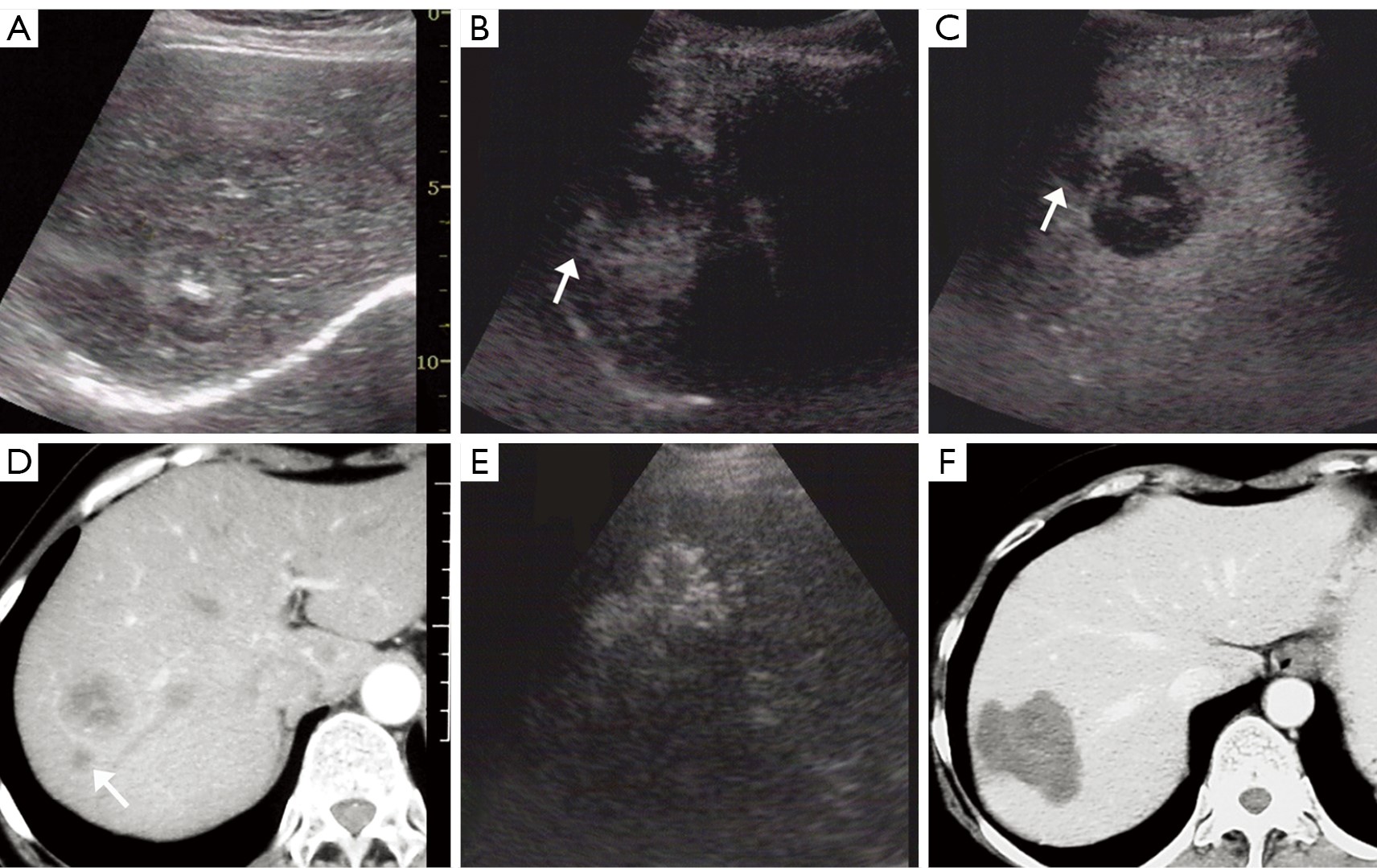
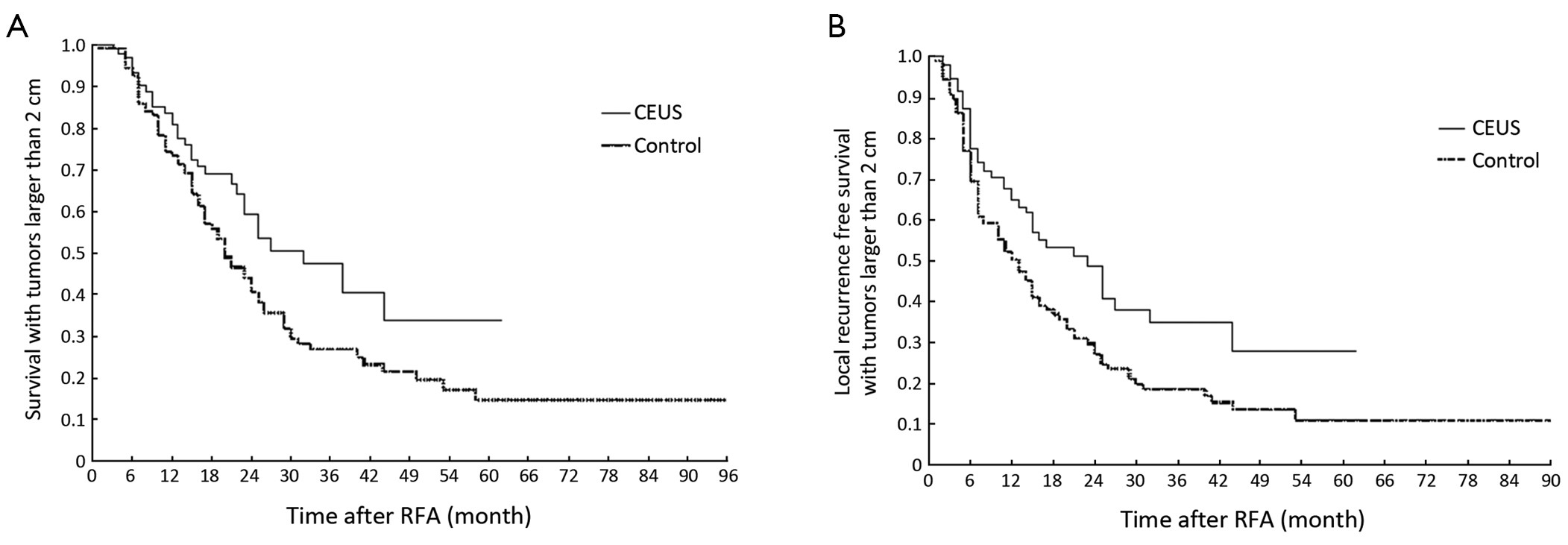
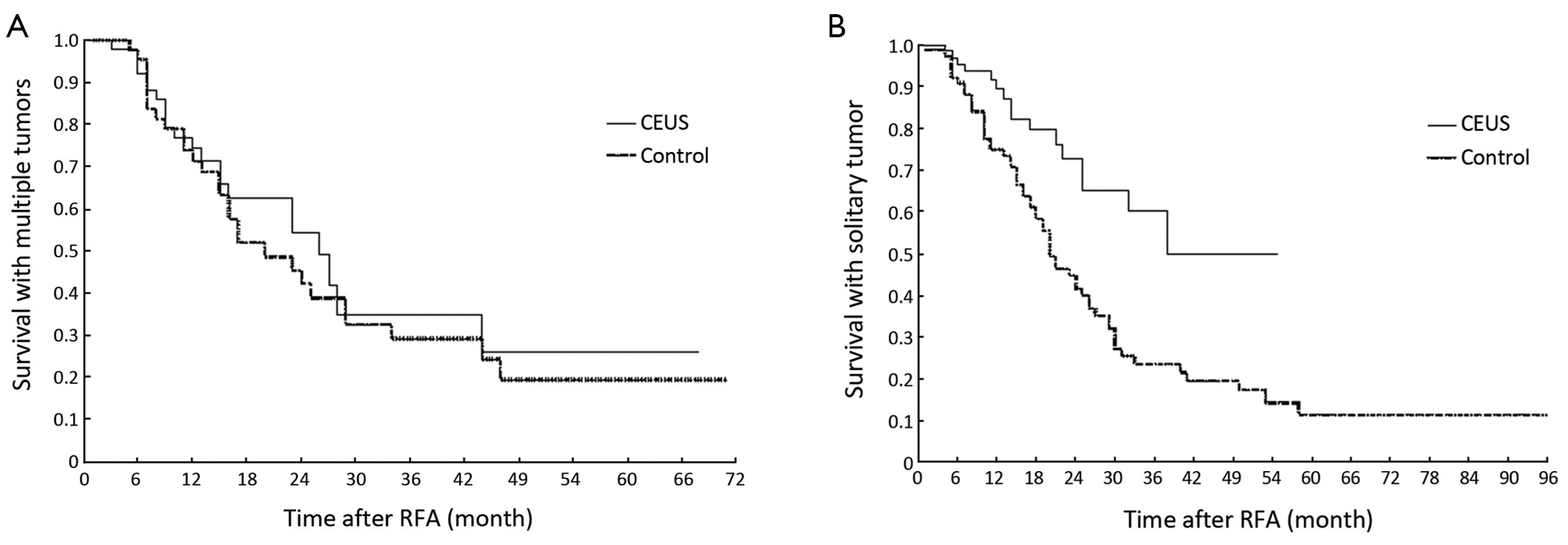
Abstract:
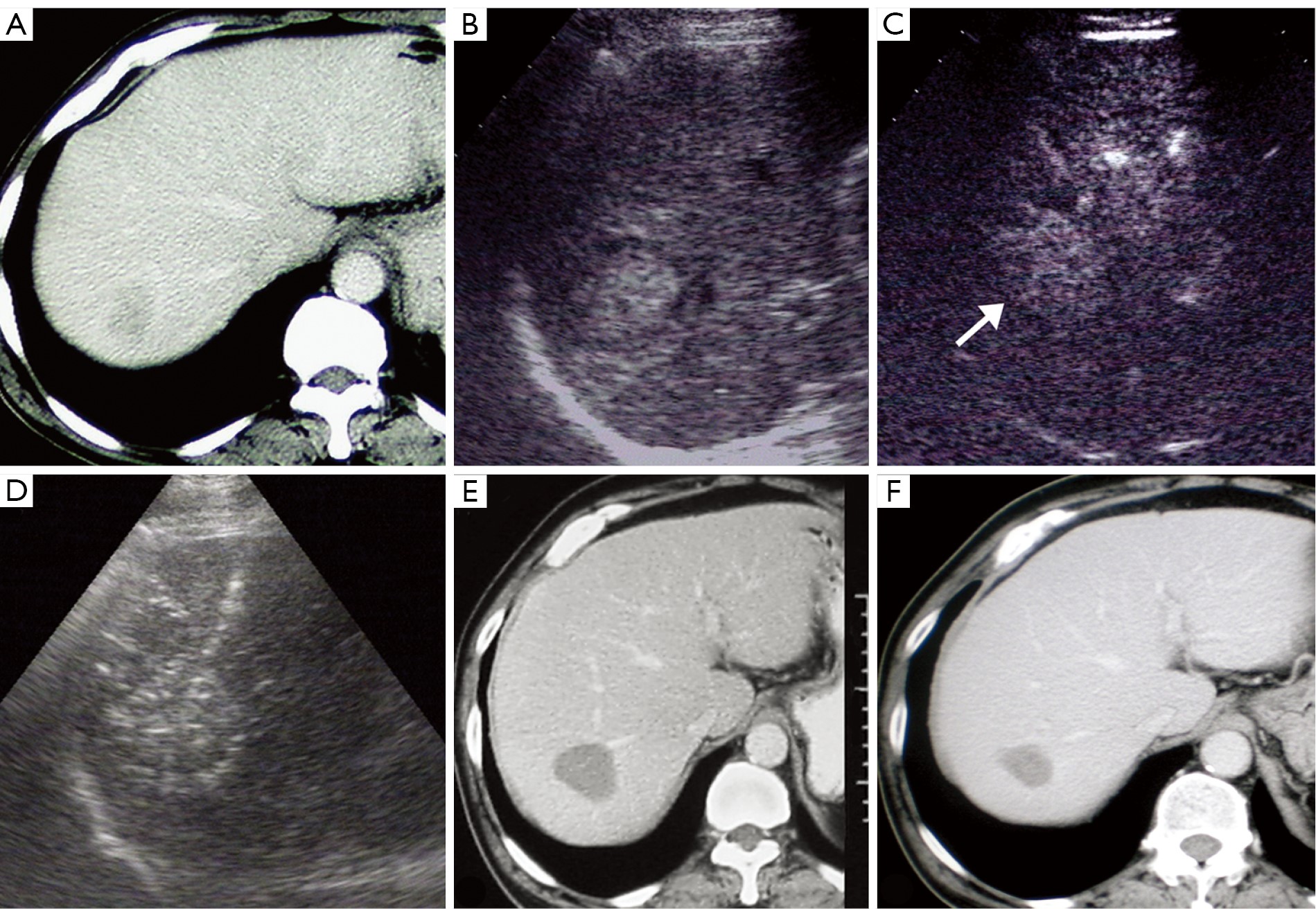
ObjectiveTo retrospectively investigate the role of contrast-enhanced ultrasonography (CEUS) in percutaneous radiofrequency ablation (RFA) in patients with liver metastases and evaluate the therapeutic efficacy of RFA assisted by CEUS. MethodsFrom May 2004 to September 2010, 136 patients with 219 liver metastatic lesions received CEUS examination 1 h before RFA (CEUS group), and other 126 patients with 216 lesions without CEUS examination in the earlier period were served as a historical control group. The mean tumor size was 3.2 cm and the mean tumor number was 1.6 in the CEUS group, while 3.4 cm and 1.7 in the control group, respectively (P>0.05). The clinical characteristics, recurrence results and survival outcomes were compared between two groups. ResultsIn the CEUS group, two isoechoic tumors were not demonstrated on unenhanced ultrasonography (US), and 63 (47%) of 134 tumors examined with CEUS were 0.3 cm larger than with unenhanced US. Furthermore, in 18.4% of 136 patients, additional 1-3 tumors were detected on CEUS. The CEUS group showed higher early tumor necrosis and lower intrahepatic recurrence than the control group. The 3-year overall survival (OS) rate and the 3-year local recurrence-free survival (LRFS) rate in the CEUS group were 50.1% and 38.3%, in contrast to 25.3% and 19.3% in the control group, respectively (P=0.002 and P<0.001). ConclusionsCEUS provides important information for RFA treatment in patients with liver metastases and better therapeutic effect could be attained.


2013, 25(2): 155-160.
doi: 10.3978/j.issn.1000-9604.2013.03.07
Abstract:
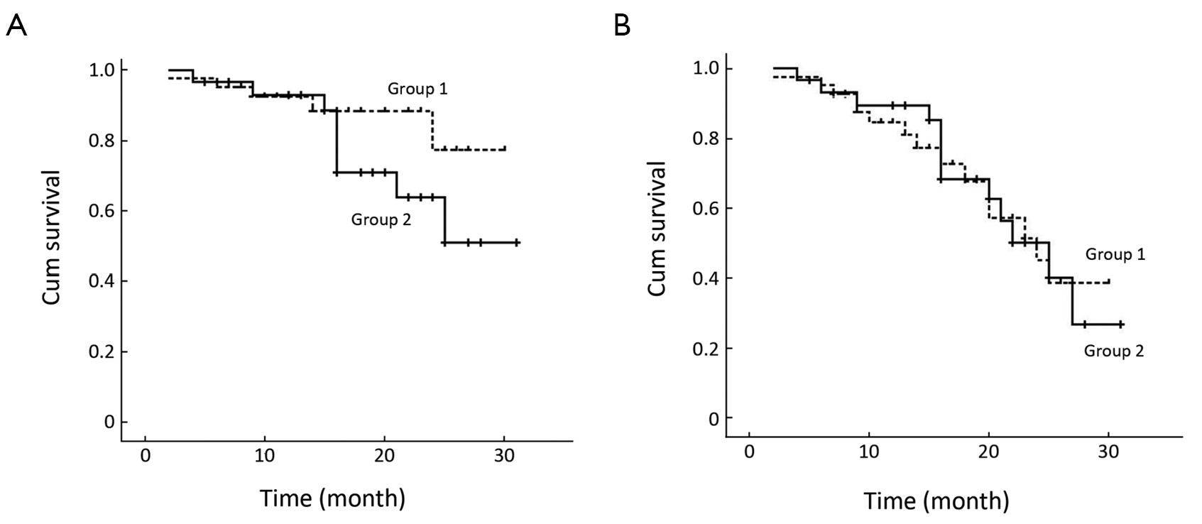
ObjectiveTo investigate the efficacy and safety of the treatment of the newly diagnosed multiple myeloma (MM) with or without renal impairment receiving the therapy of bortezomib, dexamethasone plus thalidomide (BTD) regimen in order to analyze the effects of BTD regimen on the prognosis of the MM patients with renal impairment compared with the patients without renal impairment. MethodsSeventy-two newly diagnosed MM patients entered into our study and all the patients belonged to International Stage System (ISS) 3 in which transplantation patients were excluded or the patients refused receiving transplantation therapy. According to the level of serum creatinine (Scr), the patients were divided into two groups including group 1 (n=42) (Scr <2 mg/dL) and group 2 (n=30) (Scr ≥2 mg/dL). All the patients received the therapy of BTD regimen as induction therapy, and the median treatment time was 5 (range, 2-8) cycles. The outcome was analyzed retrospectively. ResultsThe overall remission (OR) rates were 81.0% (group 1) and 80.0% (group 2). There was no statistical difference between the two groups (P>0.05). In group 2, 10 patients (33.3%) got renal function reversal, 14 patients (46.7%) got improved renal function and the median time to renal function reversal was 1.4 (range, 0.7-3.0) months. Among 12 patients with hemodialysis at diagnosis, 8 patients got rid of hemodialysis after median 4 cycles of therapy (range, 3-6 cycles). After a median follow-up period of 16 (range, 2-31) months, 5 patients (11.9%) in group 1 died and 9 patients (30.0%) in group 2 died (P=0.056). The 2-year estimate of overall survival was 77.3% in group 1 and 63.8% in group 2, respectively (P=0.188). During a median follow-up time of 13.0 months (range, 2-25 months), 15 patients (35.7%) in group 1 progressed and 13 patients (43.3%) in group 2 progressed (P=0.513). The 2-year estimate of response duration was 50.6% in group 1 and 42.1% in group 2, respectively (P=1). The main toxicities in the two groups included thrombocytopenia, peripheral neuropathy (PN), infection, herpes zoster and so on. The incidence of grade 3 and 4 adverse events was low. ConclusionsBTD regimen may become the front-line therapy for the newly diagnosed MM patients with renal impairment because BTD regimen can improve the prognosis of the patients with renal impairment as good as the patients without renal impairment.
2013, 25(2): 161-165.
doi: 10.3978/j.issn.1000-9604.2013.02.01
Abstract:
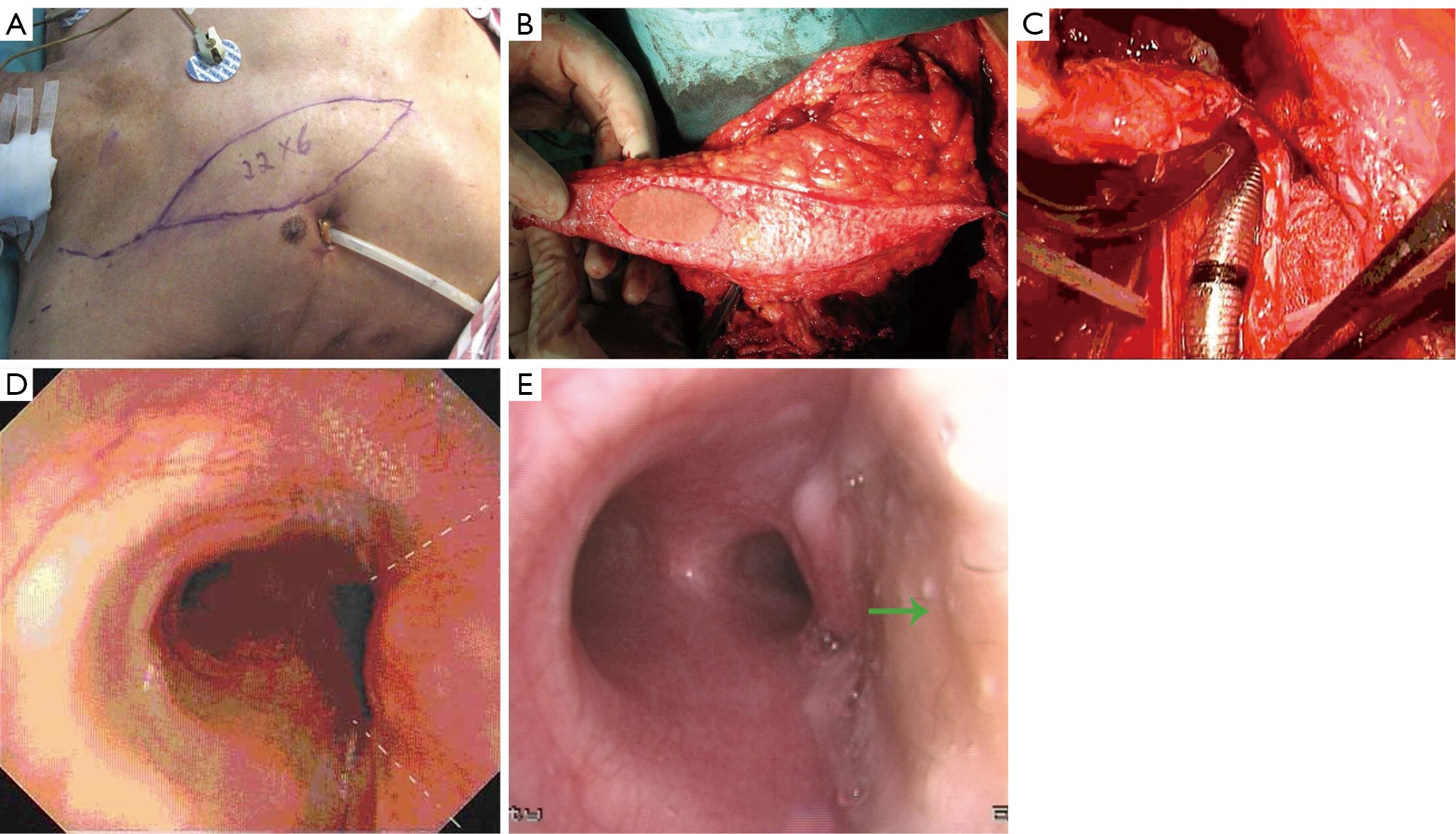
ObjectiveTo study the possibility of using portions of deepithelialized myocutaneous flaps to the reconstruction of thoracic tracheal defects after resection of a large tumor. MethodsFrom June 2007 to June 2012, five cases of defects of the thoracic trachea were reconstructed by applying portions of deepithelialized myocutaneous flaps. The patients were 27-61 years old with 4 male cases and 1 female. The cervical trachea ranged in diameter from 4-8.5 cm with circumferences of approximately 1/3-2/5 of the bronchial circumference. ResultsAll five patients with thoracic tracheal defects after resection of a large tumor were cured of portions of deepithelialized myocutaneous flaps, with no tracheal stricture remaining and vomica successfully eliminated. During the first 1 to 3 months after the operation, bronchoscopy showed that the tracheal lumens were smooth, and the visible skin of the musculocutaneous flaps became gray and exhibited a small amount of white discharge. ConclusionsDespite this being a small series and short follow-up, this thoracic tracheal reconstruction with portions of deepithelialized myocutaneous flaps shows encouraging preliminary results and could be an alternative to other methods for the treatment of carefully selected patients with thoracic tracheal defects.
2013, 25(2): 166-174.
doi: 10.3978/j.issn.1000-9604.2013.02.02
Abstract:
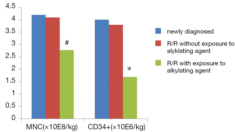
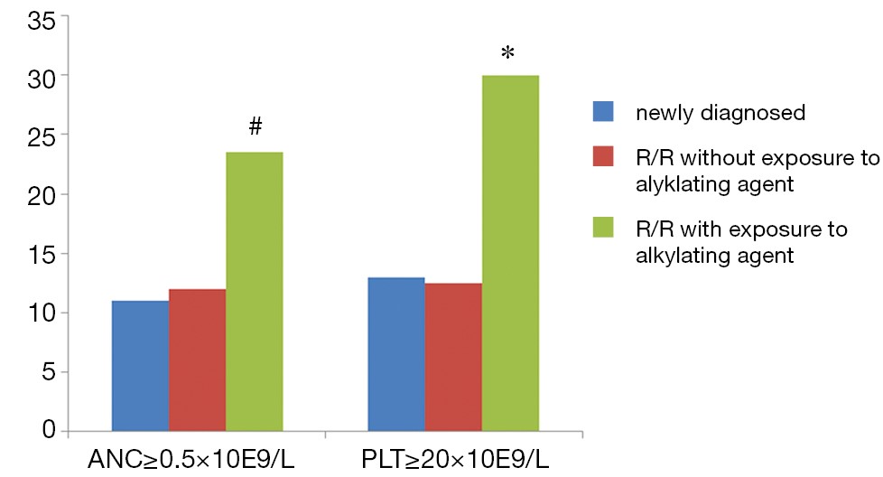
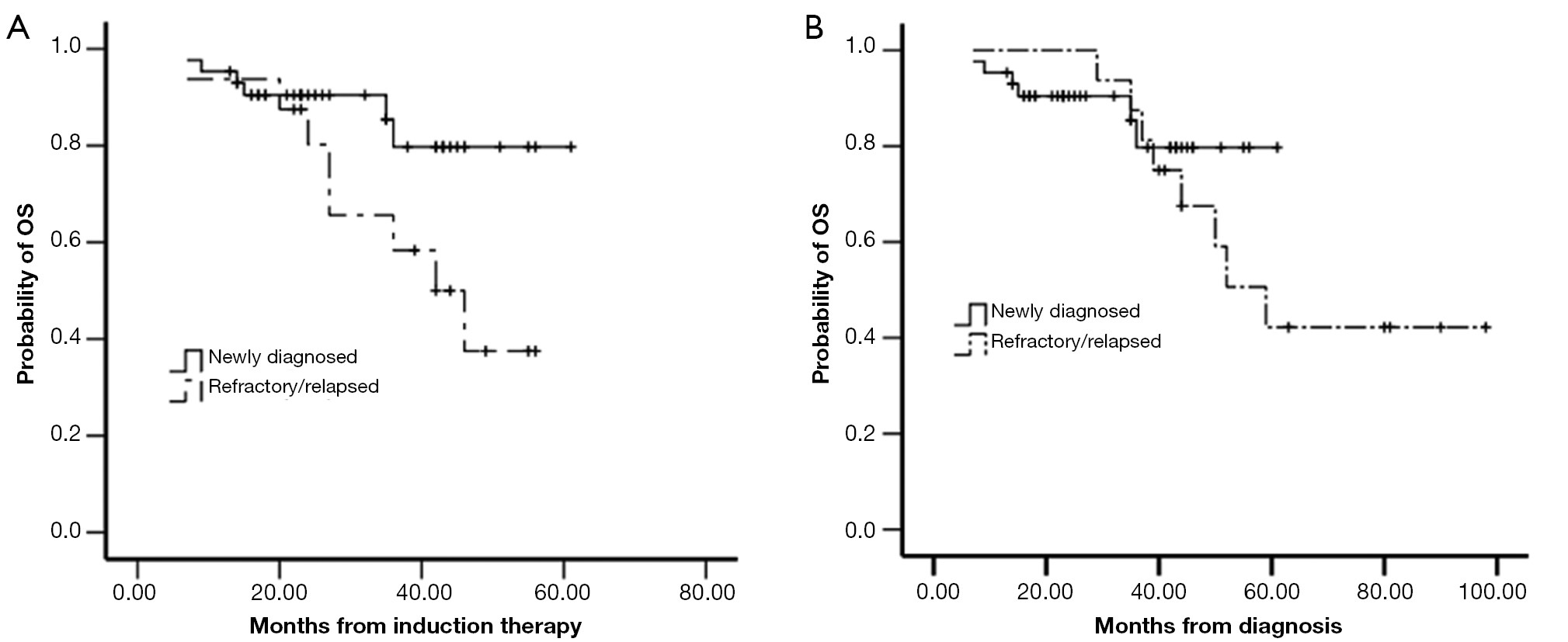
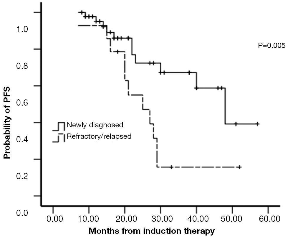
In our study, we determined the efficacy of bortezomib-based induction therapy followed by autologous stem cell transplant (ASCT) in newly diagnosed and relapsed/refractory (R/R) multiple myeloma (MM) patients and compared the advantages of early versus late transplant. We used a retrospective analysis to examine 62 patients, including 46 cases of newly diagnosed MM (early transplant group) and 16 cases of relapsed/refractory MM (late transplant group). All of these patients received bortezomib-based induction therapy followed by ASCT. The efficacy and side effects of the treatment regimen were analyzed. Patients’ overall survival (OS) and progression-free survival (PFS) times were determined. The ratio of complete remission to near-complete remission (CR/nCR) was 69.5% versus 56.2% (P=0.361), respectively, for the early transplant group versus the late transplant group, respectively, after receiving bortezomib-based induction therapy; the overall response rates of the two group were 91.3% and 81.2%, respectively (P=0.369). After receiving ASCT, the CR/nCR of the two groups increased to 84.8% and 81.3%, respectively. The median time required for neutrophil engraftment of the early transplant group and the late transplant group was 11 and 14.5 days, respectively (P=0.003); the median time required for platelet engraftment was 13 and 21.5 days (P=0.031), respectively. There were no significant differences in the toxic side effects observed during induction therapy and ASCT between the two groups. The OS of the two groups was not statistically different (P=0.058). The PFS of the early transplant group and the late transplant group was 41.6 and 26.5 months, respectively (P=0.008). Multivariate analysis demonstrated that the time of receiving ASCT, the types of M protein, and the International Staging System (ISS) stage were all independent factors that influenced PFS. In conclusion, patients in a suitable condition for ASCT should be recommended to have an early ASCT immediately after diagnosis.
In our study, we determined the efficacy of bortezomib-based induction therapy followed by autologous stem cell transplant (ASCT) in newly diagnosed and relapsed/refractory (R/R) multiple myeloma (MM) patients and compared the advantages of early versus late transplant. We used a retrospective analysis to examine 62 patients, including 46 cases of newly diagnosed MM (early transplant group) and 16 cases of relapsed/refractory MM (late transplant group). All of these patients received bortezomib-based induction therapy followed by ASCT. The efficacy and side effects of the treatment regimen were analyzed. Patients’ overall survival (OS) and progression-free survival (PFS) times were determined. The ratio of complete remission to near-complete remission (CR/nCR) was 69.5% versus 56.2% (P=0.361), respectively, for the early transplant group versus the late transplant group, respectively, after receiving bortezomib-based induction therapy; the overall response rates of the two group were 91.3% and 81.2%, respectively (P=0.369). After receiving ASCT, the CR/nCR of the two groups increased to 84.8% and 81.3%, respectively. The median time required for neutrophil engraftment of the early transplant group and the late transplant group was 11 and 14.5 days, respectively (P=0.003); the median time required for platelet engraftment was 13 and 21.5 days (P=0.031), respectively. There were no significant differences in the toxic side effects observed during induction therapy and ASCT between the two groups. The OS of the two groups was not statistically different (P=0.058). The PFS of the early transplant group and the late transplant group was 41.6 and 26.5 months, respectively (P=0.008). Multivariate analysis demonstrated that the time of receiving ASCT, the types of M protein, and the International Staging System (ISS) stage were all independent factors that influenced PFS. In conclusion, patients in a suitable condition for ASCT should be recommended to have an early ASCT immediately after diagnosis.
2013, 25(2): 175-182.
doi: 10.3978/j.issn.1000-9604.2013.02.03
Abstract:
The aims of this study were to explore whether laparoscopic surgical resections of gastric gastrointestinal stromal tumors (GISTs) would produce better perioperative and similar oncologic outcomes compared with open surgical resection in Chinese patients. Thirty-six gastric GISTs cases were divided into a minimally invasive laparoscopic group and open resection group, depending on the surgical approach that was used. The general preoperative information, operative time, incision length, intraoperative blood loss, postoperative time to first flatulence, postoperative complications, postoperative hospital stay, total hospitalization costs, and such follow-up data as recurrence, metastasis, and mortality rates were compared between two groups. Among the 36 gastric GISTs, 15 received laparoscopic surgical treatment (laparoscopy group, n=15), and 21 received routine open resection treatment (open resection group, n=21). The laparoscopy group and the open resection group showed statistically significant differences (P<0.05) in incision length (7.8±2.3 vs. 16.9±3.8 cm), postoperative time to first flatulence (3.8±1.3 vs. 5.1±2.1 d), postoperative hospitalization time (7.6±2.5 vs. 11.3±3.7 d), and total cost of hospitalization (RMB 28,239±5,521 vs. RMB 23,761±5,362). There were no statistically significant differences (P>0.05) between the laparoscopy group and the open resection group in operative time (147.8±59.3 vs. 139.2±62.1 min) and intraoperative blood loss (149.8±98.9 vs. 154.2±99.3 mL). Both groups had no postoperative complications, no recurrence and metastasis, and no postoperative mortality. There were no statistically significant differences between the two groups in postoperative complications, postoperative recurrence and metastasis, and postoperative mortality. In conclusion, compared with open resection, the laparoscopic resection of gastric GISTs offers the advantages of less trauma, faster recovery, and shorter hospital stay.
The aims of this study were to explore whether laparoscopic surgical resections of gastric gastrointestinal stromal tumors (GISTs) would produce better perioperative and similar oncologic outcomes compared with open surgical resection in Chinese patients. Thirty-six gastric GISTs cases were divided into a minimally invasive laparoscopic group and open resection group, depending on the surgical approach that was used. The general preoperative information, operative time, incision length, intraoperative blood loss, postoperative time to first flatulence, postoperative complications, postoperative hospital stay, total hospitalization costs, and such follow-up data as recurrence, metastasis, and mortality rates were compared between two groups. Among the 36 gastric GISTs, 15 received laparoscopic surgical treatment (laparoscopy group, n=15), and 21 received routine open resection treatment (open resection group, n=21). The laparoscopy group and the open resection group showed statistically significant differences (P<0.05) in incision length (7.8±2.3 vs. 16.9±3.8 cm), postoperative time to first flatulence (3.8±1.3 vs. 5.1±2.1 d), postoperative hospitalization time (7.6±2.5 vs. 11.3±3.7 d), and total cost of hospitalization (RMB 28,239±5,521 vs. RMB 23,761±5,362). There were no statistically significant differences (P>0.05) between the laparoscopy group and the open resection group in operative time (147.8±59.3 vs. 139.2±62.1 min) and intraoperative blood loss (149.8±98.9 vs. 154.2±99.3 mL). Both groups had no postoperative complications, no recurrence and metastasis, and no postoperative mortality. There were no statistically significant differences between the two groups in postoperative complications, postoperative recurrence and metastasis, and postoperative mortality. In conclusion, compared with open resection, the laparoscopic resection of gastric GISTs offers the advantages of less trauma, faster recovery, and shorter hospital stay.
2013, 25(2): 183-191.
doi: 10.3978/j.issn.1000-9604.2013.02.06
Abstract:
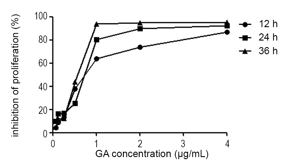
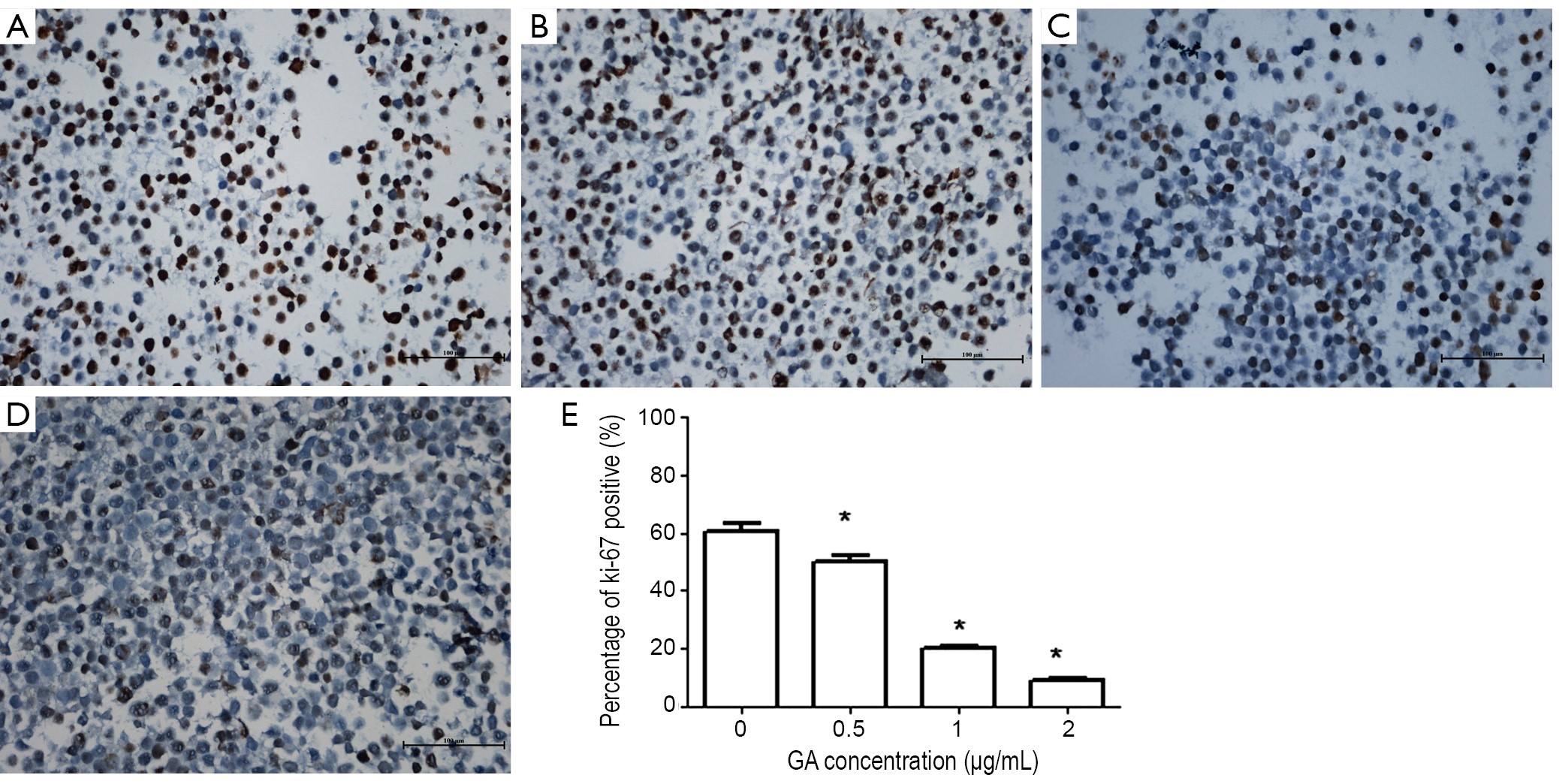
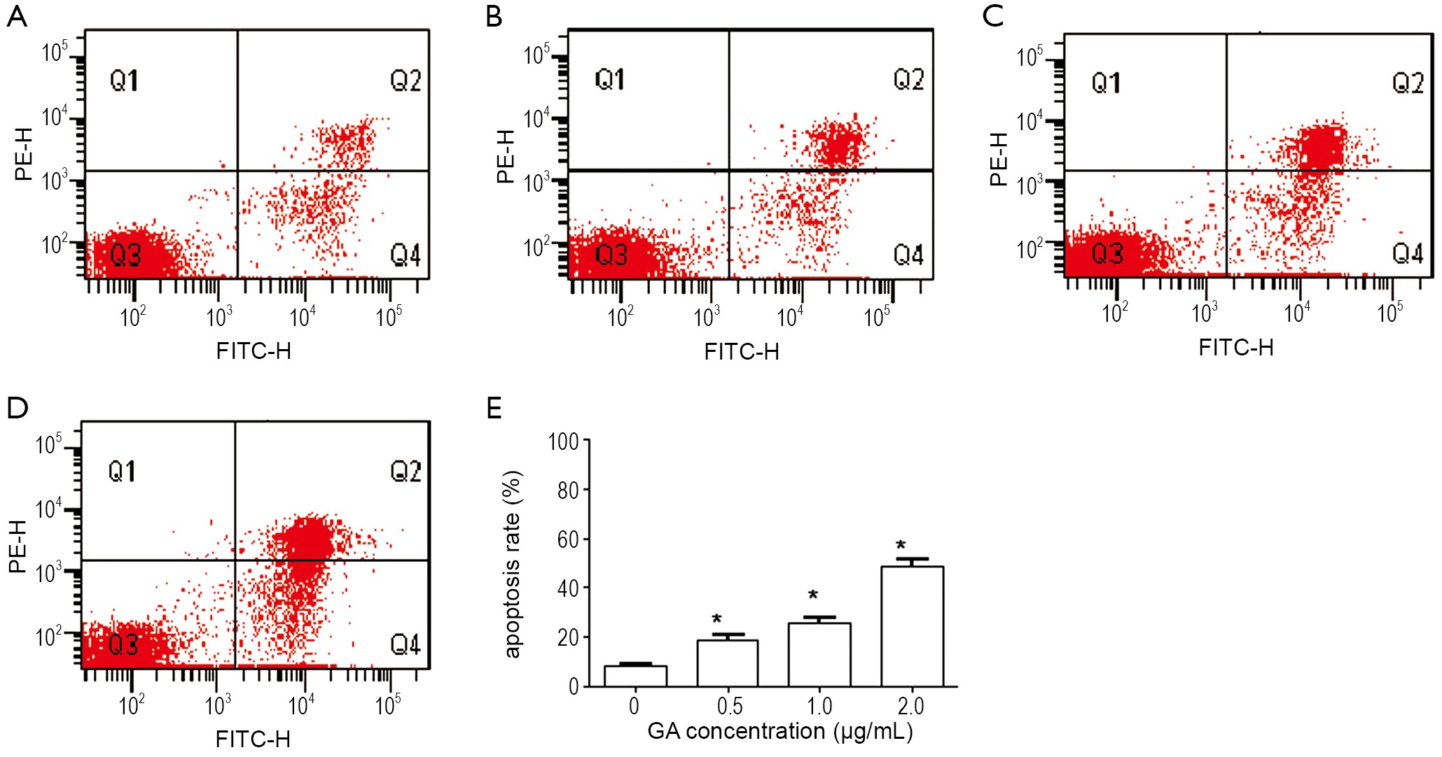
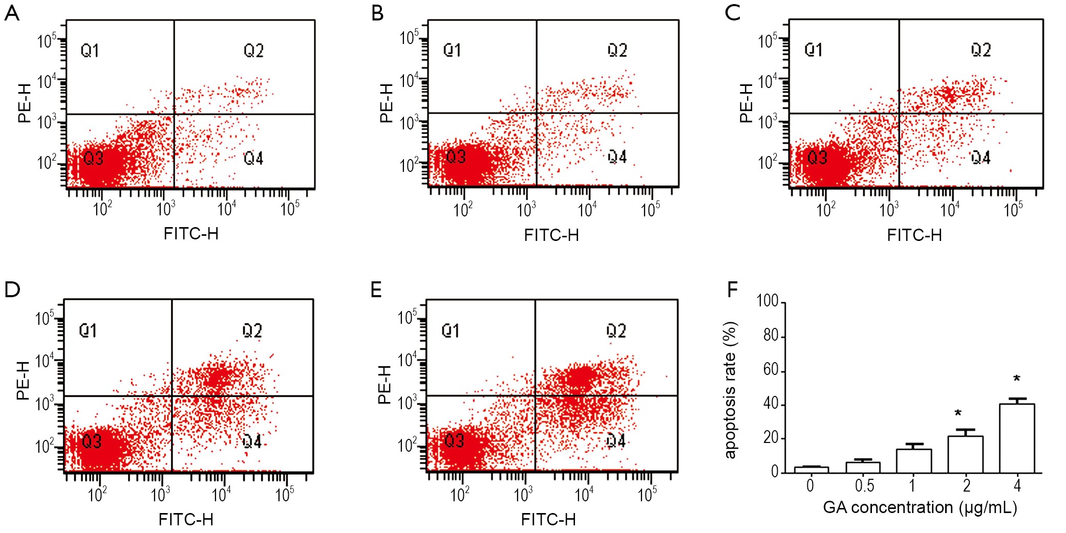
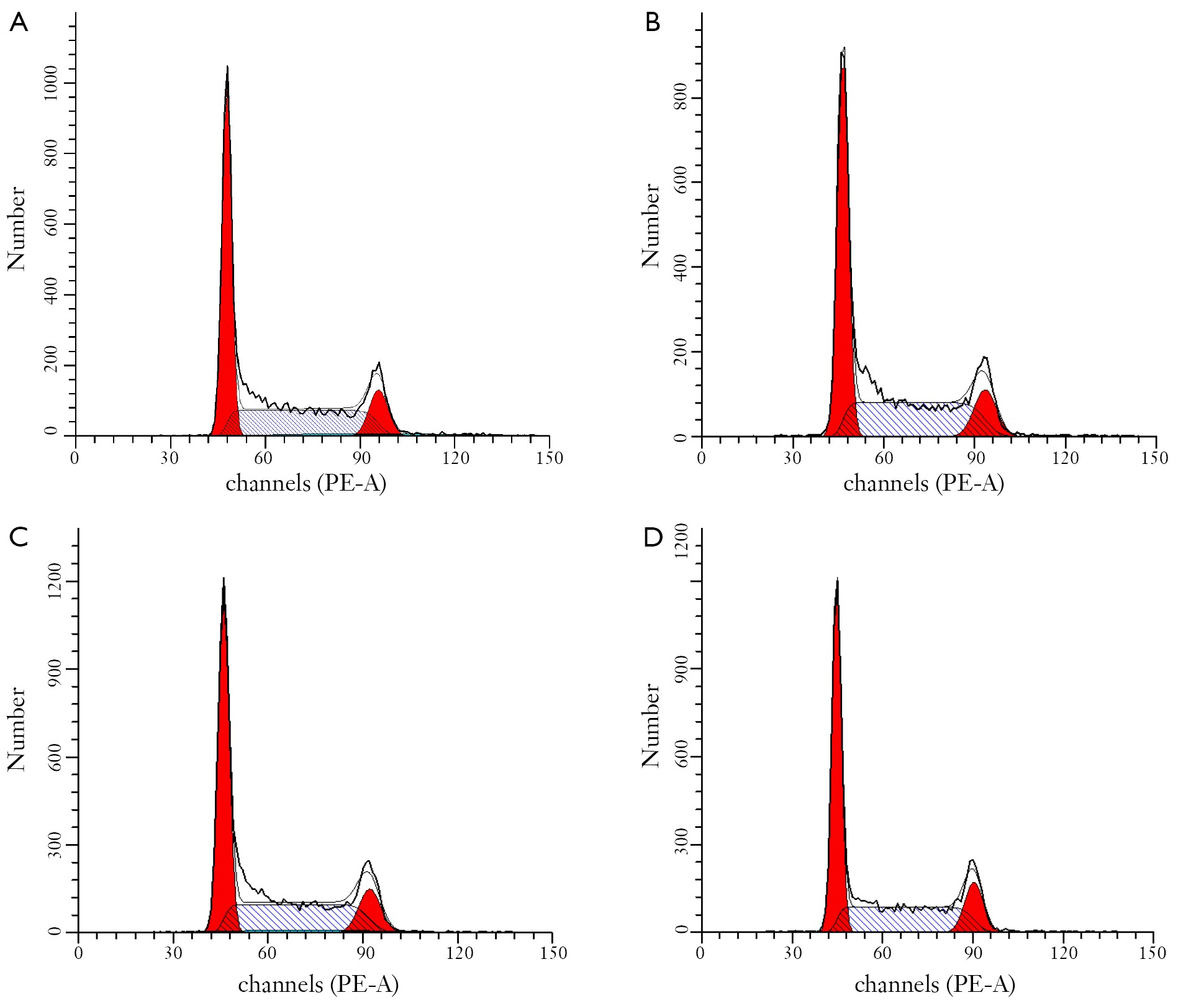
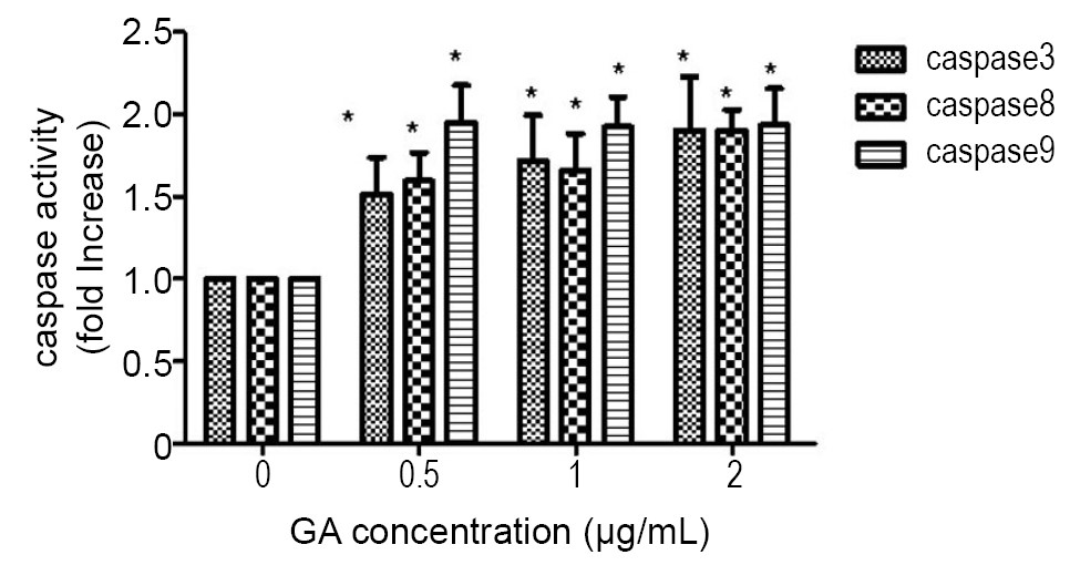
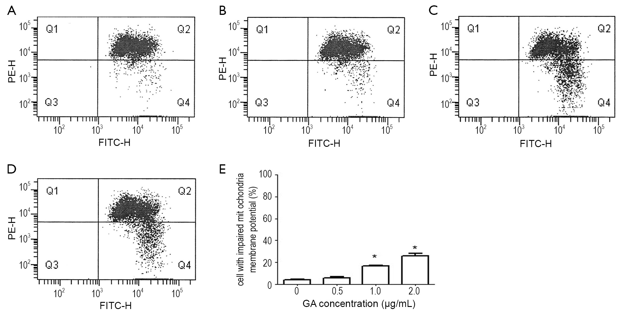
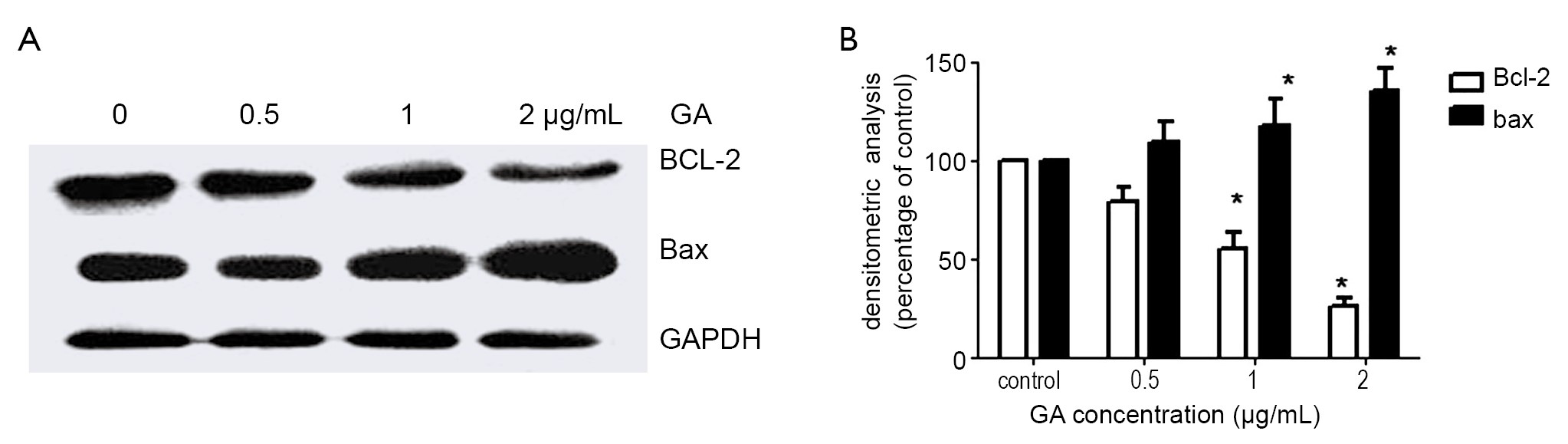
ObjectiveTo study the mechanisms in gambogic acid (GA) -induced JeKo-1 human Mantle Cell Lymphoma cell apoptosis in vitro. MethodsThe proliferation of GA-treated JeKo-1 cells was measured by CCK-8 assay and Ki-67 immunocytochemical detection. Apoptosis, cell cycle and mitochondrial membrane potential were measured by flow cytometric analysis. Caspase-3, -8 and -9 were detected by colorimetric assay. Bcl-2 and Bax were analyzed by Western blotting. ResultsGA inhibited cell growth in a time- and dose- dependent manner. GA induces apoptosis in JeKo-1 cells but not in normal bone marrow cells, which was involved in reducing the membrane potential of mitochondria, activating caspases-3, -8 and -9 and decreasing the ratio of Bcl-2 and Bax without cell cycle arresting. ConclusionsGA induced apoptosis in human MCL JeKo-1 cells by regulating Bcl-2/Bax and activating caspase-3, -8 and -9 via mitochondrial pathway without affecting cell cycle.
2013, 25(2): 192-199.
doi: 10.3978/j.issn.1000-9604.2013.03.01
Abstract:
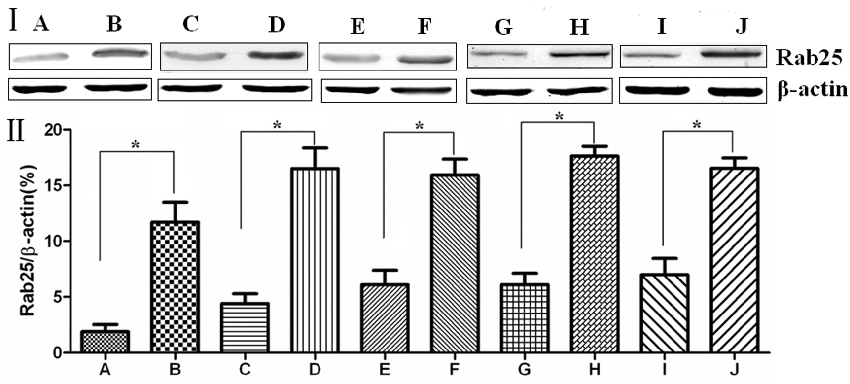
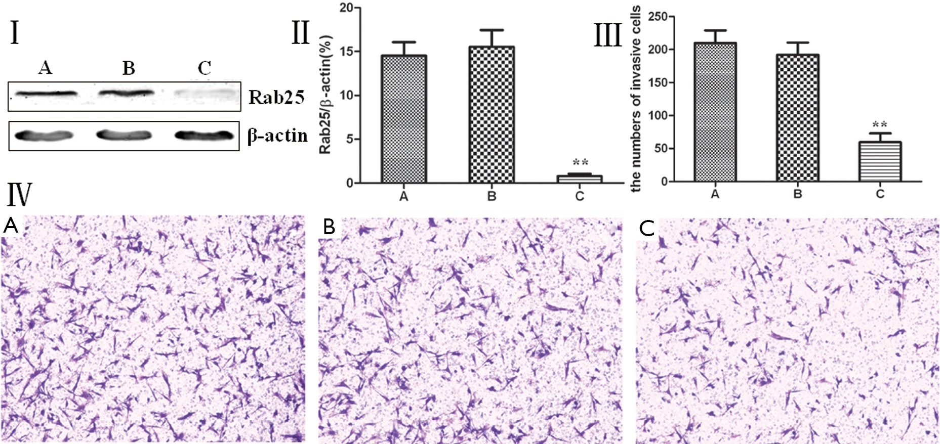
The objective of this study was to determine the expression of the important vesicle trafficking-regulating factor Rab25 in human gastric cancer tissues, to analyze the correlation between Rab25 protein expression with gastric cancer occurrence and development, and to discuss the correlation of Rab25 protein expression with gastric cancer cell metastasis. The overall aim was to provide experimental evidence that can be used to design future biological treatments of human gastric cancer. Human gastric cancer tissue and the adjacent normal gastric tissue were surgically removed, and immunohistochemistry and Western blotting were used to detect Rab25 protein expression. The correlation between Rab25 protein expression with the development and pathological characteristics of gastric cancer was analyzed. Using RNAi, Rab25 expression was reduced in the gastric cancer cell line MGC80-3, and the changes in MGC80-3 cell invasiveness were then monitored. Immunohistochemistry showed that the Rab25 protein expression rates were 78.21% and 23.08% in gastric carcinoma and the adjacent normal gastric tissue, respectively. Immunohistochemistry and Western blot results showed that Rab25 protein expression in gastric cancer was significantly higher than in adjacent normal gastric tissues (P<0.01). Less differentiated gastric cancer cells had higher expression of Rab25 protein (P<0.01). Gastric carcinomas from patients with a late pathological stage (III-IV) had significantly higher Rab25 protein expression than early stage (I-II) patients (P<0.01). Gastric carcinomas from patients with lymph node metastasis had significantly higher Rab25 protein expression than lymph node metastasis-free patients (P<0.01). Gastric carcinomas from patients with distant metastases had significantly higher Rab25 protein expression than the distant metastasis-negative patients (P<0.01). Rab25 protein expression in gastric cancer was not affected by the patients, sex, age, or tumor size (P>0.05). MGC80-3 cells transfected with Rab25 siRNA had significantly lower Rab25 protein expression (P<0.01) and a significantly lower number of cells that passed through a Transwell chamber compared with non-transfected controls and the transfected control group (P<0.01). Rab25 protein expression is associated with the development of gastric cancer. siRNA knockdown of Rab25 protein expression in MGC80-3 gastric cancer cells reduced MGC80-3 cell invasiveness and provided experimental evidence for potential future biological treatment strategies of human gastric cancer.
The objective of this study was to determine the expression of the important vesicle trafficking-regulating factor Rab25 in human gastric cancer tissues, to analyze the correlation between Rab25 protein expression with gastric cancer occurrence and development, and to discuss the correlation of Rab25 protein expression with gastric cancer cell metastasis. The overall aim was to provide experimental evidence that can be used to design future biological treatments of human gastric cancer. Human gastric cancer tissue and the adjacent normal gastric tissue were surgically removed, and immunohistochemistry and Western blotting were used to detect Rab25 protein expression. The correlation between Rab25 protein expression with the development and pathological characteristics of gastric cancer was analyzed. Using RNAi, Rab25 expression was reduced in the gastric cancer cell line MGC80-3, and the changes in MGC80-3 cell invasiveness were then monitored. Immunohistochemistry showed that the Rab25 protein expression rates were 78.21% and 23.08% in gastric carcinoma and the adjacent normal gastric tissue, respectively. Immunohistochemistry and Western blot results showed that Rab25 protein expression in gastric cancer was significantly higher than in adjacent normal gastric tissues (P<0.01). Less differentiated gastric cancer cells had higher expression of Rab25 protein (P<0.01). Gastric carcinomas from patients with a late pathological stage (III-IV) had significantly higher Rab25 protein expression than early stage (I-II) patients (P<0.01). Gastric carcinomas from patients with lymph node metastasis had significantly higher Rab25 protein expression than lymph node metastasis-free patients (P<0.01). Gastric carcinomas from patients with distant metastases had significantly higher Rab25 protein expression than the distant metastasis-negative patients (P<0.01). Rab25 protein expression in gastric cancer was not affected by the patients, sex, age, or tumor size (P>0.05). MGC80-3 cells transfected with Rab25 siRNA had significantly lower Rab25 protein expression (P<0.01) and a significantly lower number of cells that passed through a Transwell chamber compared with non-transfected controls and the transfected control group (P<0.01). Rab25 protein expression is associated with the development of gastric cancer. siRNA knockdown of Rab25 protein expression in MGC80-3 gastric cancer cells reduced MGC80-3 cell invasiveness and provided experimental evidence for potential future biological treatment strategies of human gastric cancer.

2013, 25(2): 200-205.
doi: 10.3978/j.issn.1000-9604.2013.03.04
Abstract:
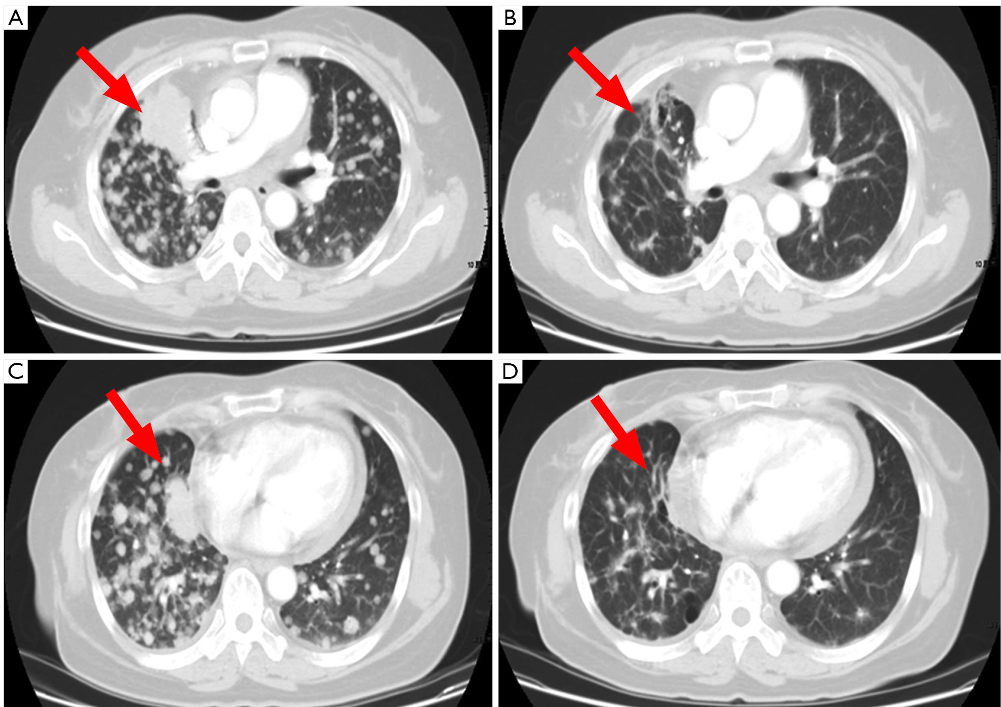
ObjectiveTo observe the efficacy and safety of albumin-bound paclitaxel (ABP) monotherapy in treating recurrent advanced non-small-cell lung cancer (NSCLC). MethodsWe retrospectively analyzed the short-term efficacy and toxicities of ABP monotherapy in treating 21 patients who had previously undergone multiple cycles of therapy for their advanced NSCLC in our hospital since 2010. The treatment-related survival was also analyzed. ResultsOf these 21 patients, the best overall response was partial response (PR) in 6 patients (28.6%), stable disease (SD) in 10 patients (47.6%), and progressive disease (PD) in 5 patients (23.8%). The overall response rate (ORR) was 28.6% and the disease control rate (DCR) (PR + SD) was 76.2%. The median progression-free survival (PFS) was 4.0 months (95% CI, 5.0-7.0 months). The main grade 3/4 toxicities included neutropenia (11.1%), peripheral nerve toxicity (5.6%), muscle and joint aches (5.6%), and fatigue (5.6%). ConclusionsThe ABP monotherapy can achieve good objective response in advanced NSCLC patients who have previously received multiple cycles of treatment and be well tolerated.

2013, 25(2): 206-211.
doi: 10.3978/j.issn.1000-9604.2013.03.10
Abstract:
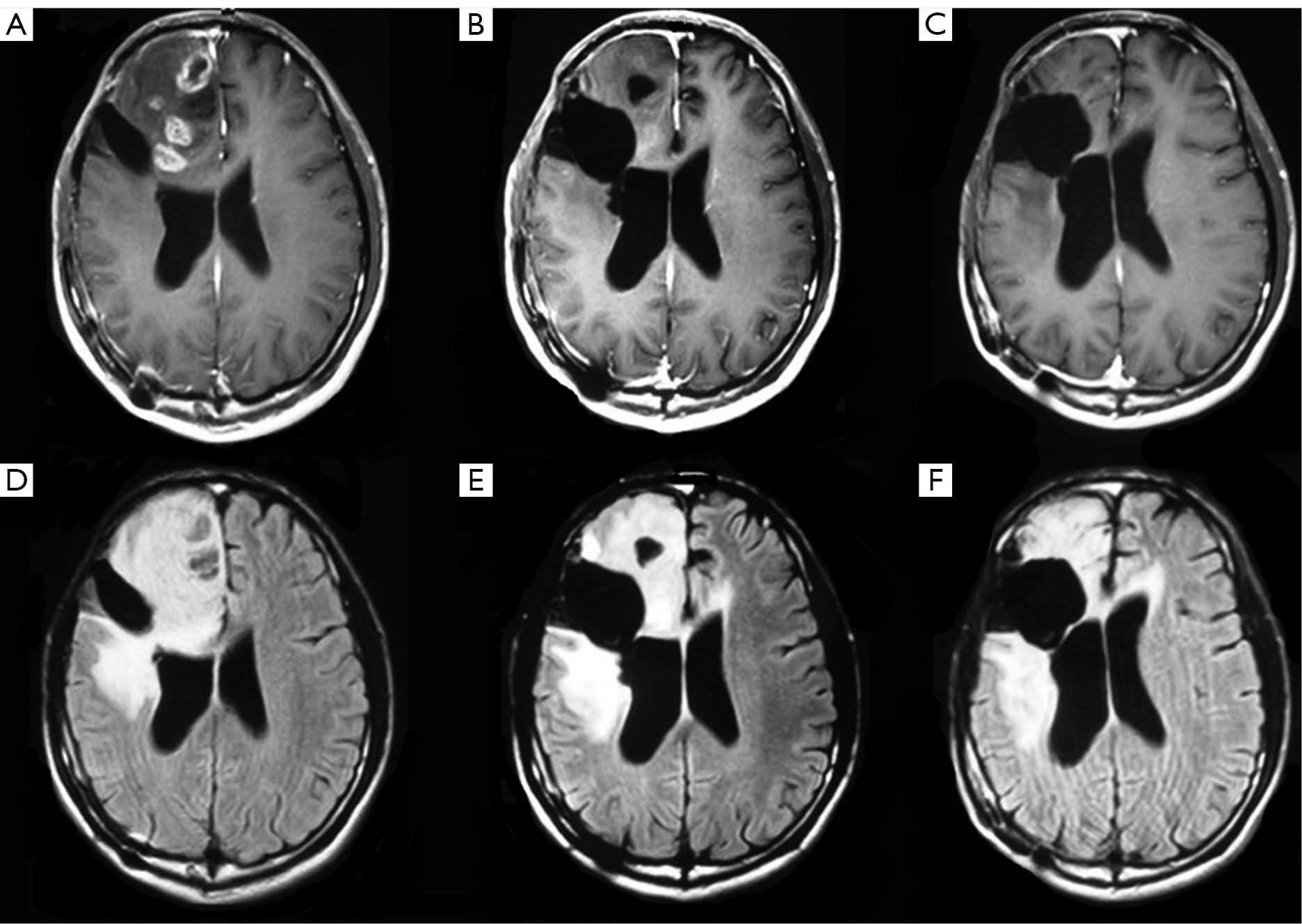
ObjectiveWe retrospectively studied the efficacy of bevacizumab as salvage therapy for recurrent malignant glioma with a focus on the overall survival (OS). MethodsPatients who received a therapy other than surgery for recurrent malignant glioma were included. Efficacy was evaluated using MRI. Neurological function was evaluated using the Response Assessment in Neuro-Oncology (RANO). The survival rate was calculated using the Kaplan-Meier method. ResultsFifty-one patients with recurrent glioma (31 grade III, 20 grade IV) were included. Among them, 22 subjects (43.1%) received bevacizumab. The median OS was 10.2 months (range, 1 to 27 months). Patients receiving bevacizumab had comparable OS (a median of 9.9 vs. 10.0 months) and similar 6-month survival rate (43% vs. 34%) to those who did not receive bevacizumab. A subgroup analysis failed to notice any significant difference in grade III glioma patients receiving bevacizumab vs. those who did not. The median survival was significantly longer at 8.9 months (range, 4 to 13 months) in grade IV glioma patients receiving bevacizumab than in those who did not (5.6 months, range, 2 to 7 months, P=0.042). The 6-month survival rate was higher (83%) in those who received bevacizumab than in those who did not (47%, P=0.046). No grade 3/4 adverse events were observed in any patient. ConclusionsBevacizumab, as a rescue therapy, provides a survival benefit for recurrent grade IV glioma.
2013, 25(2): 212-222.
doi: 10.3978/j.issn.1000-9604.2013.04.01
Abstract:
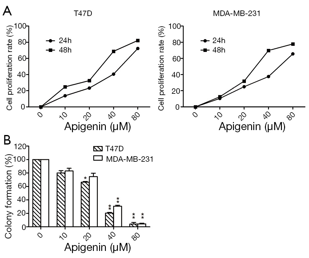
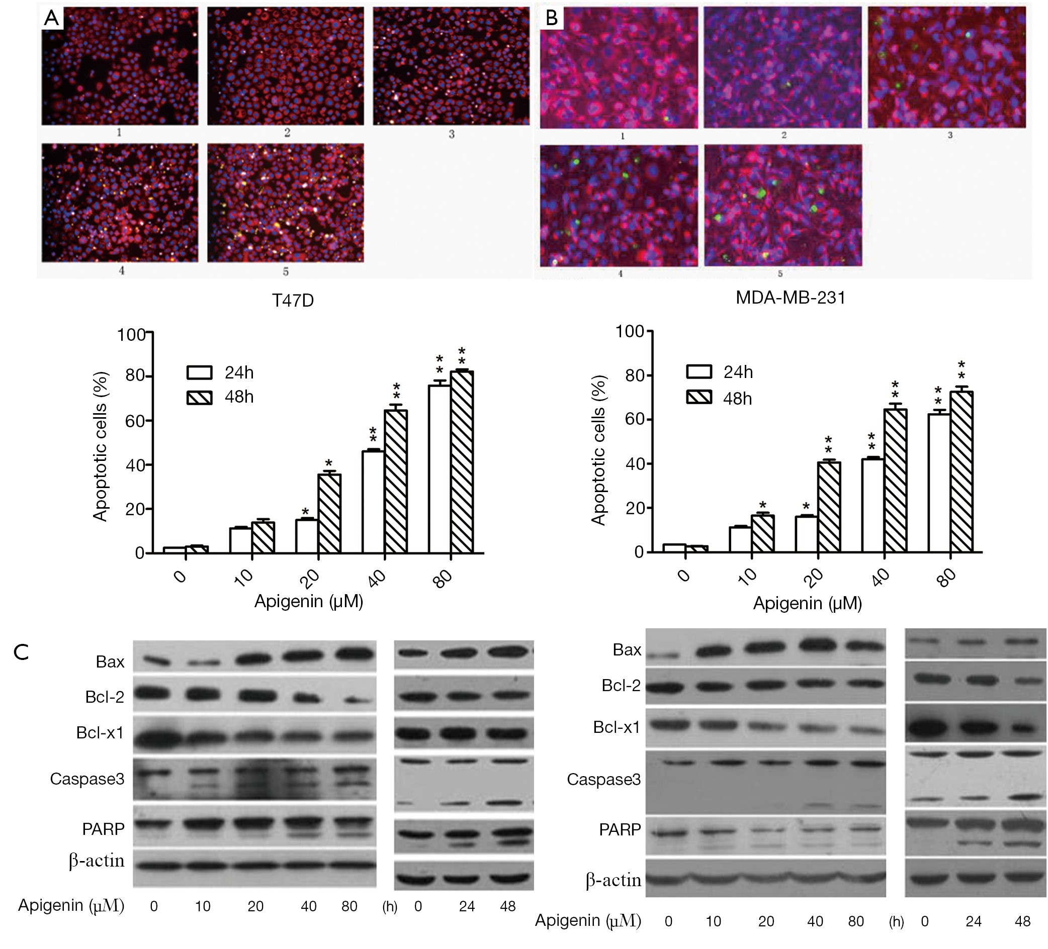
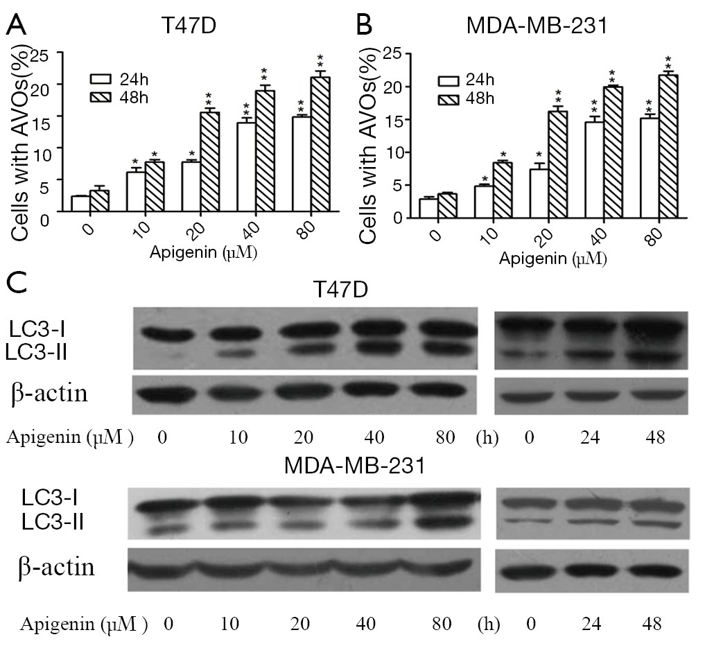
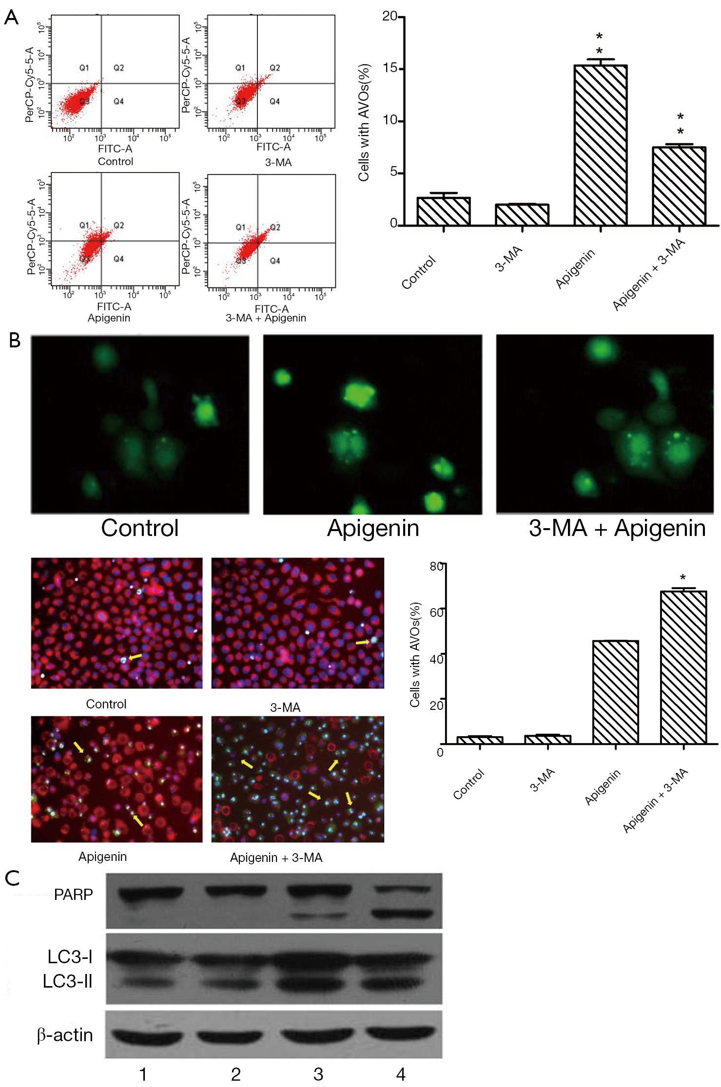
Apigenin (4',5,7-trihydroxyflavone) is a member of the flavone subclass of flavonoids present in fruits and vegetables. The involvement of autophagy in the apigenin-induced apoptotic death of human breast cancer cells was investigated. Cell proliferation and viability were assessed by 3-(4,5-dimethylthiazol-2-yl)-2,5-diphenyltetrazolium bromide (MTT) and clonogenic assays. Flow cytometry, fluorescent staining and Western blot analysis were employed to detect apoptosis and autophagy, and the role of autophagy was assessed using autophagy inhibitors. Apigenin dose- and time-dependently repressed the proliferation and clonogenic survival of the human breast cancer T47D and MDA-MB-231 cell lines. The death of T47D and MDA-MB-231 cells was due to apoptosis associated with increased levels of Caspase3, PARP cleavage and Bax/Bcl-2 ratios. The results from flow cytometry and fluorescent staining also verified the occurrence of apoptosis. In addition, the apigenin-treated cells exhibited autophagy, as characterized by the appearance of autophagosomes under fluorescence microscopy and the accumulation of acidic vesicular organelles (AVOs) by flow cytometry. Furthermore, the results of the Western blot analysis revealed that the level of LC3-II, the processed form of LC3-I, was increased. Treatment with the autophagy inhibitor, 3-methyladenine (3-MA), significantly enhanced the apoptosis induced by apigenin, which was accompanied by an increase in the level of PARP cleavage. Similar results were also confirmed by flow cytometry and fluorescence microscopy. These results indicate that apigenin has apoptosis- and autophagy-inducing effects in breast cancer cells. Autophagy plays a cyto-protective role in apigenin-induced apoptosis, and the combination of apigenin and an autophagy inhibitor may be a promising strategy for breast cancer control.
Apigenin (4',5,7-trihydroxyflavone) is a member of the flavone subclass of flavonoids present in fruits and vegetables. The involvement of autophagy in the apigenin-induced apoptotic death of human breast cancer cells was investigated. Cell proliferation and viability were assessed by 3-(4,5-dimethylthiazol-2-yl)-2,5-diphenyltetrazolium bromide (MTT) and clonogenic assays. Flow cytometry, fluorescent staining and Western blot analysis were employed to detect apoptosis and autophagy, and the role of autophagy was assessed using autophagy inhibitors. Apigenin dose- and time-dependently repressed the proliferation and clonogenic survival of the human breast cancer T47D and MDA-MB-231 cell lines. The death of T47D and MDA-MB-231 cells was due to apoptosis associated with increased levels of Caspase3, PARP cleavage and Bax/Bcl-2 ratios. The results from flow cytometry and fluorescent staining also verified the occurrence of apoptosis. In addition, the apigenin-treated cells exhibited autophagy, as characterized by the appearance of autophagosomes under fluorescence microscopy and the accumulation of acidic vesicular organelles (AVOs) by flow cytometry. Furthermore, the results of the Western blot analysis revealed that the level of LC3-II, the processed form of LC3-I, was increased. Treatment with the autophagy inhibitor, 3-methyladenine (3-MA), significantly enhanced the apoptosis induced by apigenin, which was accompanied by an increase in the level of PARP cleavage. Similar results were also confirmed by flow cytometry and fluorescence microscopy. These results indicate that apigenin has apoptosis- and autophagy-inducing effects in breast cancer cells. Autophagy plays a cyto-protective role in apigenin-induced apoptosis, and the combination of apigenin and an autophagy inhibitor may be a promising strategy for breast cancer control.
2013, 25(2): 223-234.
doi: 10.3978/j.issn.1000-9604.2013.03.03
Abstract:
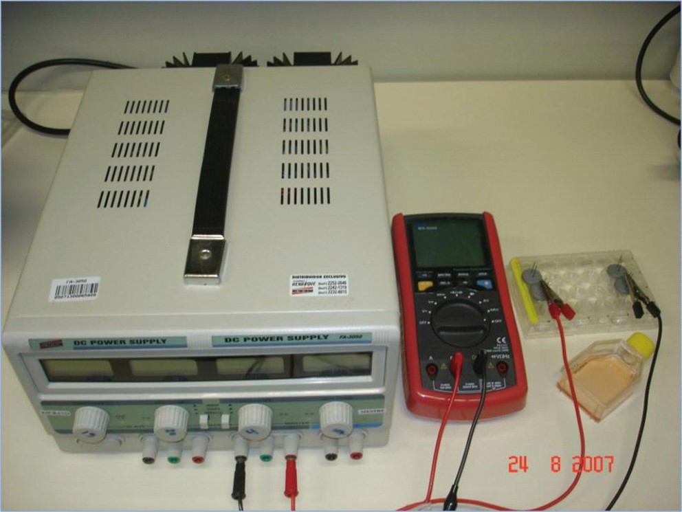
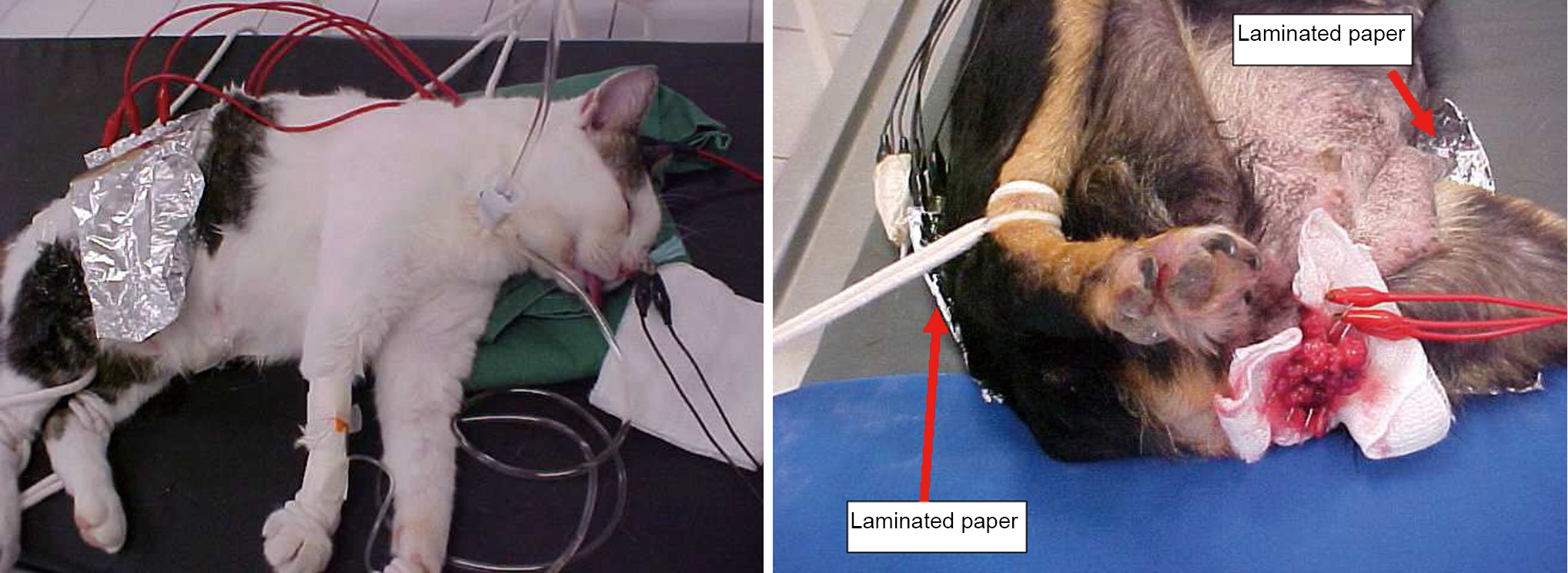
Electrochemical treatment is an alternative modality for tumor treatment based on the application of a low intensity direct electric current to the tumor tissue through two or more platinum electrodes placed within the tumor zone or in the surrounding areas. This treatment is noted for its great effectiveness, minimal invasiveness and local effect. Several studies have been conducted worldwide to evaluate the antitumoral effect of this therapy. In all these studies a variety of biochemical and physiological responses of tumors to the applied treatment have been obtained. By this reason, researchers have suggested various mechanisms to explain how direct electric current destroys tumor cells. Although, it is generally accepted this treatment induces electrolysis, electroosmosis and electroporation in tumoral tissues. However, action mechanism of this alternative modality on the tumor tissue is not well understood. Although the principle of Electrochemical treatment is simple, a standardized method is not yet available. The mechanism by which Electrochemical treatment affects tumor growth and survival may represent more complex process. The present work analyzes the latest and most important research done on the electrochemical treatment of tumors. We conclude with our point of view about the destruction mechanism features of this alternative therapy. Also, we suggest some mechanisms and strategies from the thermodynamic point of view for this therapy. In the area of Electrochemical treatment of cancer this tool has been exploited very little and much work remains to be done. Electrochemical treatment constitutes a good therapeutic option for patients that have failed the conventional oncology methods.
Electrochemical treatment is an alternative modality for tumor treatment based on the application of a low intensity direct electric current to the tumor tissue through two or more platinum electrodes placed within the tumor zone or in the surrounding areas. This treatment is noted for its great effectiveness, minimal invasiveness and local effect. Several studies have been conducted worldwide to evaluate the antitumoral effect of this therapy. In all these studies a variety of biochemical and physiological responses of tumors to the applied treatment have been obtained. By this reason, researchers have suggested various mechanisms to explain how direct electric current destroys tumor cells. Although, it is generally accepted this treatment induces electrolysis, electroosmosis and electroporation in tumoral tissues. However, action mechanism of this alternative modality on the tumor tissue is not well understood. Although the principle of Electrochemical treatment is simple, a standardized method is not yet available. The mechanism by which Electrochemical treatment affects tumor growth and survival may represent more complex process. The present work analyzes the latest and most important research done on the electrochemical treatment of tumors. We conclude with our point of view about the destruction mechanism features of this alternative therapy. Also, we suggest some mechanisms and strategies from the thermodynamic point of view for this therapy. In the area of Electrochemical treatment of cancer this tool has been exploited very little and much work remains to be done. Electrochemical treatment constitutes a good therapeutic option for patients that have failed the conventional oncology methods.
2013, 25(2): 235-239.
doi: 10.3978/j.issn.1000-9604.2013.03.08
Abstract:
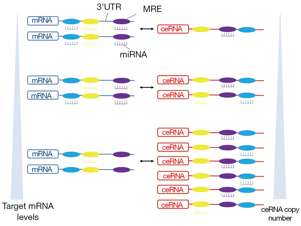
microRNAs (miRNAs) are a class of endogenous, single-stranded non-coding RNAs of 20-23 nucleotides in length, functioning as negative regulators of gene expression at the post-transcriptional level. The dysregulation of miRNAs has been demonstrated to play critical roles in tumorigenesis, either through inhibiting tumor suppressor genes or activating oncogenes inappropriately. Besides their promising clinical applications in cancer diagnosis and treatment, recent studies have uncovered that miRNAs could act as a regulatory language, through which messenger RNAs, transcribed pseudogenes, and long noncoding RNAs crosstalk with each other and form a novel regulatory network. RNA transcripts involved in this network have been termed as competing endogenous RNAs (ceRNAs), since they influence each other’s level by competing for the same pool of miRNAs through miRNA response elements (MREs) on their target transcripts. The discovery of miRNA-ceRNA network not only provides the possibility of an additional level of post-transcriptional regulation, but also dictates a reassessment of the existing regulatory pathways involved in cancer initiation and progression.
microRNAs (miRNAs) are a class of endogenous, single-stranded non-coding RNAs of 20-23 nucleotides in length, functioning as negative regulators of gene expression at the post-transcriptional level. The dysregulation of miRNAs has been demonstrated to play critical roles in tumorigenesis, either through inhibiting tumor suppressor genes or activating oncogenes inappropriately. Besides their promising clinical applications in cancer diagnosis and treatment, recent studies have uncovered that miRNAs could act as a regulatory language, through which messenger RNAs, transcribed pseudogenes, and long noncoding RNAs crosstalk with each other and form a novel regulatory network. RNA transcripts involved in this network have been termed as competing endogenous RNAs (ceRNAs), since they influence each other’s level by competing for the same pool of miRNAs through miRNA response elements (MREs) on their target transcripts. The discovery of miRNA-ceRNA network not only provides the possibility of an additional level of post-transcriptional regulation, but also dictates a reassessment of the existing regulatory pathways involved in cancer initiation and progression.
2013, 25(2): 240-249.
doi: 10.3978/j.issn.1000-9604.2013.03.06
Abstract:
As prostate cancer is a biologically heterogeneous disease for which a variety of treatment options are available, the major objective of prostate cancer imaging is to achieve more precise disease characterization. In clinical practice, magnetic resonance imaging (MRI) is one of the imaging tools for the evaluation of prostate cancer, the fusion of MRI or dynamic contrast-enhanced MRI (DCE-MRI) with magnetic resonance spectroscopic imaging (MRSI) is improving the evaluation of cancer location, size, and extent, while providing an indication of tumor aggressiveness. This review summarizes the role of MRI in the application of prostate cancer and describes molecular MRI techniques (including MRSI and DCE-MRI) for aiding prostate cancer management.
As prostate cancer is a biologically heterogeneous disease for which a variety of treatment options are available, the major objective of prostate cancer imaging is to achieve more precise disease characterization. In clinical practice, magnetic resonance imaging (MRI) is one of the imaging tools for the evaluation of prostate cancer, the fusion of MRI or dynamic contrast-enhanced MRI (DCE-MRI) with magnetic resonance spectroscopic imaging (MRSI) is improving the evaluation of cancer location, size, and extent, while providing an indication of tumor aggressiveness. This review summarizes the role of MRI in the application of prostate cancer and describes molecular MRI techniques (including MRSI and DCE-MRI) for aiding prostate cancer management.
2013, 25(2): 250-253.
doi: 10.3978/j.issn.1000-9604.2012.12.05
Abstract:
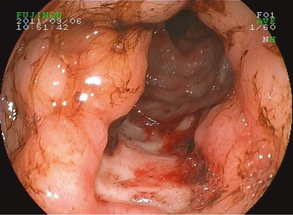
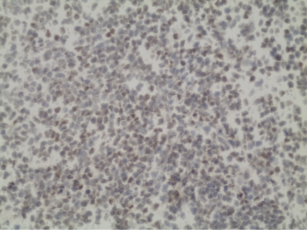
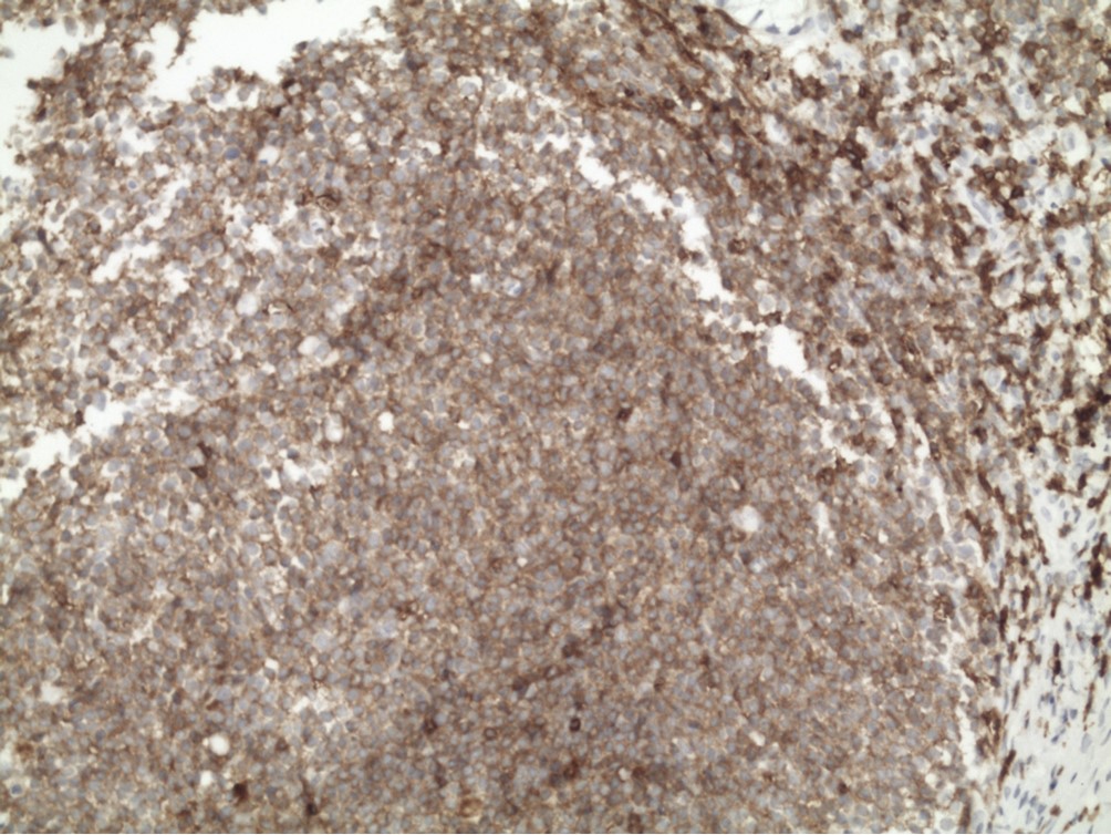
The incidence of primary gastrointestinal lymphomas (PGILs) has been increasing. The clinical presentation and treatment of PGIL are distinct from those of nodular lymphomas. Symptoms include abdominal pain, abdominal mass, changes in bowel habits, obstruction, and bleeding. Less life-threatening gastrointestinal bleeding occurs after chemotherapy and few reports have focused on the bleeding of PGILs. We report a case of severe gastrointestinal bleeding caused by low-dose chemotherapy, which was dramatically improved by rituximab monotherapy treatment. The prevention and treatment of gastrointestinal bleeding in PGIL should be given much attention.
The incidence of primary gastrointestinal lymphomas (PGILs) has been increasing. The clinical presentation and treatment of PGIL are distinct from those of nodular lymphomas. Symptoms include abdominal pain, abdominal mass, changes in bowel habits, obstruction, and bleeding. Less life-threatening gastrointestinal bleeding occurs after chemotherapy and few reports have focused on the bleeding of PGILs. We report a case of severe gastrointestinal bleeding caused by low-dose chemotherapy, which was dramatically improved by rituximab monotherapy treatment. The prevention and treatment of gastrointestinal bleeding in PGIL should be given much attention.

2013, 25(2): 254-258.
doi: 10.3978/j.issn.1000-9604.2013.03.05
Abstract:
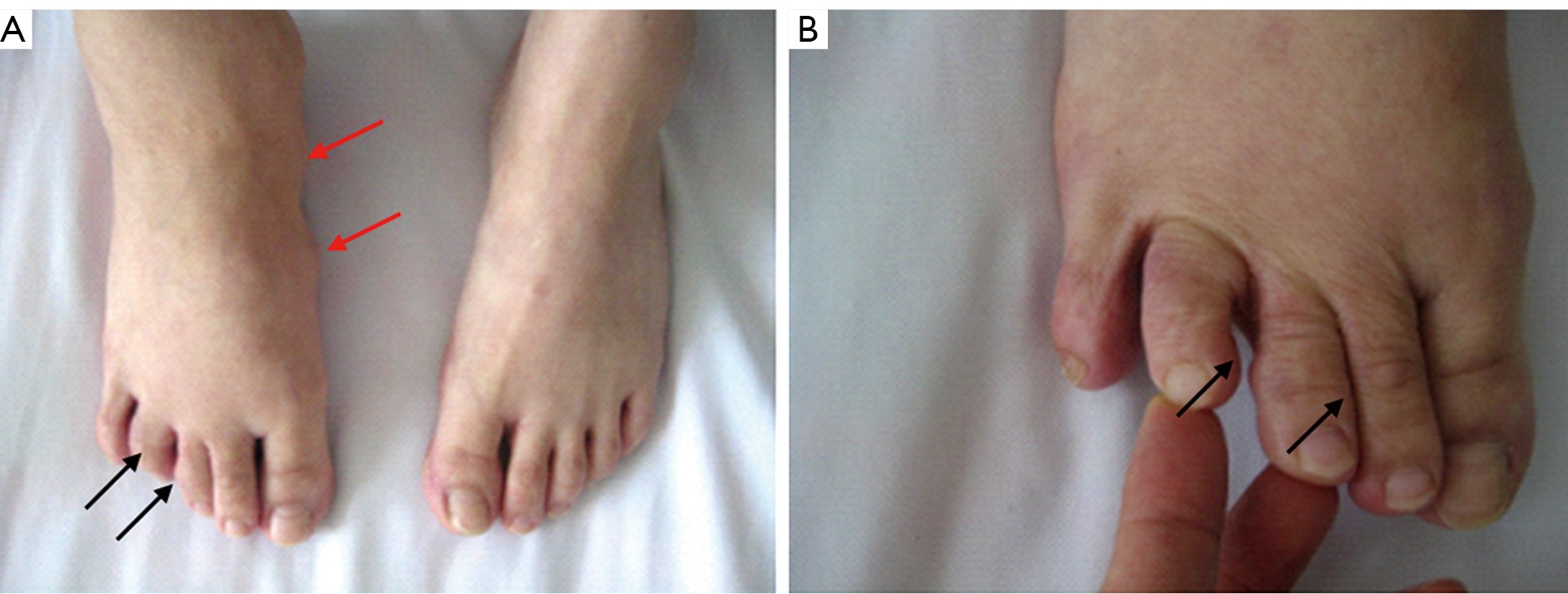
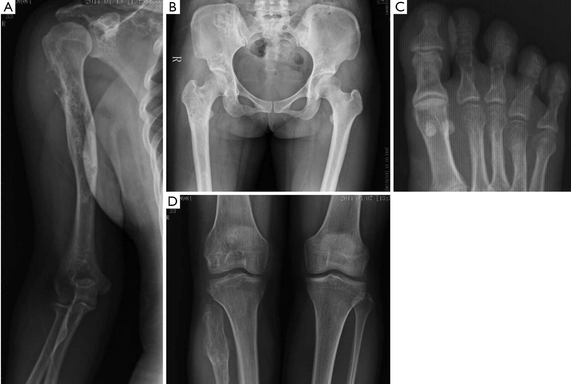
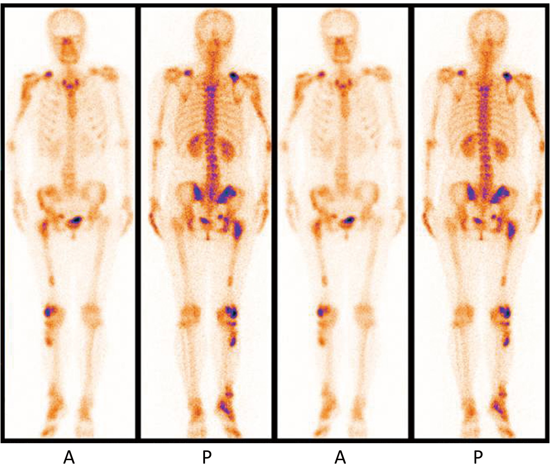
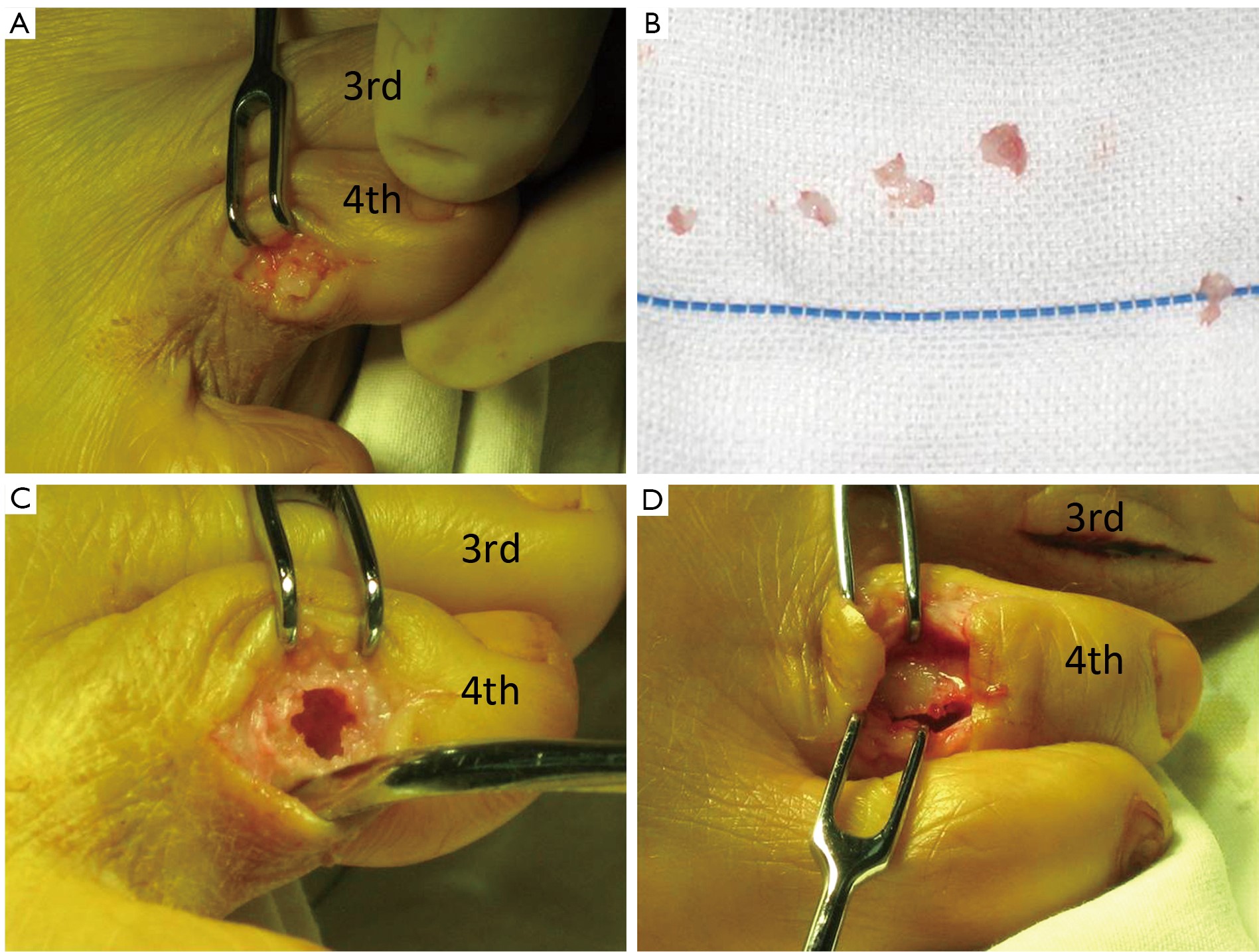
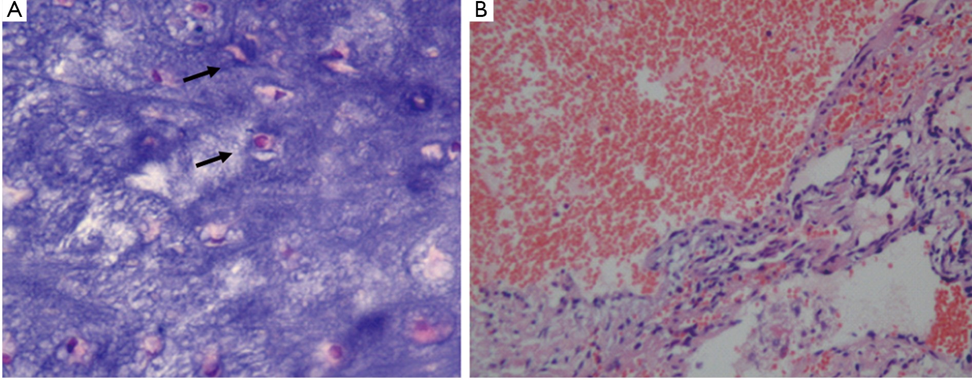
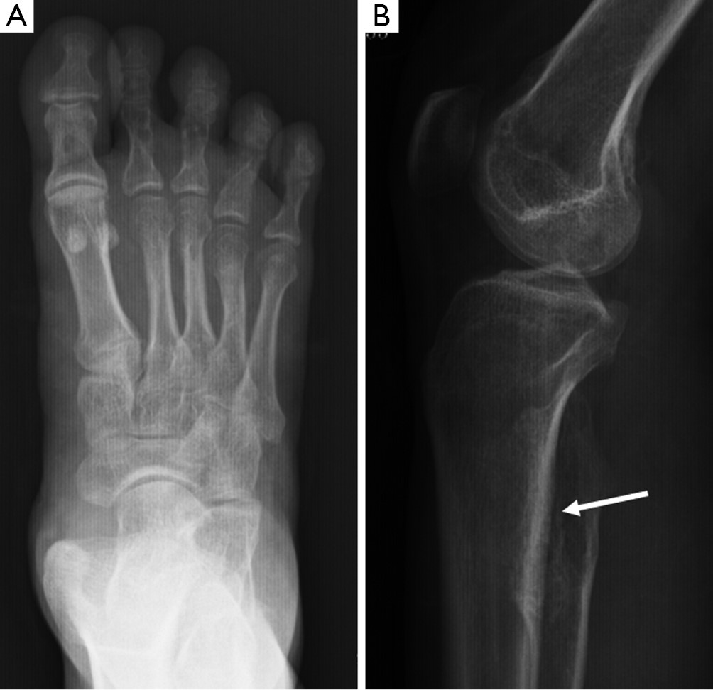
Maffucci syndrome is a congenital, non-hereditary mesodermal dysplasia manifested by multiple enchondromas and hemangiomas. It is associated with diverse secondary musculoskeletal deformities, which is exceedingly rare. We report a case of hemangiomas and enchondromas localized in the unilateral limb in a patient with Maffucci syndrome. Treatment consists of orthopedic and surgical intervention to minimize deformities and for cosmetic purpose. Careful surveillance for malignant degeneration of both skeletal and non-skeletal tumors, especially in the brain and abdomen, is essential.
Maffucci syndrome is a congenital, non-hereditary mesodermal dysplasia manifested by multiple enchondromas and hemangiomas. It is associated with diverse secondary musculoskeletal deformities, which is exceedingly rare. We report a case of hemangiomas and enchondromas localized in the unilateral limb in a patient with Maffucci syndrome. Treatment consists of orthopedic and surgical intervention to minimize deformities and for cosmetic purpose. Careful surveillance for malignant degeneration of both skeletal and non-skeletal tumors, especially in the brain and abdomen, is essential.
2013, 25(2): 259-262.
doi: 10.3978/j.issn.1000-9604.2013.03.09
Abstract:
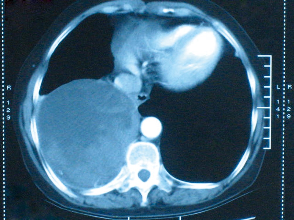
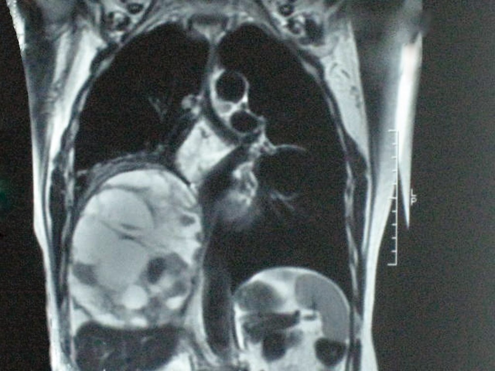
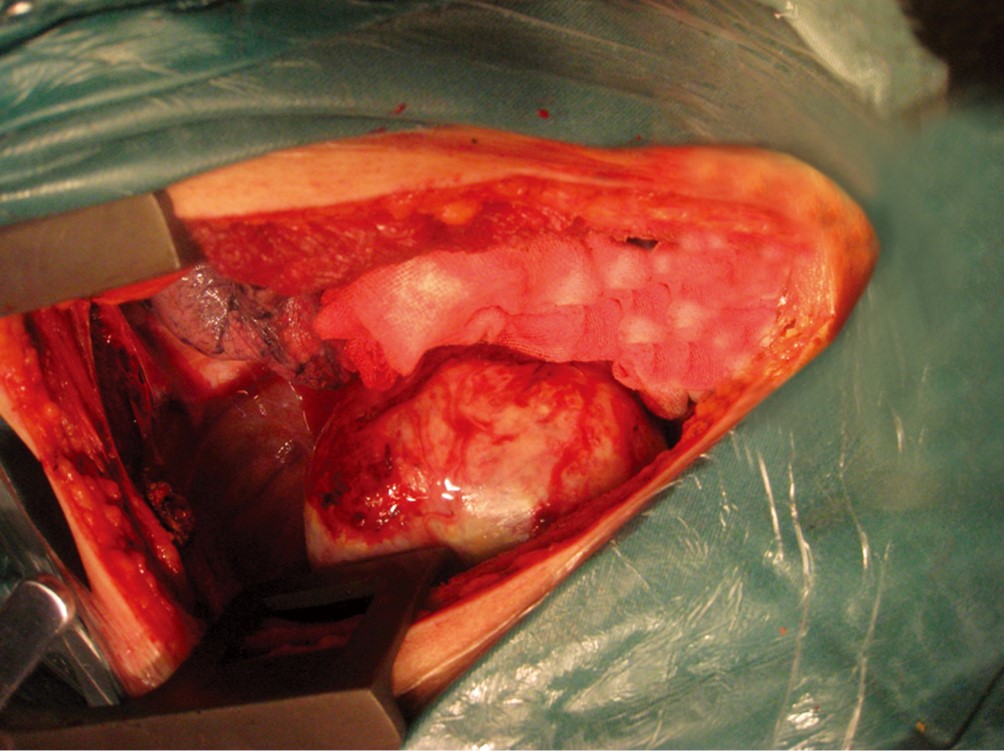
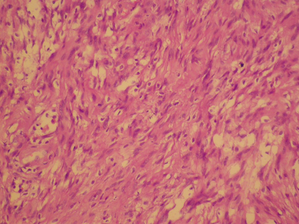
Neurogenic tumors are commonly found in the mediastinum, especially in the posterior mediastinum or in the chest wall, neurogenic tumors may reach large size before becoming symptomatic. If the neurogenic tumor occupied more than half size of the chest wall accompanied by mediastinal shift, tracheal compression, or superior vena reflux disorder, it may be called giant intrathoracic neurogenic tumors. Giant intrathoracic neurogenic tumors are relatively rare. Most of intrathoracic neurogenic tumors were benign or low-grade malignant tumors in nature. Complete surgical excision should be the rule for these patients. We report two cases of giant neurogenic tumors, and study the clinical manifestations, diagnostic methods, surgical management, and prognosis in the light of the most important published data.
Neurogenic tumors are commonly found in the mediastinum, especially in the posterior mediastinum or in the chest wall, neurogenic tumors may reach large size before becoming symptomatic. If the neurogenic tumor occupied more than half size of the chest wall accompanied by mediastinal shift, tracheal compression, or superior vena reflux disorder, it may be called giant intrathoracic neurogenic tumors. Giant intrathoracic neurogenic tumors are relatively rare. Most of intrathoracic neurogenic tumors were benign or low-grade malignant tumors in nature. Complete surgical excision should be the rule for these patients. We report two cases of giant neurogenic tumors, and study the clinical manifestations, diagnostic methods, surgical management, and prognosis in the light of the most important published data.