
2013 Vol.25(5)
Display Mode: |
2013, 25(5): 486-487.
doi: 10.3978/j.issn.1000-9604.2013.10.13
 Abstract
 FullText HTML
 PDF 85KB
Abstract:
2013, 25(5): 488-489.
doi: 10.3978/j.issn.1000-9604.2013.10.12
Abstract:
2013, 25(5): 490-492.
doi: 10.3978/j.issn.1000-9604.2013.10.15
Abstract:
2013, 25(5): 493-499.
doi: 10.3978/j.issn.1000-9604.2013.09.02
Abstract:
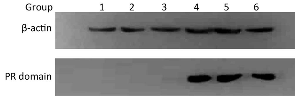
ObjectivePR domain is responsible for the tumor suppressing activity of RIZ1. The study aimed to construct human PR domain eukaryotic expression vectors, transfect human esophageal cancer cells (TE13), and evaluate the anticancer activity of PR domain on human esophageal cancer TE13 cells. MethodsFirst, mRNA was extracted from human esophageal cancer tissue by RT-PCR, then reverse-transcribed to cDNA. After amplifying from the DNA template, PR domain was linked to T vector. Second, after extraction, PR domain was cut using enzyme and linked to pcDNA3.1(+). Then, the plasmid was transfered to Trans1-T1 phage resistant competent cells, following by extracting the ultrapure plasmid, and transfecting into TE13 cells. In the end, the protein expression of pcDNA3.1(+)/PR domain in TE13 was detected by Western blot, and the apoptosis of TE13 by technique of flow cytometry. ResultsMore than 5,000 bp purposed band of pcDNA3.1(+)/PR domain plasmid was found by agarose gel electrophoresis. After transfection, the PR domain (molecular weight of about 28 Da) was found only in 3, 4 and 5 groups by Western blot. Flow cytometry assay showed apoptosis in experimental group was significantly more than that in the control group (P<0.05). ConclusionsThe PR domain eukaryotic expression vector was constructed successfully. The protein of the PR domain could be expressed in esophageal cancer TE13 cells firmly after transfection, and a single PR domain could promote apoptosis of TE13 cells.


2013, 25(5): 500-504.
doi: 10.3978/j.issn.1000-9604.2013.09.01
Abstract:
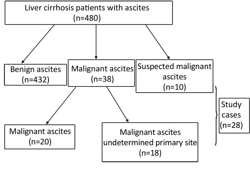
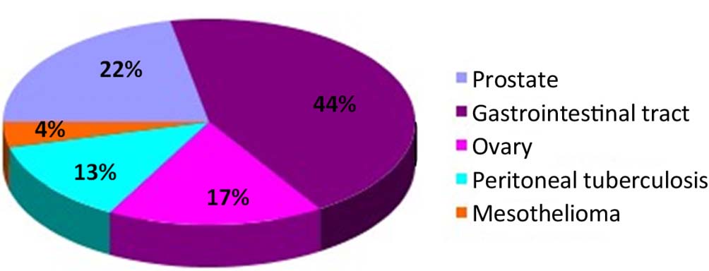
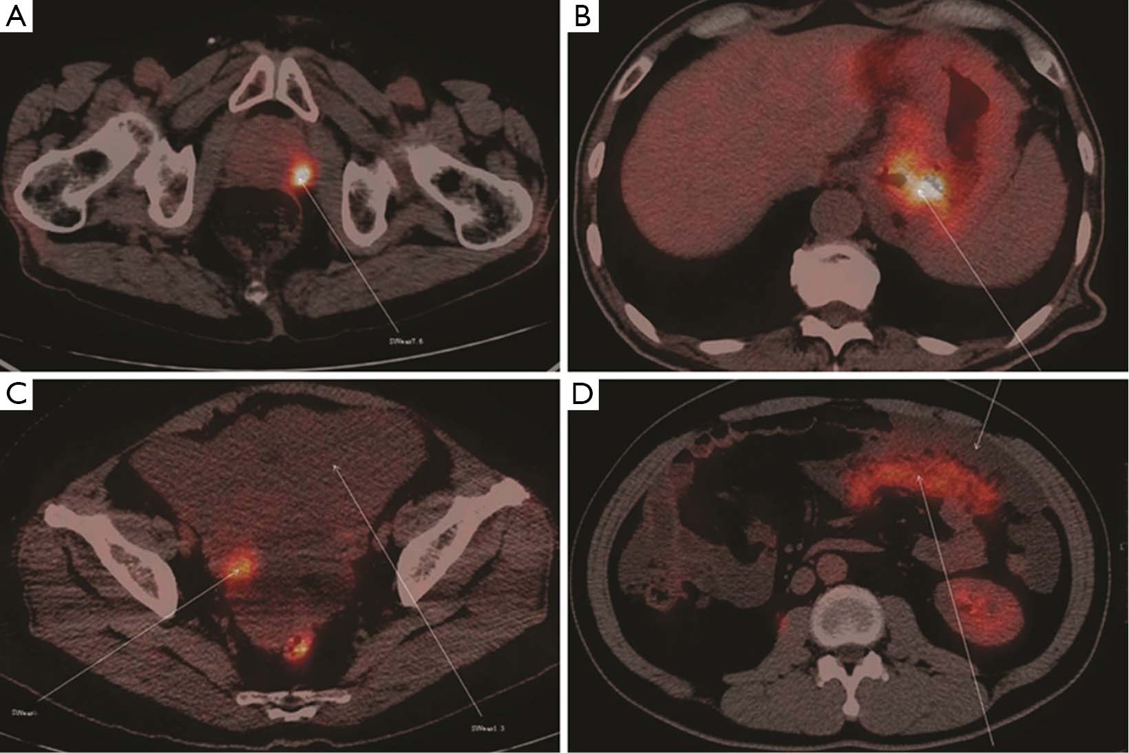
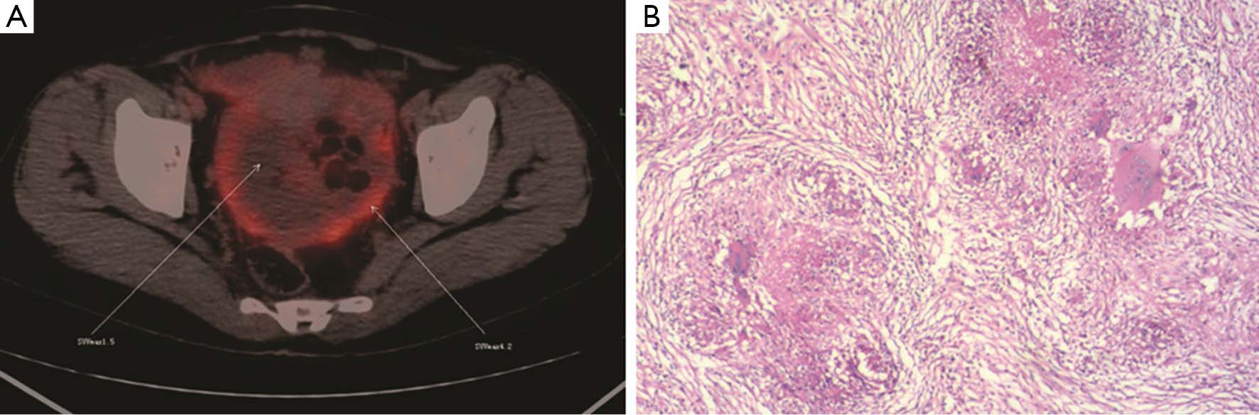
ObjectiveAscites in patients with hepatic cirrhosis is caused by cirrhosis in most cases. For most malignant ascites, the primary malignancy could be readily identified using conventional imaging methods, e.g., computed tomography (CT) and magnetic resonance imaging (MRI). However, in a small fraction of the patients, the primary malignancy remains occult even with these examinations. In this retrospective study, we assessed the usefulness of 18F-FDG PET/CT in patients with hepatic cirrhosis and malignant ascites of otherwise unknown origin. MethodsTwenty-eight patients with malignant ascites of unknown primary sites after CT, MRI and ultrasound during the period of five years between January 2008 and December 2012 had received 18F-FDG PET/CT. Medical records of these patients were reviewed and analyzed. ResultsElevated 18F-FDG absorption was found in 23 of 28 cases in the following sites: gastrointestinal tract (n=10, 43.5%), prostate (n=5, 21.7%), peritoneum (n=4, 13.3%), and ovary (n=4, 13.3%). Cancer was confirmed by pathology in 20 cases after open or laparoscopic surgeries. Five patients were found to have benign ascites, among which, 3 were found to be false positive due to tuberculosis. SUV values were significantly higher for tumors than for benign lesions (mean values, 6.95 vs. 2.94; P=0.005). ConclusionsThe 18F-FDG PET/CT can be as a powerful imaging tool in identifying tissue origin in liver cirrhosis patients suspected of cancers or with cancers of unknown primary sites.
2013, 25(5): 505-513.
doi: 10.3978/j.issn.1000-9604.2013.08.14
Abstract:
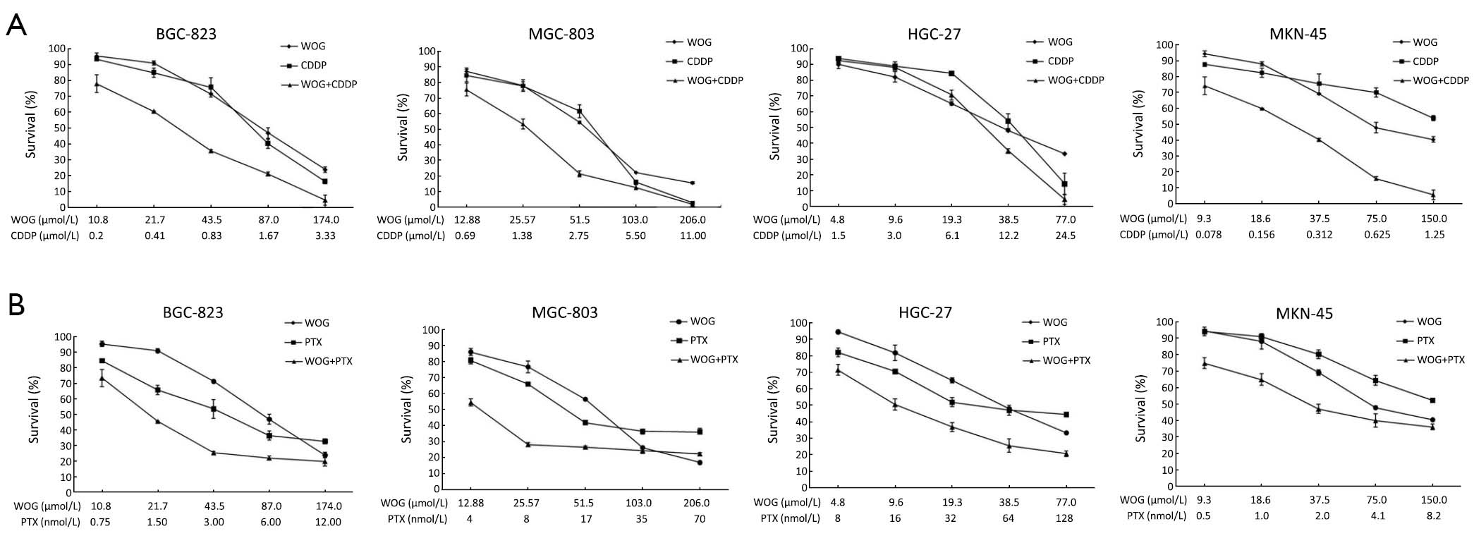
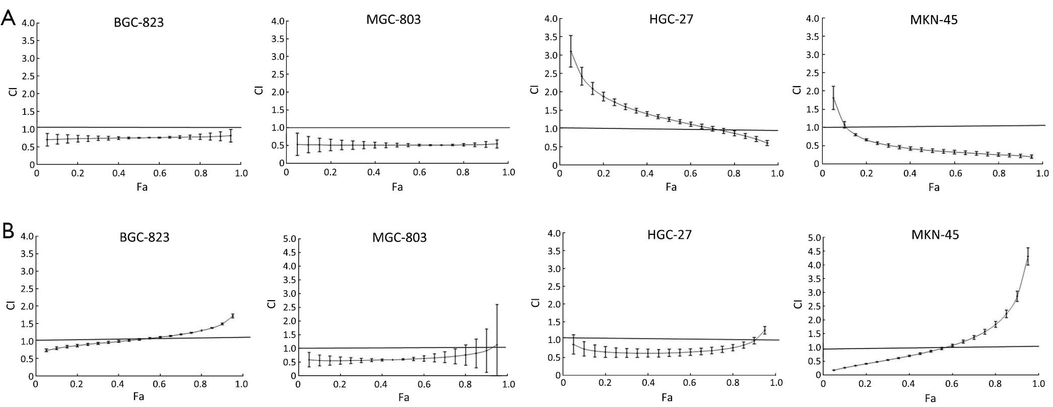
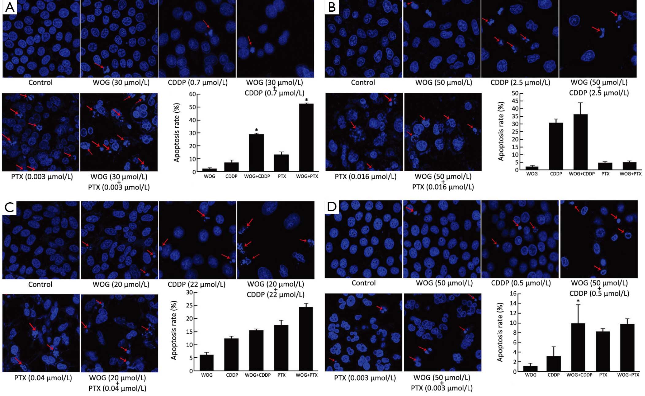
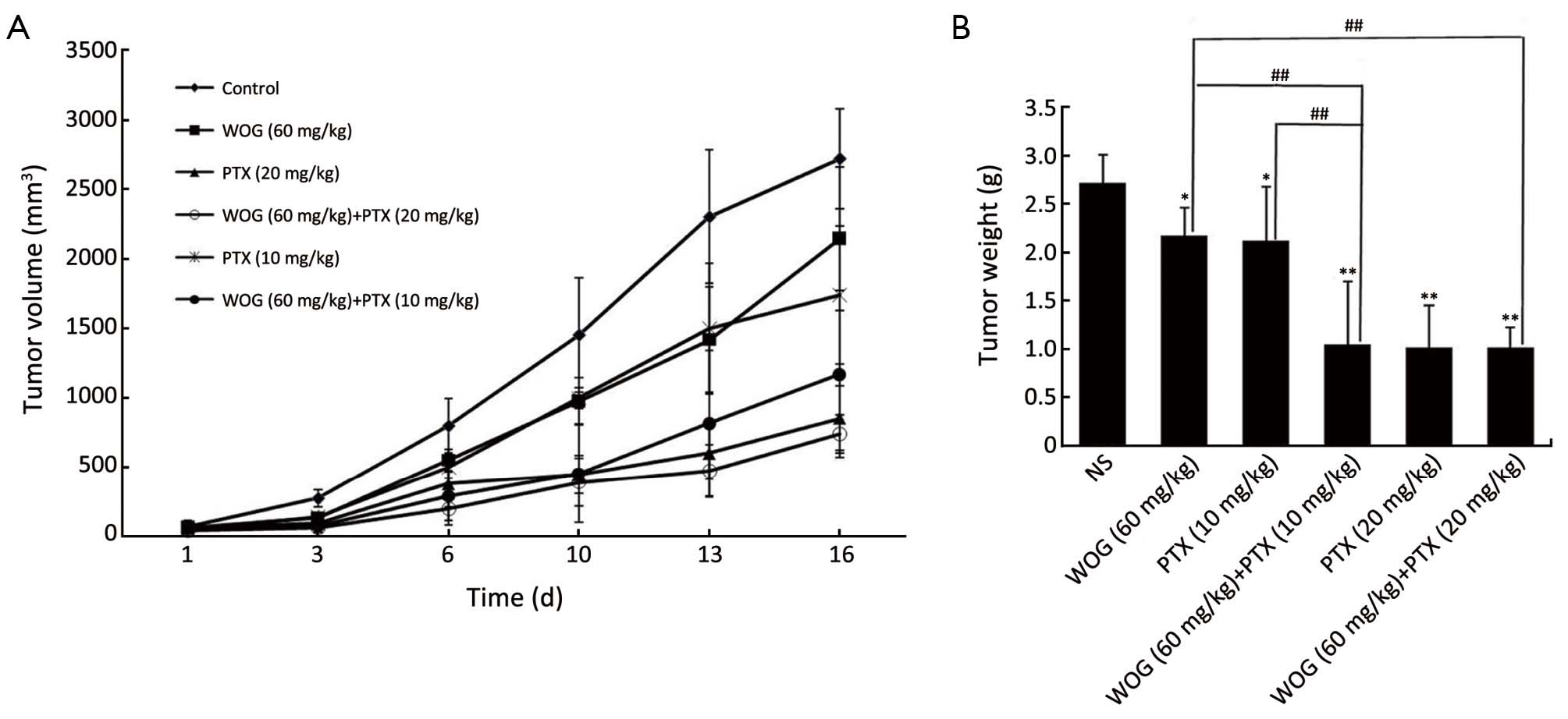
ObjectiveTo investigate the synergistic inhibitory effects of wogonin (WOG) and chemotherapeutic drugs on growth of gastric cancer cells and tumor xenografts. MethodsThe IC50 values of WOG, cisplatin (CDDP) and paclitaxel (PTX) in four gastric cancer cell lines were determined by MTS assay. Hoechst staining and the median effect method of Chou-Talalay were used to assess the apoptosis of cells and the interaction of two drugs, respectively. BGC-823-derived xenografts in nude mice were established to investigate the effects of WOG combined with chemotherapeutic drugs in vivo. ResultsWOG, CDDP and PTX inhibited the growth of BGC-823, MGC-803, MKN-45 and HGC-27 gastric cancer cells in a dose-dependent manner. WOG combined with CDDP or PTX synergistically inhibited the growth of all gastric cancer cell lines in vitro. In BGC-823, MGC-803, HGC-27 and MKN-45 cell lines, synergisms between WOG and PTX were shown when the fraction affected (Fa) values were <0.45, <0.90, <0.85 and <0.60. While WOG and CDDP had a synergistic inhibitory effect when the Fa values were >0, >0, >0.65 and >0.10. From the results of in vivo experiments using tumor xenografts, WOG and low-dose PTX showed better efficacy than either drug alone. The inhibitory percentages of tumor weight were 61.58%, 20.29%, and 22.28% for the combination, WOG-alone, and low-dose PTX-alone groups, respectively. Notably, WOG combined with CDDP displayed very high toxicity. ConclusionsA synergistic inhibitory effect on growth was observed when WOG was combined with low-dose PTX in gastric cancer cells and tumor xenografts. These findings provide evidence for the design of a clinical trial to test the combination of WOG with low-dose PTX in human gastric cancer.
2013, 25(5): 514-519.
doi: 10.3978/j.issn.1000-9604.2013.09.03
Abstract:
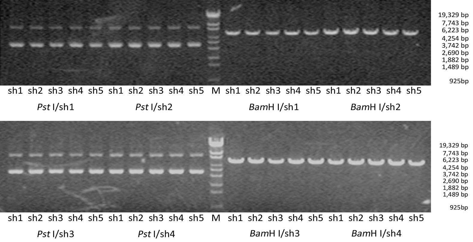
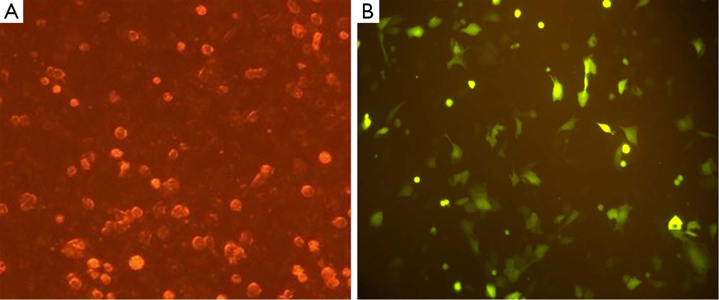
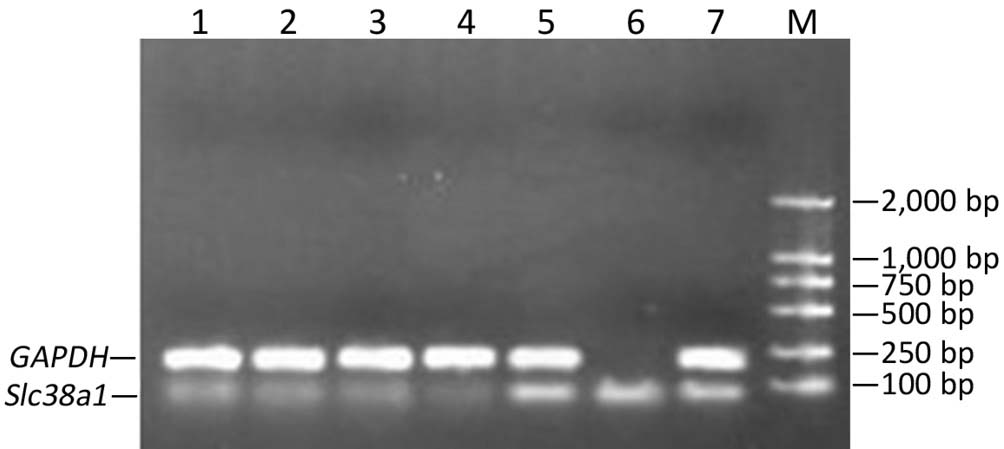
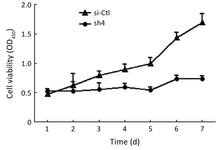
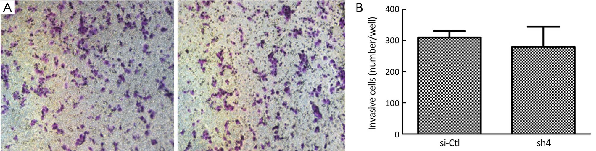
ObjectiveEarly metastasis is a major biological feature of pancreatic cancer. The current study examined whether silencing Slc38a1, a gene involved in energy metabolism, using short hairpin RNA (shRNA) could inhibit the growth, migration, and invasiveness of pancreatic cancer cells. MethodsA series of Slc38a1 shRNAs were designed and cloned into the pGPU6/GFP/Neo vectors. An shRNA with the most efficacious inhibitory action on SCL38A1 expression (65% inhibition) upon screening in DH5α bacteria was used to transfect SW1990 human pancreatic cancer cells. Cell growth, migration, and invasiveness were examined using cell counting kit-8, Boyden chamber without and with Matrigel, respectively. ResultsTransfection of SW1990 cells with the SLCs38A1 shRNA significantly decreased the proliferation (P<0.0001) and migratory potential (by 46.7%, P=0.0399) of the cancer cells. Invasiveness, however, was not affected. ConclusionsInhibiting Slc38a1 using shRNA technology could decrease the growth and migration of representative pancreatic cancer cells. However, the fact that invasiveness was not affected suggested that SLC38A1 is unlikely to be responsible for early metastasis.

2013, 25(5): 520-526.
doi: 10.3978/j.issn.1000-9604.2013.10.01
Abstract:
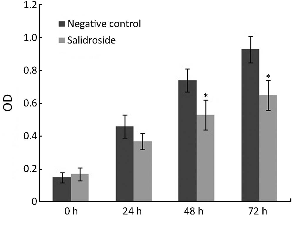
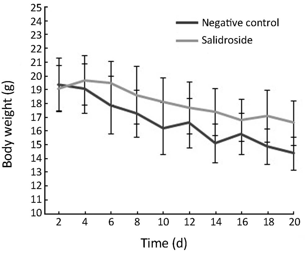
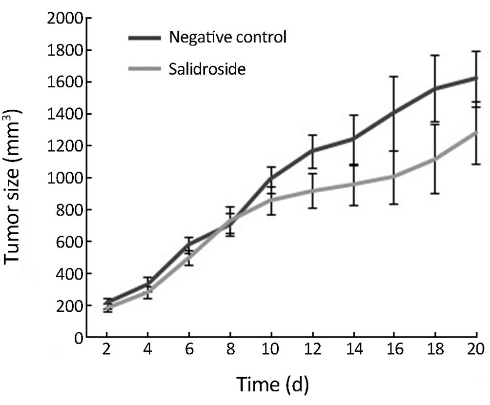
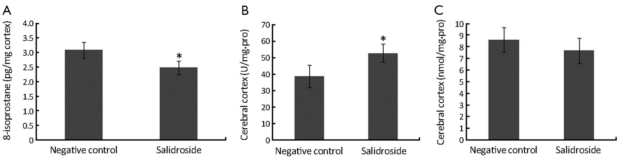
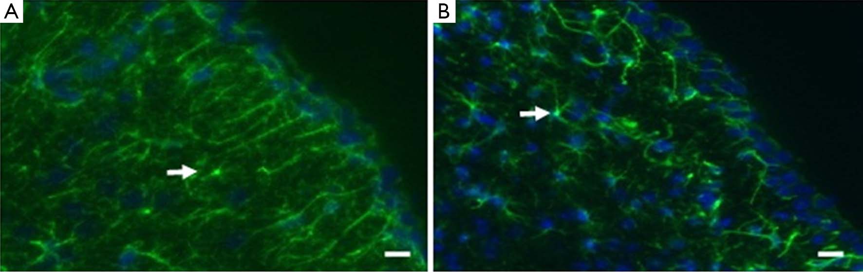
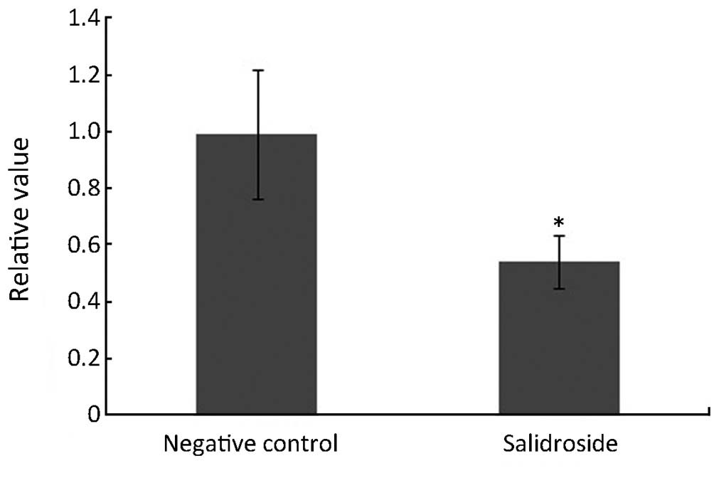
ObjectiveTo test the effects of salidroside on formation and growth of glioma together with tumor microenvironment. MethodsSalidroside extracted from Rhodiola rosea was purified and treated on human glioma cells U251 at the concentration of 20 µg/mL. 3-(4,5-dimethylthiazol-2-yl)-2,5-dephenyltetrazolium bromide (MTT) assay for cytotoxicity and flow cytometry (FCM) for cell cycle analysis were performed. Then for in vivo study, xenotransplantation tumor model in nude mice was generated and treated with salidroside at the concentration of 50 mg/kg.d for totally 20 d. Body weight and tumor size were detected every 2 d after the treatment. The levels of 8-isoprostane, superoxide dismutase (SOD) and malondialdehyde (MDA), special markers for oxidative stress, were detected while immunofluoresence staining was performed for astrocyte detection. ResultsFor in vitro study, salidroside could decrease the viability of human glioma cells U251 and the growth of U251 cells at G0/G1 checkpoint during the cell cycle. For in vivo study, salidroside could also inhibit the growth of human glioma tissue in nude mice. The body weight of these nude mice treated with salidroside did not decrease as quickly as control group. In the tumor xenotransplantation nude mice model, mice were found of inhibition of oxidative stress by detection of biomarkers. Furthermore, overgrowth of astrocytes due to the stimulation of oxidative stress in the cortex of brain was inhibited after the treatment of salidroside. ConclusionsSalidroside could inhibit the formation and growth of glioma both in vivo and in vitro and improve the tumor microenvironment via inhibition of oxidative stress and astrocytes.
2013, 25(5): 527-535.
doi: 10.3978/j.issn.1000-9604.2013.09.04
Abstract:
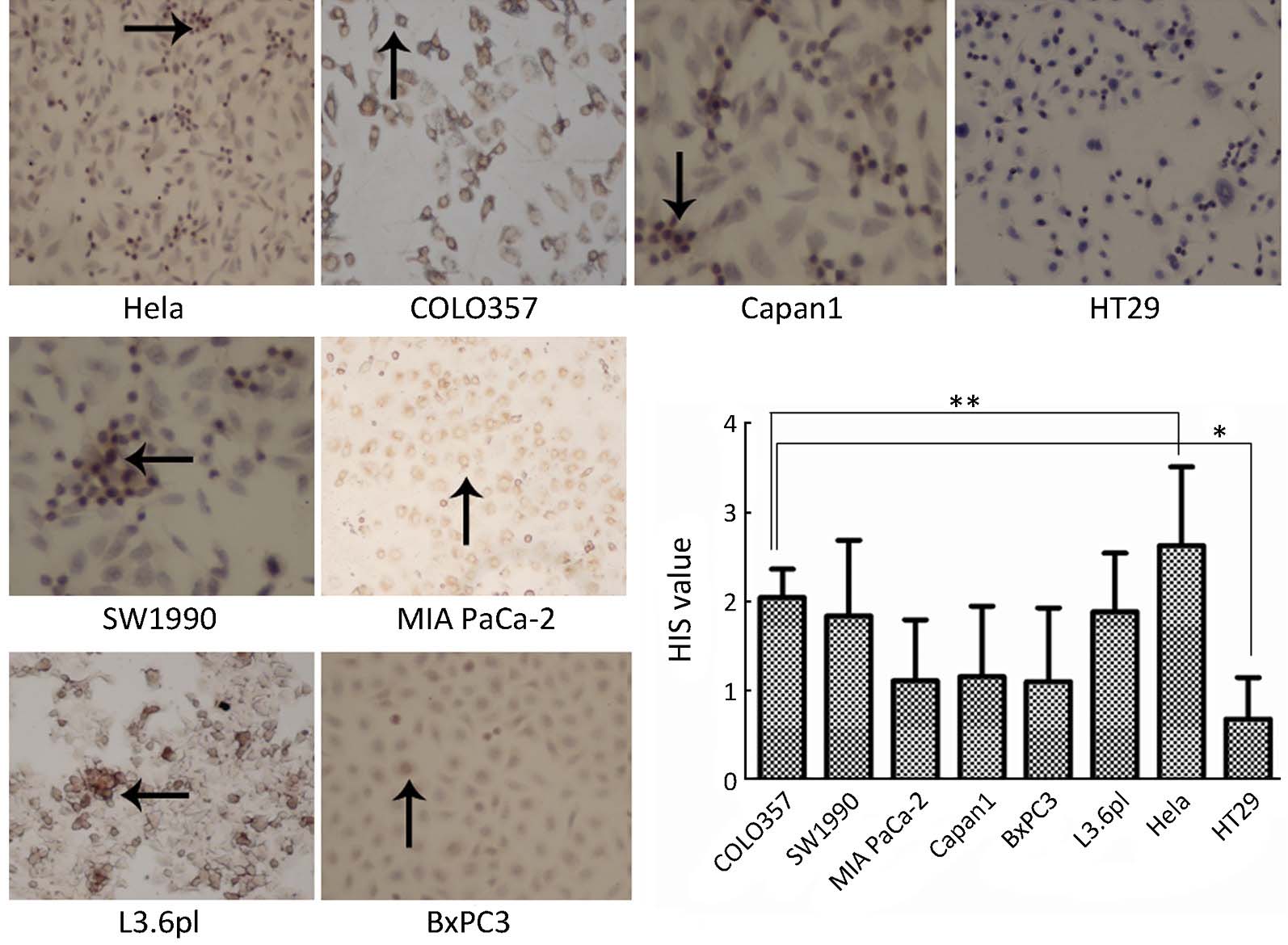
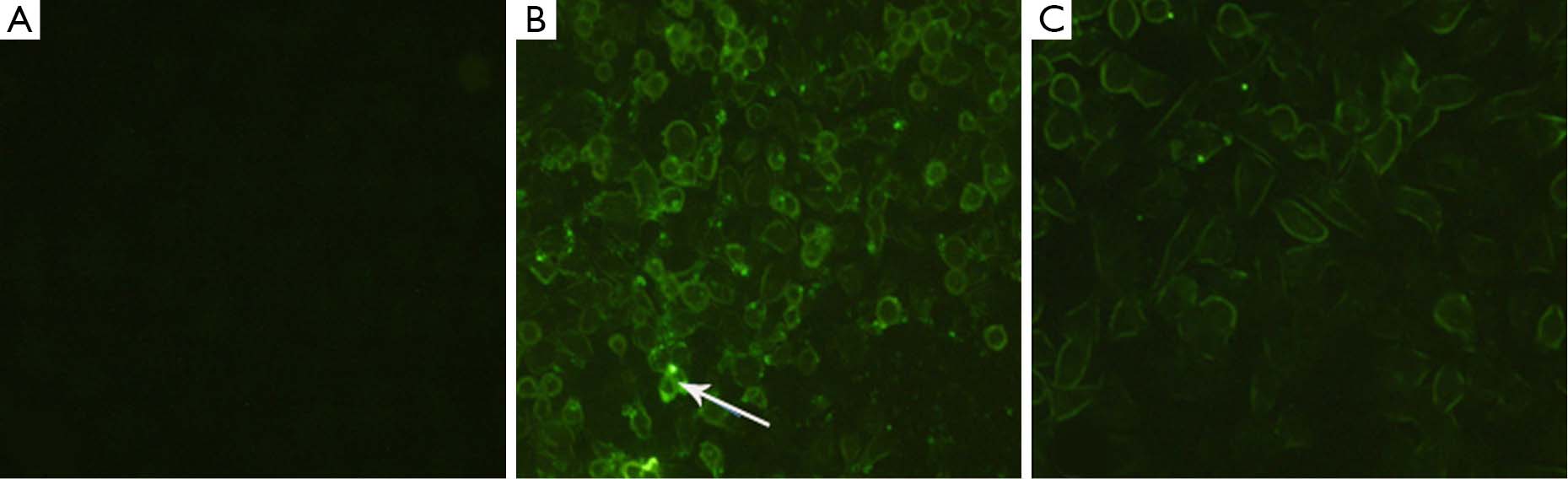
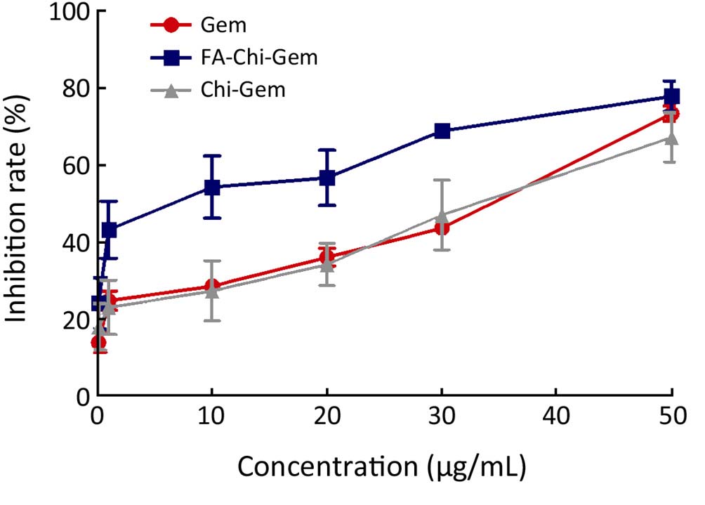
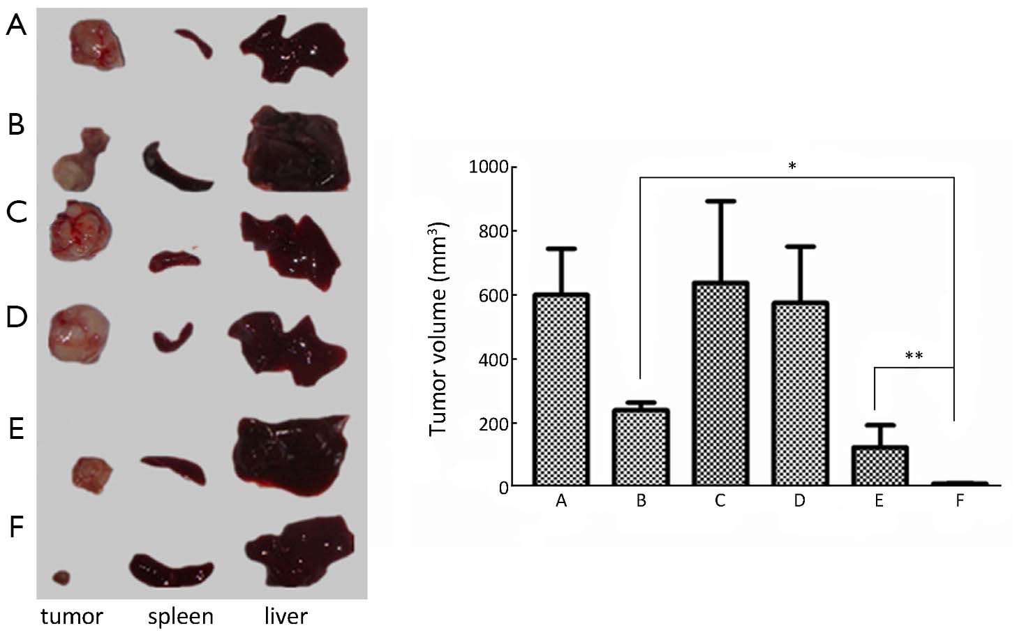
ObjectiveHuman pancreatic cancer is one of the most common clinical malignancies. The effect of comprehensive treatment based on surgery is general. The effects of chemotherapy were not obvious mainly because of lack of targeting and chemoresistance in pancreatic cancer. This study aimed to investigate the effects of folate receptor (FR)-mediated gemcitabine FA-Chi-Gem nanoparticles with a core-shell structure by electrostatic spray on pancreatic cancer. MethodsIn this study, the levels of expression of FR in six human pancreatic cancer cell lines were studied by immunohistochemical analysis. The uptake rate of isothiocyanate-labeled FA-Chi nanoparticles in FR high expression cell line COLO357 was assessed by fluorescence microscope and the inhibition rate of FA-Chi-Gem nanoparticles on COLO357 cells was evaluated by MTT assay. Moreover, the biodistribution of PEG-FA-ICGDER02-Chi in the orthotopic pancreatic tumor model was observed using near-infrared imaging and the human pancreatic cancer orthotopic xenografts were treated with different nanoparticles and normal saline control. ResultsThe expression of FR in COLO357 was the highest among the six pancreatic cancer cell lines. The FR mainly distributed on cell membrane and fewer in the cytoplasm in pancreatic cancer. Moreover, the absorption rate of the FA-Chi-Gem nanoparticles was more than the Chi nanoparticles without FA modified. The proliferation of COLO357 was significantly inhibited by FA-Chi-Gem nanoparticles. The PEG-FA-ICGDER02-Chi nanoparticles were enriched in tumor tissue in human pancreatic cancer xenografts, while non-targeted nanoparticles were mainly in normal liver tissue. PEG-FA-Gem-Chi significantly inhibited the growth of human pancreatic cancer xenografts (PEG-FA-Gem-Chi vs. Gem, t=22.950, P=0.000). ConclusionsPEG-FA-FITC-Chi nanoparticles might be an effective targeted drug for treating human FR-positive pancreatic cancer.


2013, 25(5): 536-543.
doi: 10.3978/j.issn.1000-9604.2013.10.02
Abstract:
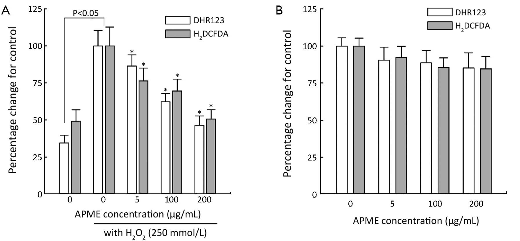
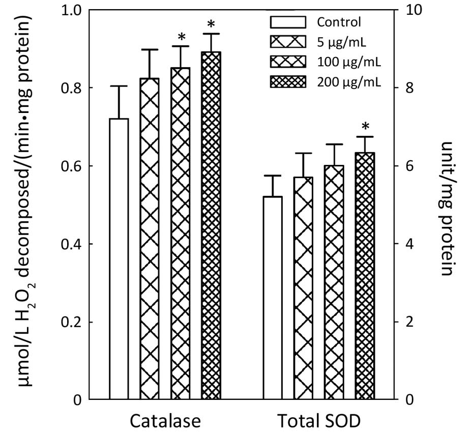
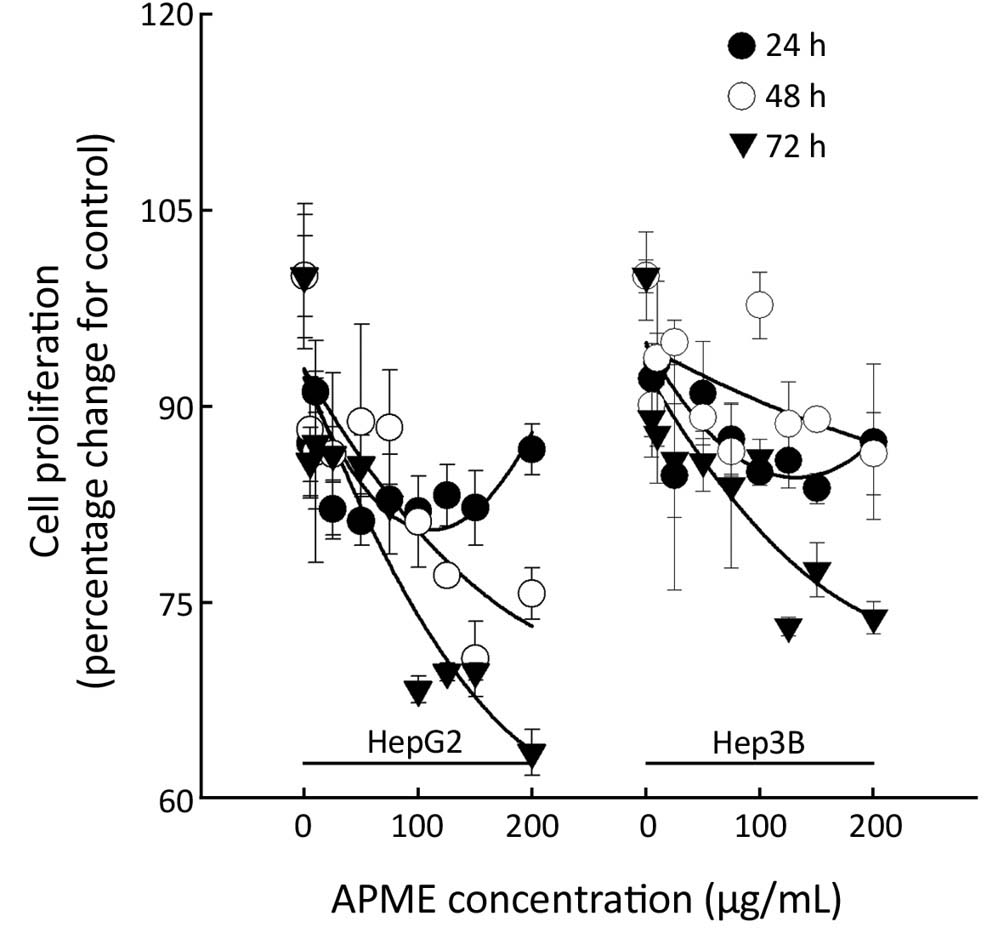
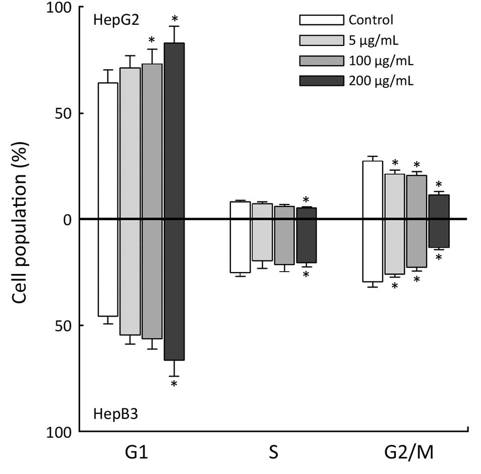
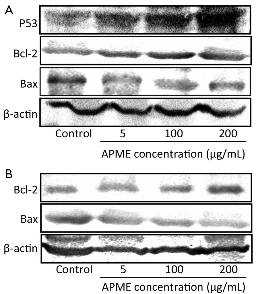
ObjectiveThe aim of the present study was to investigate antioxidant and the anticancerigen activity of a methanol extract from Artemisia princeps var. orientalis (APME), a well-known traditional herbal medicine in Asia, in hepatocellular cancer cells. MethodsTo evaluate the antioxidant activity of APME, reactive oxygen species (ROS) and the antioxidant enzymes, superoxide dismutase (SOD) and catalase were investigated in HepG2 cells exposed to APME (5, 100, and 200 µg/mL) for 72 h. Then, to evaluate the anticancer activity of APME, we investigated the proliferation and apoptosis induction of HepG2 and Hep3B cells exposed to APME (1-200 µg/mL) for 24, 48, and 72 h. ResultsAPME dose-dependently reduced the generation of ROS in the presence of H2O2 compared with control cells. Furthermore, it increased catalase and SOD activity. Moreover, APME inhibited cell proliferation in a dose- and time-dependent manner, but at concentrations lower than 100 µg/mL, the inhibition was less dose-dependent than time-dependent. HepG2 and Hep3B cells exposed to 5, 100, and 200 µg/mL APME for 72 h underwent cell cycle arrest and apoptosis. Exposure to APME resulted in a significant increase in the number of cells in G1 phase and a decrease in the G2/M phase cell population. In addition, APME induced P53 expression of HepG2 cells in a dose-dependent manner, and played a role in the downregulation of Bcl-2 and upregulation of Bax in both HepG2 and Hep3B cells. ConclusionsThese results indicate the potential role of APME as an antioxidant and anticancerigen agent in hepatocarcinoma cell lines.

2013, 25(5): 544-548.
doi: 10.3978/j.issn.1000-9604.2013.10.04
Abstract:
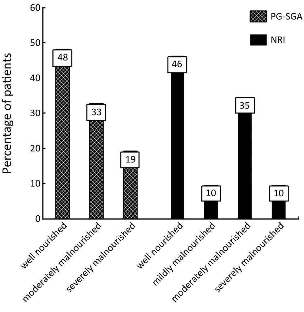
ObjectiveTo validate malnutrition screening tool of nutrition risk index (NRI) against patient-generated subjective global assessment (PG-SGA) as a gold standard tool in colorectal cancer patients before radiotherapy. MethodsNutritional status of 52 volunteer colorectal cancer patients with a mean age of 54.1±16.8 years who referred to radiotherapy center were assessed by PG-SGA (gold standard method) and NRI. Serum albumin levels of patients were determined by colorimetric method. A contingency table was used to determine the sensitivity, specificity, and predictive value of the NRI in screening patients at risk of malnutrition, in comparison with the PG-SGA in patients before radiotherapy. ResultsThe findings of PG-SGA and NRI showed that 52% and 45% of patients in our study were moderately or severely malnourished respectively. The NRI had a sensitivity of 66% and a specificity of 60% against PG-SGA. The positive predictive value was 64% and the negative predicative value was 62%. The agreement between NRI and PG-SGA was statistically insignificant (kappa =0.267; P>0.05). ConclusionsThe findings of present study showed that the prevalence of malnutrition was high in patients with colorectal cancer. Moreover, NRI method had low sensitivity and specificity in assessing nutritional status of patients with cancer. It seems that the combination of anthropometric, laboratory parameters and a subjective scoring system may be helpful tools in screening of malnutrition in cancer patients.
2013, 25(5): 549-555.
doi: 10.3978/j.issn.1000-9604.2013.10.05
Abstract:
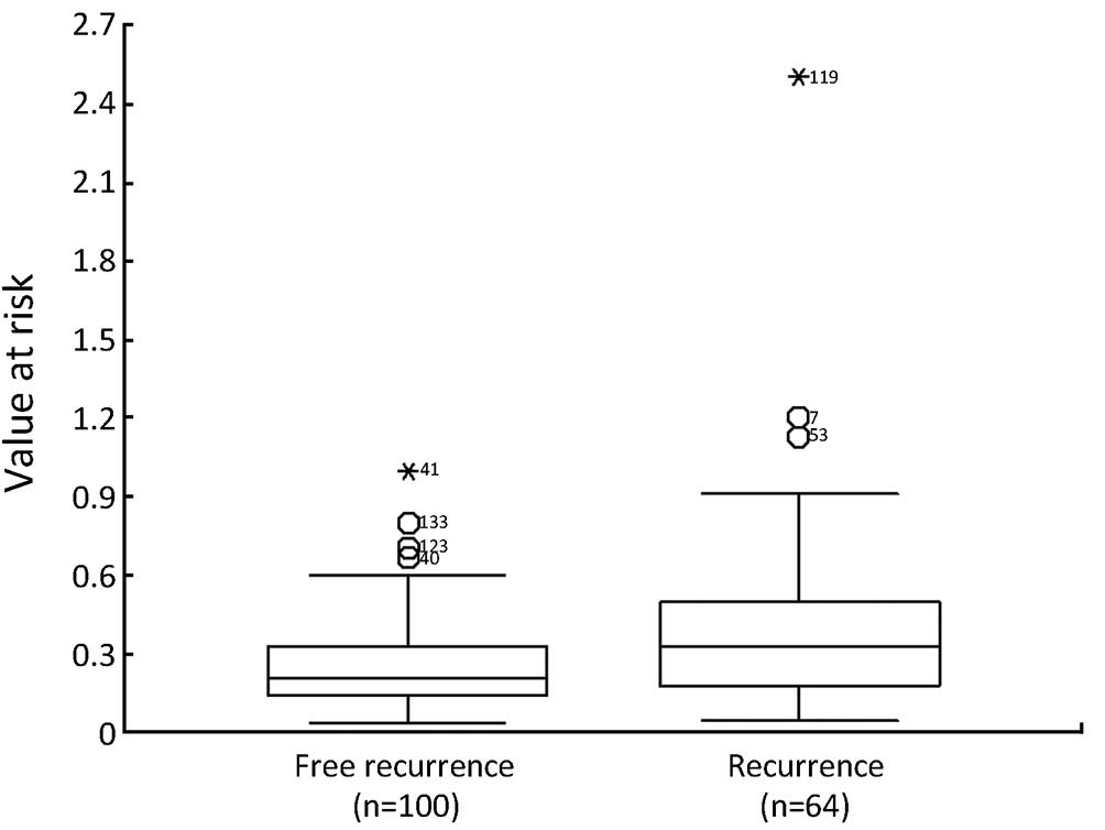
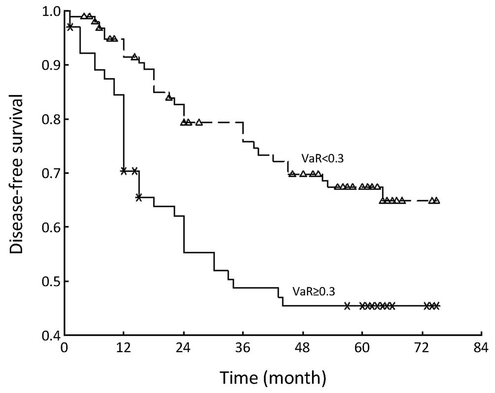
ObjectiveThe aim of the present study was to construct a risk assessment model which was tested by disease-free survival (DFS) of esophageal cancer after radical surgery. MethodsA total of 164 consecutive esophageal cancer patients who had undergone radical surgery between January 2005 and December 2006 were retrospectively analyzed. The cutpoint of value at risk (VaR) was inferred by stem-and-leaf plot, as well as by independent-samples t-test for recurrence-free time, further confirmed by crosstab chi-square test, univariate analysis and Cox regression analysis for DFS. ResultsThe cutpoint of VaR was 0.3 on the basis of our model. The rate of recurrence was 30.3% (30/99) and 52.3% (34/65) in VaR <0.3 and VaR ≥0.3 (chi-square test, χ2 =7.984, P=0.005), respectively. The 1-, 3-, and 5-year DFS of esophageal cancer after radical surgery was 70.4%, 48.7%, and 45.3%, respectively in VaR ≥0.3, whereas 91.5%, 75.8%, and 67.3%, respectively in VaR <0.3 (Log-rank test, χ2 =9.59, P=0.0020), and further confirmed by Cox regression analysis [hazard ratio =2.10, 95% confidence interval (CI): 1.2649-3.4751; P=0.0041]. ConclusionsThe model could be applied for integrated assessment of recurrence risk after radical surgery for esophageal cancer.
2013, 25(5): 556-564.
doi: 10.3978/j.issn.1000-9604.2013.10.06
Abstract:
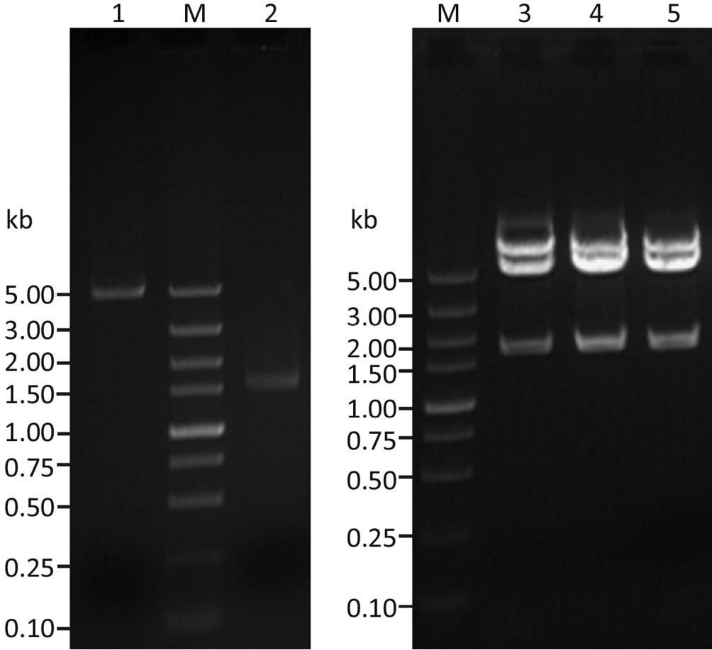
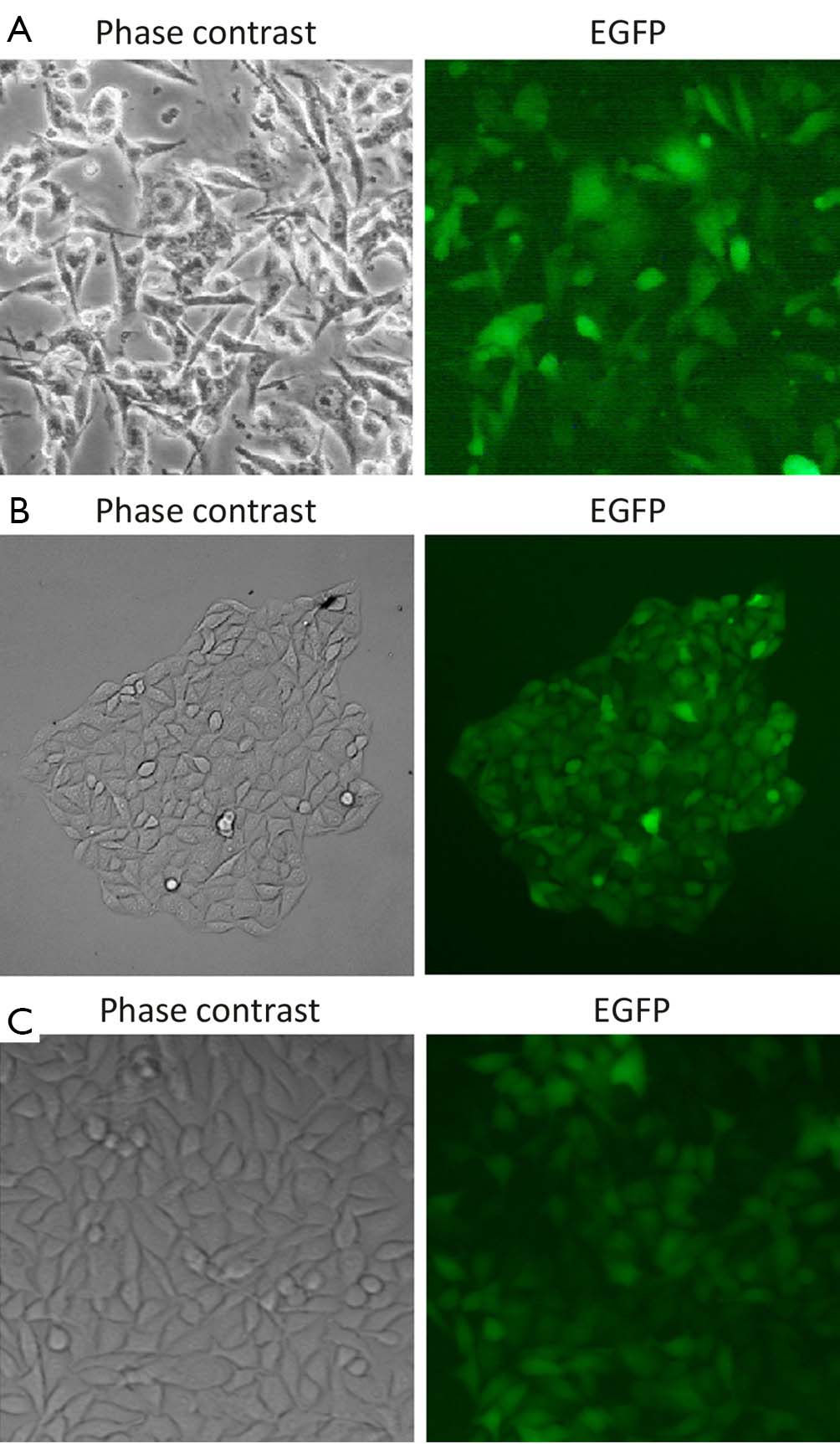
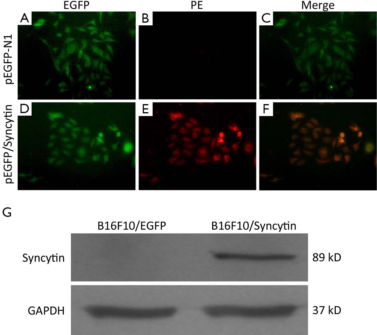
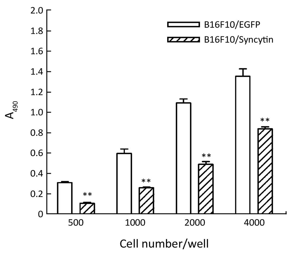
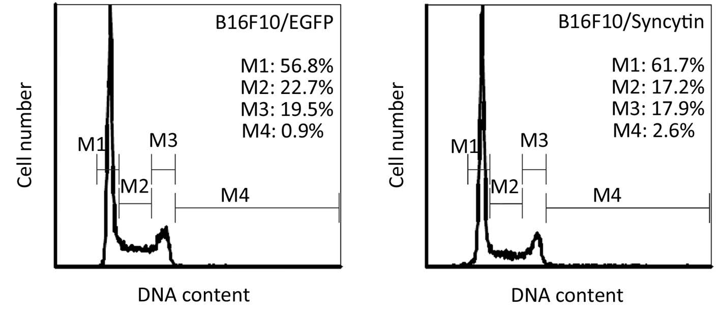
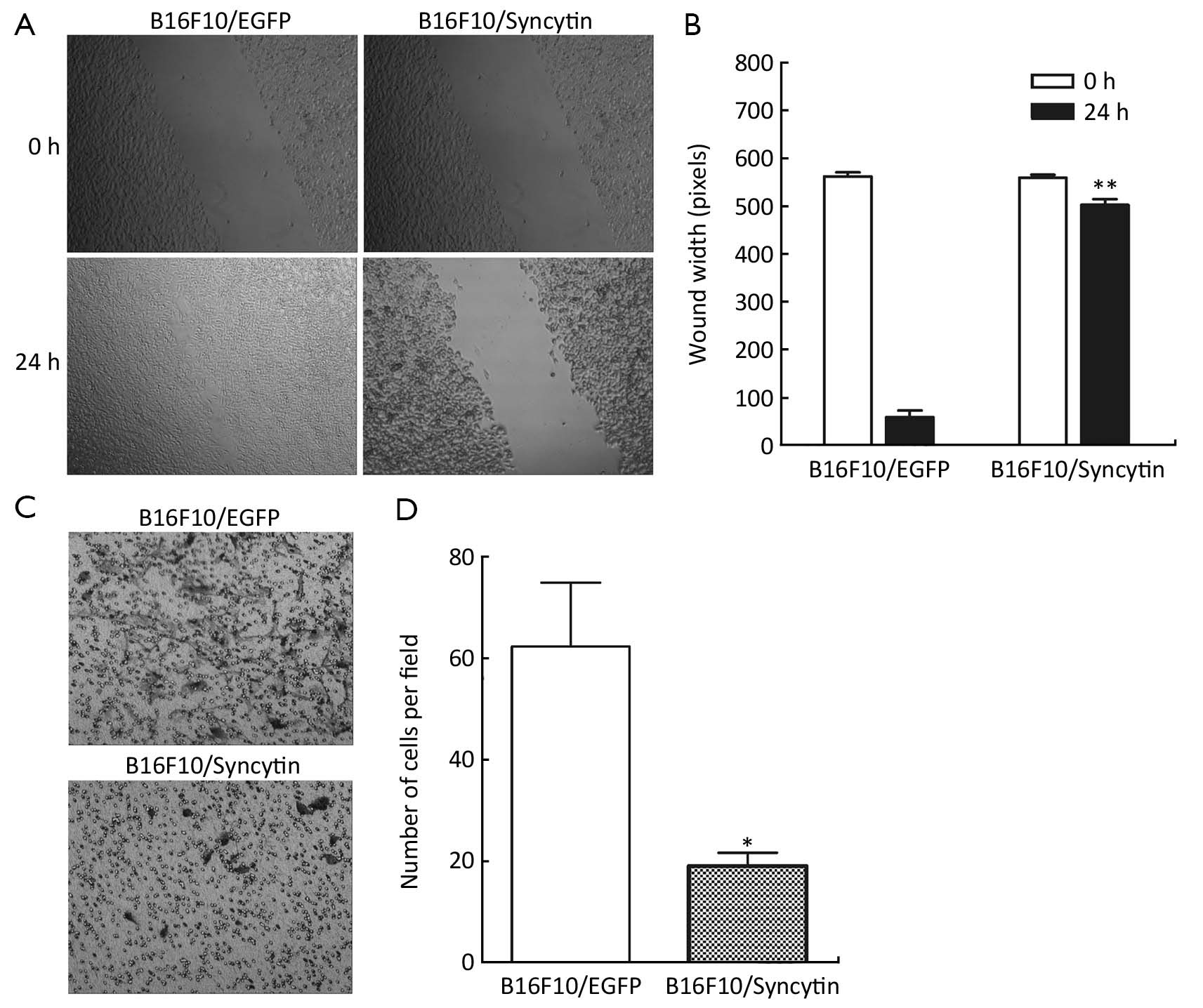
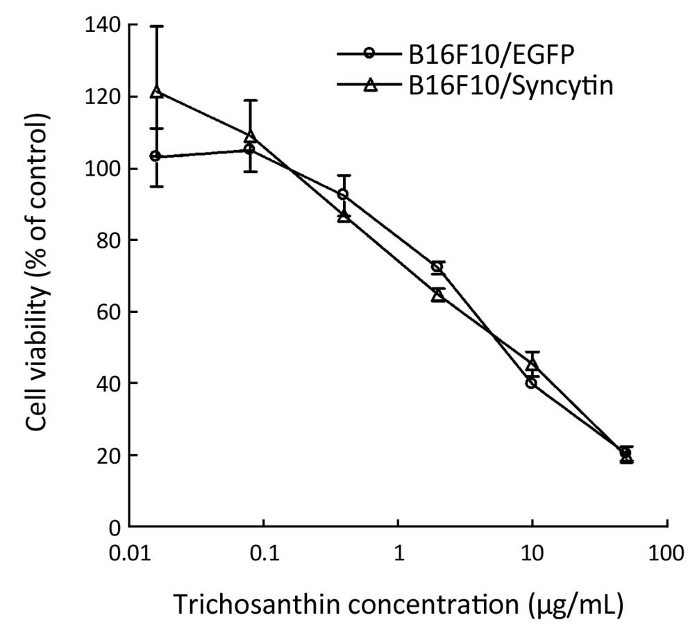
ObjectiveFusogenic endogenous retroviral syncytin plays an important role in the formation of syncytiotrophoblasts in human placenta. Apart from its expression in placenta, brain and testis, syncytin has also been found in many cancers. Although syncytin has been proposed to serve as a positive prognostic marker in some cancers, the underlying mechanism is unclear. The aim of this study is to evaluate the effects of syncytin expression on the invasive phenotype of melanoma cells. MethodsThe eukaryotic expression plasmid for syncytin-EGFP was constructed and transfected into B16F10 melanoma cells. The effect of syncytin on the invasion potential of tumor cells was evaluated in B16F10 subline cells that stably expressed syncytin-EGFP fusion protein or EGFP alone. ResultsThe B16F10 sublines that stably expressed syncytin-EGFP or EGFP alone were established respectively and confirmed by immunofluorescent and immunoblotting assay. Syncytin expression in B16F10 cells was associated with decreased cell proliferation, migration and invasion. Multinucleated giant cells that contained as many as five nuclei were induced in syncytin-expressing cells. In addition, syncytin expression did not alter the sensitivity of B16F10 cells to trichosanthin, a toxin that damages syncytiotrophoblasts more efficiently than other tissues. ConclusionsThese results suggest that syncytin expression in some cancers may confine their invasion potential and thus serve as a positive prognostic factor.

2013, 25(5): 565-571.
doi: 10.3978/j.issn.1000-9604.2013.10.03
Abstract:
ObjectiveRecent evidence indicates that dysregulation of microRNA (miRNA) biogenesis is implicated in cancer development and progression. Based on the important role of miRNA biogenesis genes in carcinogenesis, we hypothesized that genetic variations of the miRNA biogenesis genes may modulate susceptibility to cervical cancer. MethodsWe identified three single nucleotide polymorphisms (SNPs) located in the 3'-untranslated regions (3'-UTR) of of miRNA biogenesis key genes (rs1057035 in DICER, rs3803012 in RAN and rs10773771 in HIWI) and genotyped these SNPs in a case-control study of 1,486 cervical cancer cases and 1,549 cancer-free controls in Chinese women. ResultsLogistic regression analyses showed that no significant associations were observed between the three SNPs and cervical cancer risk [rs3803012 in RAN AG/GG vs. AA adjusted OR =1.104, 95% confidence interval (CI): 0.859-1.419; rs1057035 in DICER CT/CC vs. TT adjusted OR =0.962, 95% CI: 0.805-1.149; rs10773771 in HIWI CT/CC vs. TT adjusted OR =0.963, 95% CI: 0.826-1.122]. ConclusionsThe findings did not suggest that genetic variants in the 3'-UTR of RAN, DICER and HIWI of miRNA biogenesis genes were associated with the risk of cervical cancer in this Chinese population.
2013, 25(5): 572-584.
doi: 10.3978/j.issn.1000-9604.2013.10.10
Abstract:
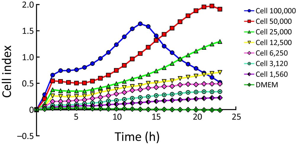
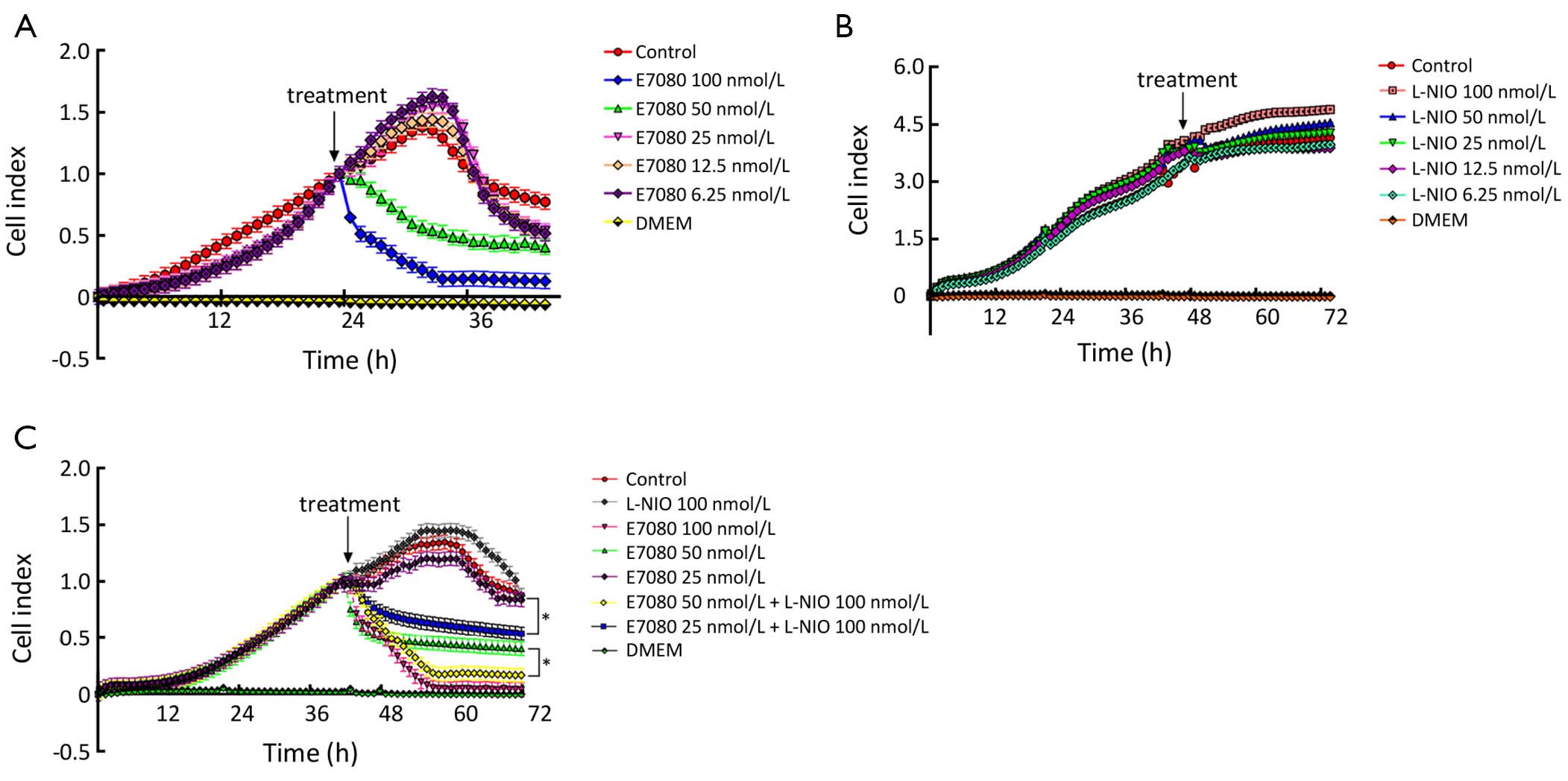
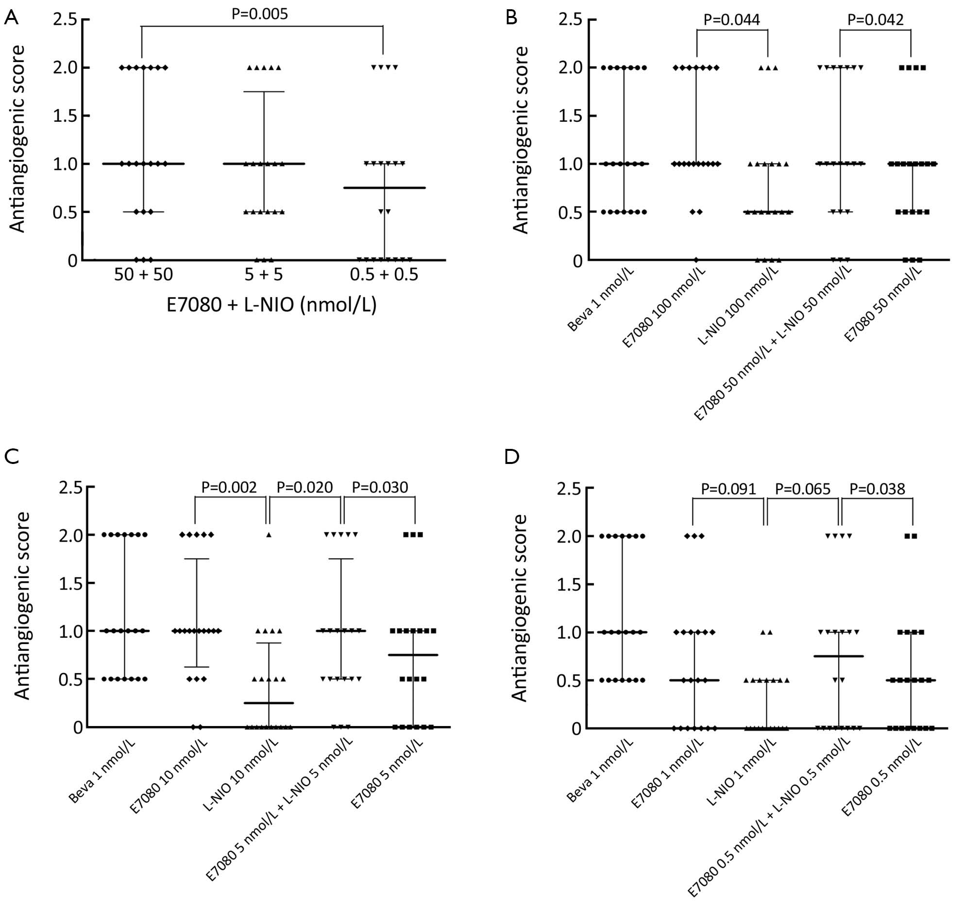
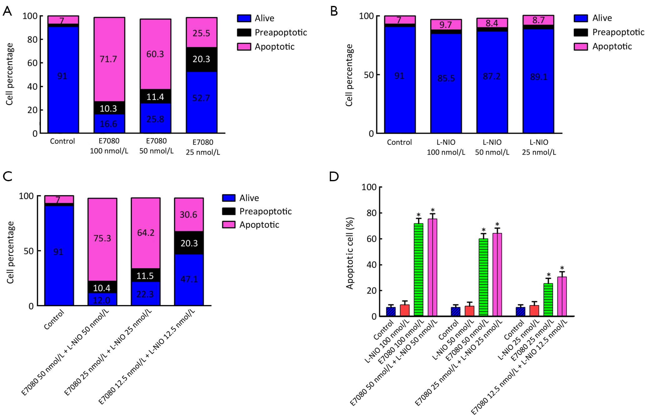
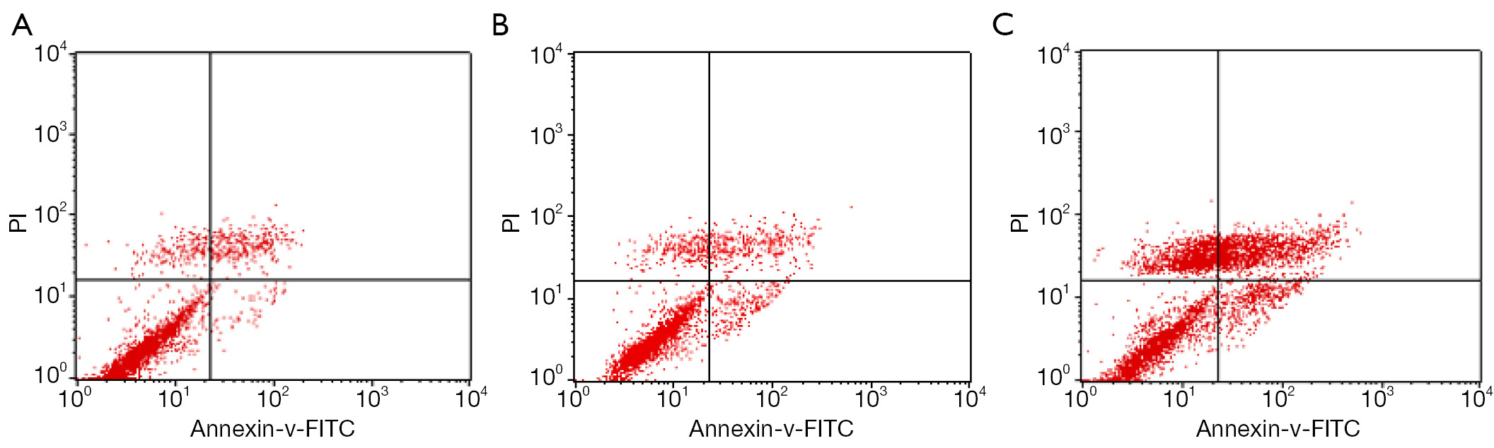
ObjectiveTo investigate the effects of E7080 and N5-(1-iminoethyl)-L-ornithine dihydrochloride (L-NIO) on colorectal cancer alone and in combination. MethodsHT29 colorectal cancer cell line from Sap Institute was used. Real-time cell analysis (xCELLigence system) was performed to determine the effects of E7080 and L-NIO on colorectal cell proliferation. While apoptosis was determined with Annexin V staining, and the effect of agents on angiogenesis was determined with chorioallantoic membrane (CAM) model. ResultsWe found that E7080 has a strong antiproliferative effect with an half maximum inhibition of concentration (IC50) value of 5.60×10⁻⁸ mol/L. Also it has been observed that E7080 showed antiangiogenic and apoptotic effects on HT29 colorectal cancer cells. Antiangiogenic scores of E7080 were 1.2, 1.0 and 0.6 for 100, 10 and 1 nmol/L E7080 concentrations, respectively. Furthermore, apoptosis has been detected in 71% of HT29 colorectal cancer cells after administration of 100 nmol/L E7080 which may indicate strong apoptotic effect. Meanwhile administration of L-NIO alone did not show any effect, but the combination of E7080 with L-NIO increased the antiproliferative, antiangiogenic and apoptotic effects of E7080. ConclusionsResults of this study indicate that E7080 may be a good choice in treatment of colorectal tumors. Furthermore the increased effects of E7080 when combined with L-NIO raise the possibility to use a lower dose of E7080 and therefore avoid/minimize the side effects observed with E7080.


2013, 25(5): 585-592.
doi: 10.3978/j.issn.1000-9604.2013.10.16
Abstract:
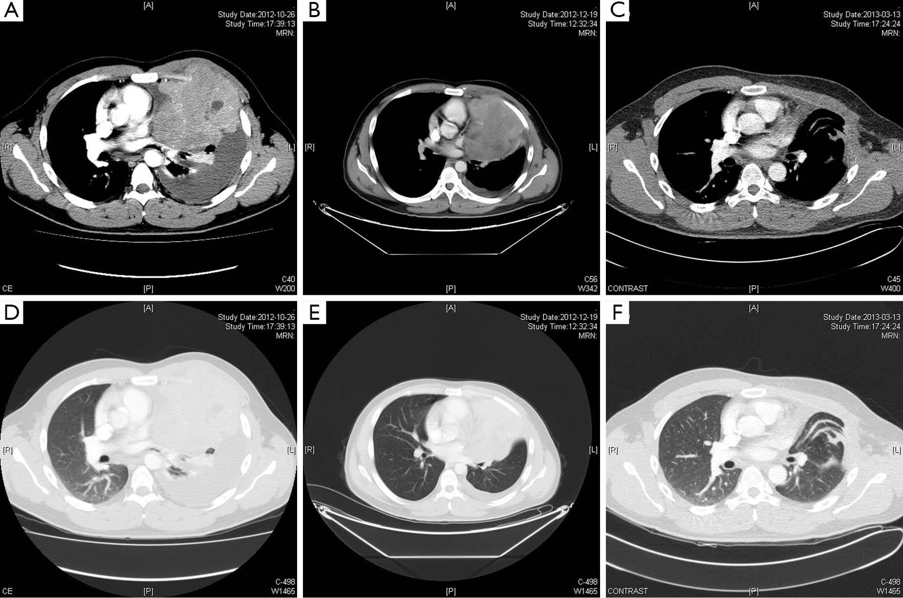
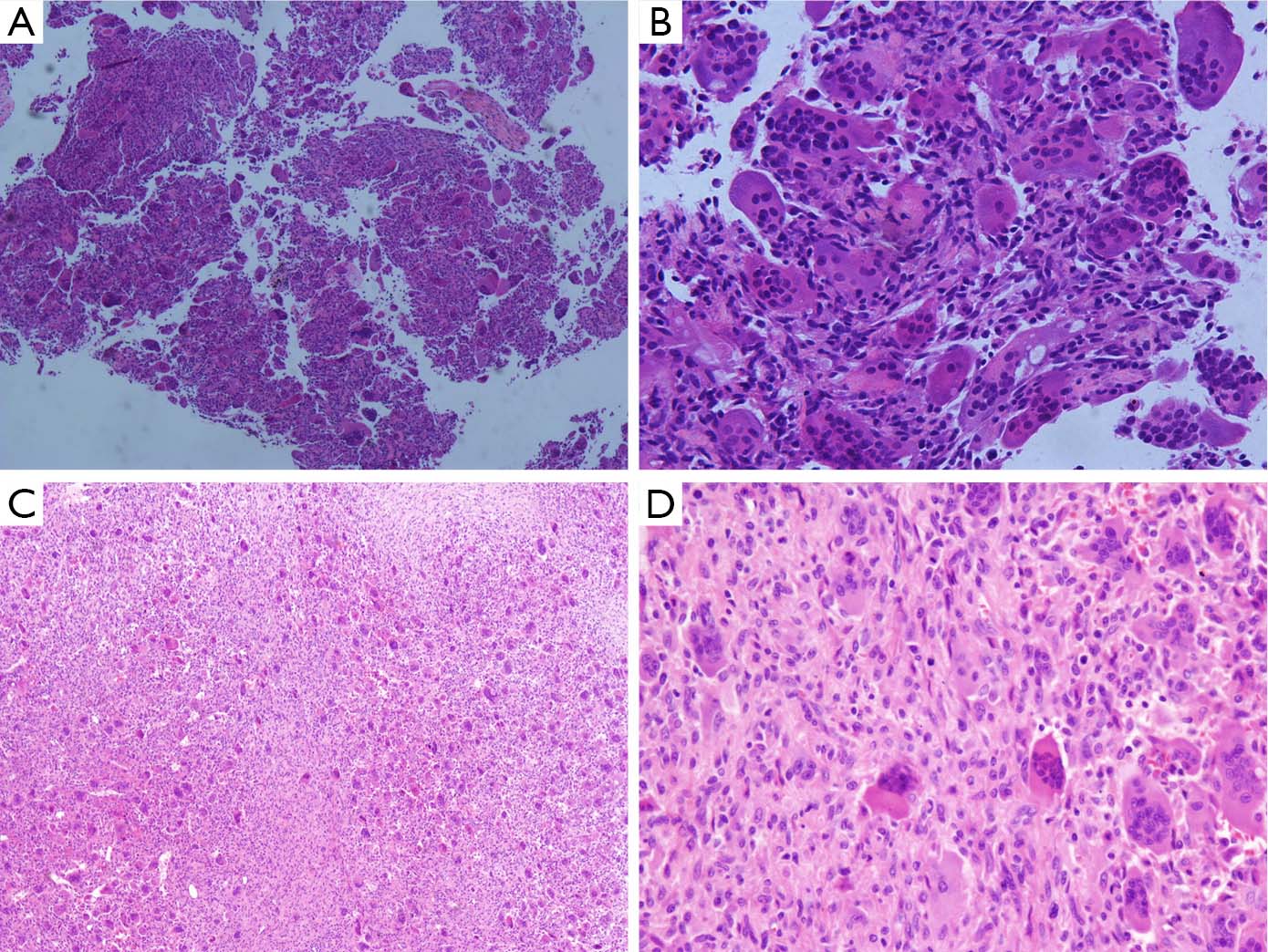
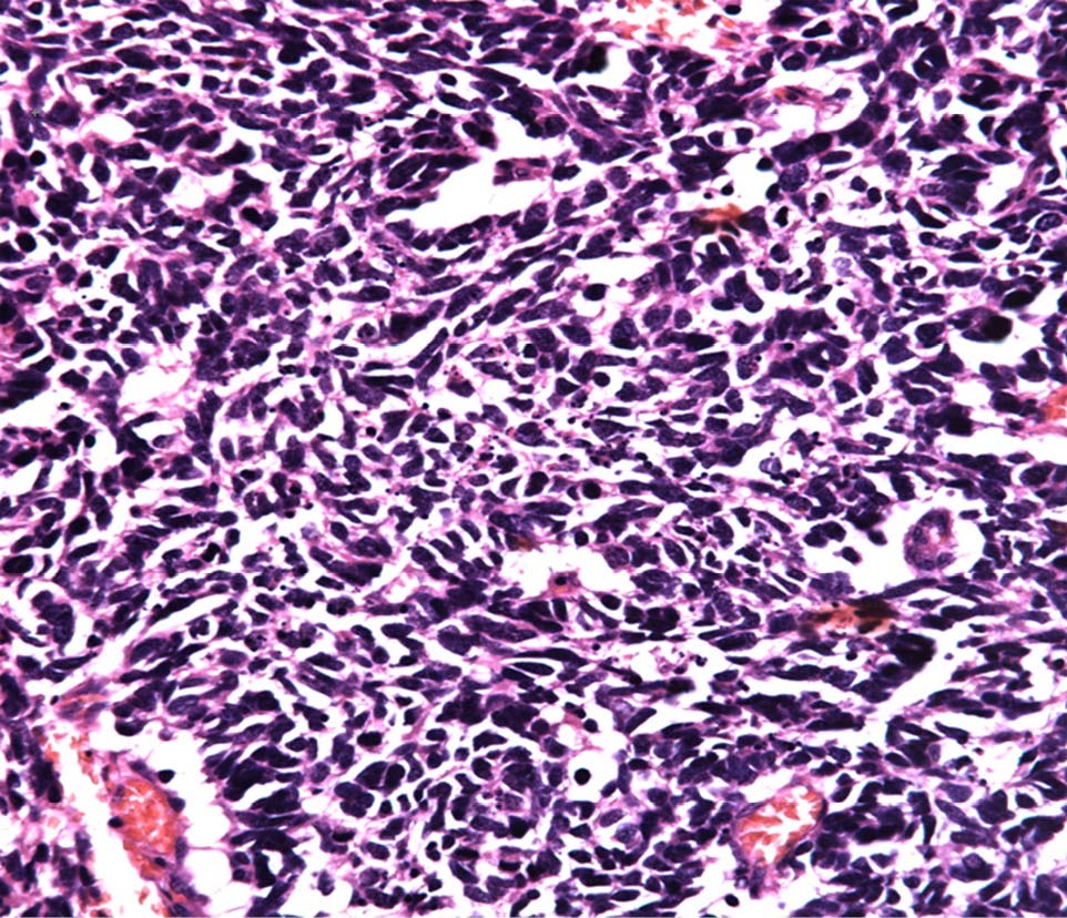
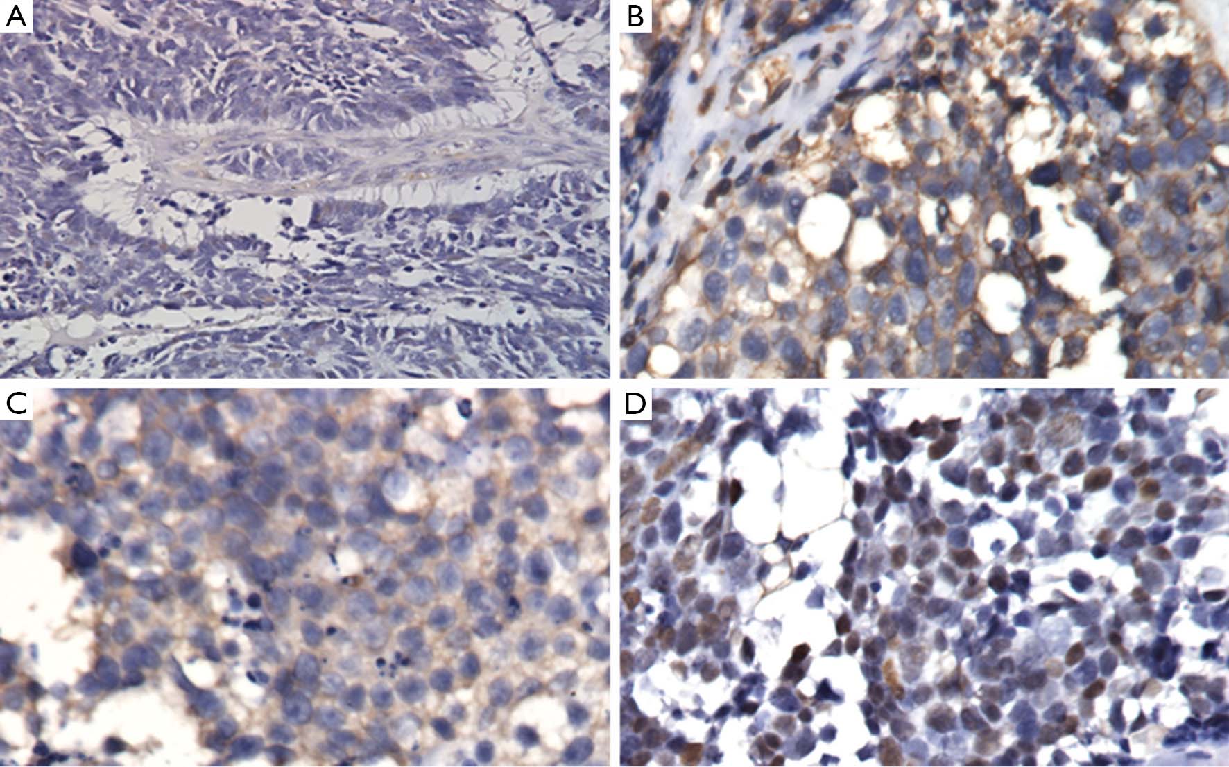
A giant cell tumor occurs mainly in the proximal tibia, humerus, distal radius bone and the pelvic bone. It is rarely observed in such sites as the ribs and the temporal bone. The condition is primarily treated with surgical excision and functional reconstruction. The effect of chemotherapy on lung metastases and locally advanced giant cell tumors has remained unknown. We collected and analyzed the data of six patients with rare giant cell tumors located in the head and neck patients. After an average follow-up of 42.6 months after surgery (14 to 90 months), no local recurrence or metastasis was observed. We also collected and analyzed the data of five patients with metastatic giant cell tumors who were undergoing surgery for the primary tumor before; of three patients who had experienced multiple chemotherapy cycles, one had spontaneous regression, and one survived for long timer despite progression. The other two patients had their major metastatic lesions resected by surgery, and presented long-term survival during the follow up. In addition, this study reports one patient with locally advanced giant cell tumor of the rib, who has undergone successful surgical resection following two cycles of chemotherapy with ifosfamide and liposomal doxorubicin. Complete resection of the lesion at the head and neck is the key to relapse-free survival. The prognosis of lung metastases in patients with giant cell tumors is relatively satisfying. Neoadjuvant chemotherapy is also conducive to the surgery for locally advanced lesions and improvement of the quality of life.
A giant cell tumor occurs mainly in the proximal tibia, humerus, distal radius bone and the pelvic bone. It is rarely observed in such sites as the ribs and the temporal bone. The condition is primarily treated with surgical excision and functional reconstruction. The effect of chemotherapy on lung metastases and locally advanced giant cell tumors has remained unknown. We collected and analyzed the data of six patients with rare giant cell tumors located in the head and neck patients. After an average follow-up of 42.6 months after surgery (14 to 90 months), no local recurrence or metastasis was observed. We also collected and analyzed the data of five patients with metastatic giant cell tumors who were undergoing surgery for the primary tumor before; of three patients who had experienced multiple chemotherapy cycles, one had spontaneous regression, and one survived for long timer despite progression. The other two patients had their major metastatic lesions resected by surgery, and presented long-term survival during the follow up. In addition, this study reports one patient with locally advanced giant cell tumor of the rib, who has undergone successful surgical resection following two cycles of chemotherapy with ifosfamide and liposomal doxorubicin. Complete resection of the lesion at the head and neck is the key to relapse-free survival. The prognosis of lung metastases in patients with giant cell tumors is relatively satisfying. Neoadjuvant chemotherapy is also conducive to the surgery for locally advanced lesions and improvement of the quality of life.
2013, 25(5): 593-599.
doi: 10.3978/j.issn.1000-9604.2013.10.11
Abstract:
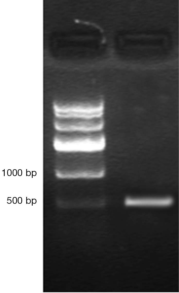
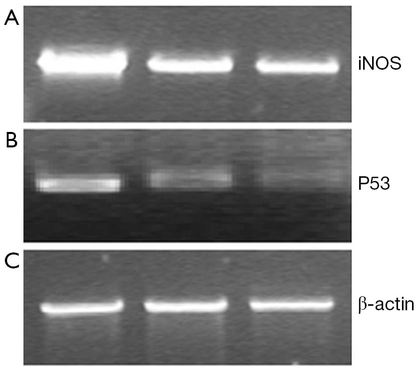
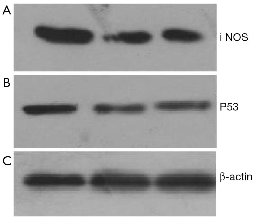
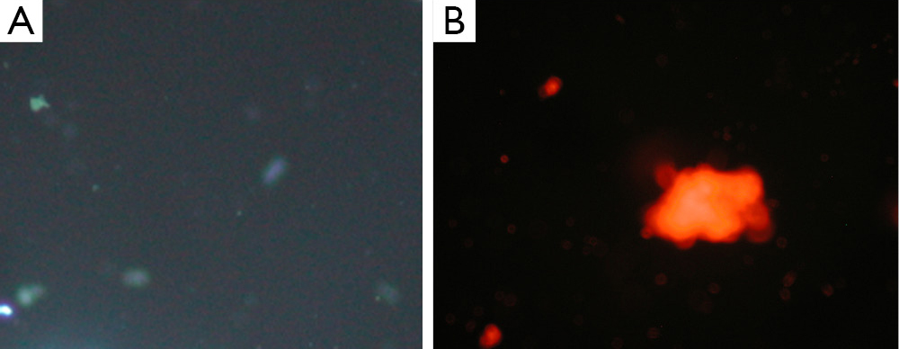
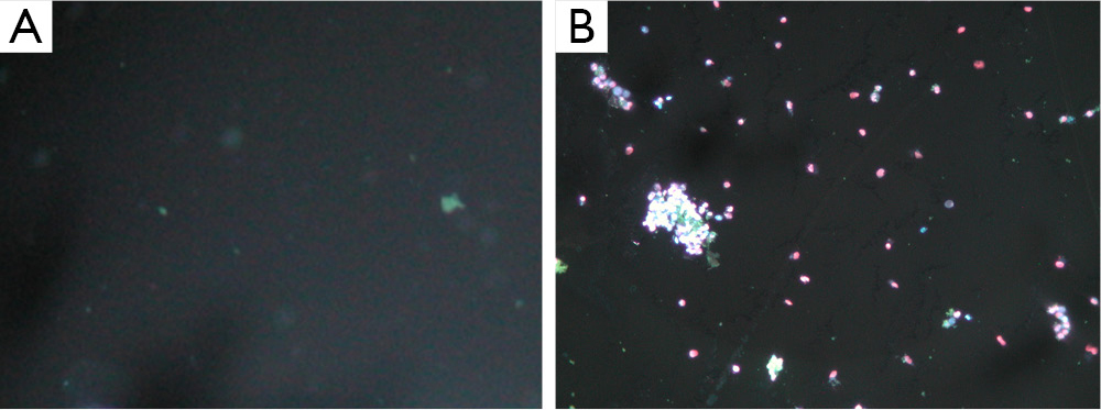
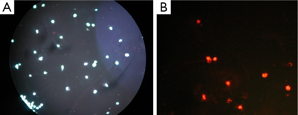
ObjectivesTo investigate the effects of adenovirus-mediated inducible nitric oxide synthase gene transfection on bladder transitional cell carcinoma T24 cells, and to provide novel insights and approaches to clinical therapies against bladder transitional cell carcinoma. MethodsFirstly, construct recombinant adenovirus vector pAd-iNOS of iNOS, followed by transfection of pAd-iNOS into HECK293 packaging cells. Thirdly, harvest recombinant adenovirus rAd-iNOS after amplification and purification procedures. Finally, transfect the recombinant adenovirus rAd-iNOS into human bladder carcinoma T24 cells and examine the effect of rAd-iNOS transfection on apoptosis of T24 and possible mechanism. ResultsAs shown by this study, the recombinant adenovirus rAd-iNOS was constructed successfully. The virus titer was 5.8×10⁸ PFU/mL and recombinant was verified by PCR analysis. Transfection of adenovirus rAd-iNOS into T24 cells could induce secretion of high NO concentration, P53 protein expression up-regulation, as well as promotion of T24 cell apoptosis. ConclusionsThe transfection of human bladder carcinoma T24 cells from recombinant adenovirus rAd-iNOS was confirmed to induce intracellular iNOS over-expression, high production of NO, up-regulation of intracellular P53 expression and promotion of cell apoptosis.

2013, 25(5): 600-602.
doi: 10.3978/j.issn.1000-9604.2013.10.14
Abstract:
Gastrointestinal stromal tumors (GISTs) occur most frequently in the stomach. Diagnosis of gastric GIST is not always clear before surgery. Flexible endoscopy may suggest the nature of the lesion (a bulky tumor with preserved mucosa); however, biopsy is rarely diagnostic. Therefore, diagnostic medication with safe drugs may provide a feasible way under such conditions after an informed consent is obtained. Based on the excellent efficacy of imatinib mesylate (IM) in the treatment of GIST, we successfully applied it in the diagnostic medication of two patients with clinically suspected gastric stromal tumors. In conclusion, the diagnostic medication with IM can be an alternative option for patients with suspected GIST that can not be confirmed pathologically.
Gastrointestinal stromal tumors (GISTs) occur most frequently in the stomach. Diagnosis of gastric GIST is not always clear before surgery. Flexible endoscopy may suggest the nature of the lesion (a bulky tumor with preserved mucosa); however, biopsy is rarely diagnostic. Therefore, diagnostic medication with safe drugs may provide a feasible way under such conditions after an informed consent is obtained. Based on the excellent efficacy of imatinib mesylate (IM) in the treatment of GIST, we successfully applied it in the diagnostic medication of two patients with clinically suspected gastric stromal tumors. In conclusion, the diagnostic medication with IM can be an alternative option for patients with suspected GIST that can not be confirmed pathologically.

2013, 25(5): 603-607.
doi: 10.3978/j.issn.1000-9604.2013.09.05
Abstract:
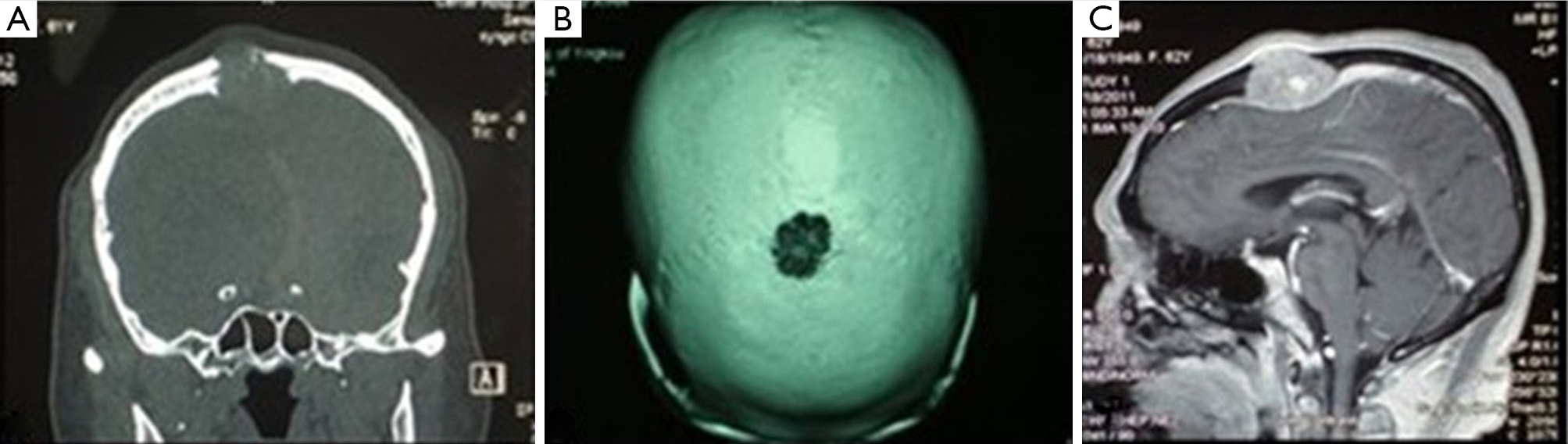
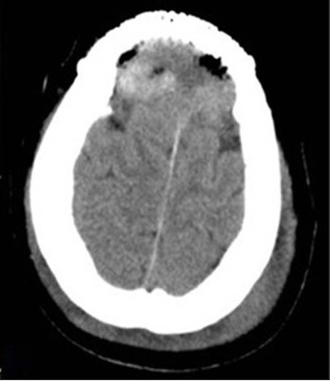
Although thyroid carcinoma is a relatively common form of malignancy, metastatic spread to the skull is rare. Here, we report a case of papillary thyroid carcinoma with frontal and parietal metastasis. A 61-year-old Chinese woman presented with a one year history of a growing mass on the center of the frontal and parietal bone, initially thought to be meningioma. Biopsy of the skull base mass after intracalvarium excision, indicated a tumor of thyroid origin. One month later the patient underwent a total thyroidectomy. Pathological examination confirmed a diagnosis of papillary thyroid carcinoma with frontal and parietal bone metastasis. Based on this experience, the key to successful management of the skull metastasis of thyroid carcinoma is prompt diagnosis and appropriate treatment. Skull metastasis should be considered at the outset of the clinical course of papillary thyroid cancer. To facilitate this, patients should be meticulously investigated by a multidisciplinary team to improve quality of life.
Although thyroid carcinoma is a relatively common form of malignancy, metastatic spread to the skull is rare. Here, we report a case of papillary thyroid carcinoma with frontal and parietal metastasis. A 61-year-old Chinese woman presented with a one year history of a growing mass on the center of the frontal and parietal bone, initially thought to be meningioma. Biopsy of the skull base mass after intracalvarium excision, indicated a tumor of thyroid origin. One month later the patient underwent a total thyroidectomy. Pathological examination confirmed a diagnosis of papillary thyroid carcinoma with frontal and parietal bone metastasis. Based on this experience, the key to successful management of the skull metastasis of thyroid carcinoma is prompt diagnosis and appropriate treatment. Skull metastasis should be considered at the outset of the clinical course of papillary thyroid cancer. To facilitate this, patients should be meticulously investigated by a multidisciplinary team to improve quality of life.


2013, 25(5): 608-611.
doi: 10.3978/j.issn.1000-9604.2013.10.07
Abstract:
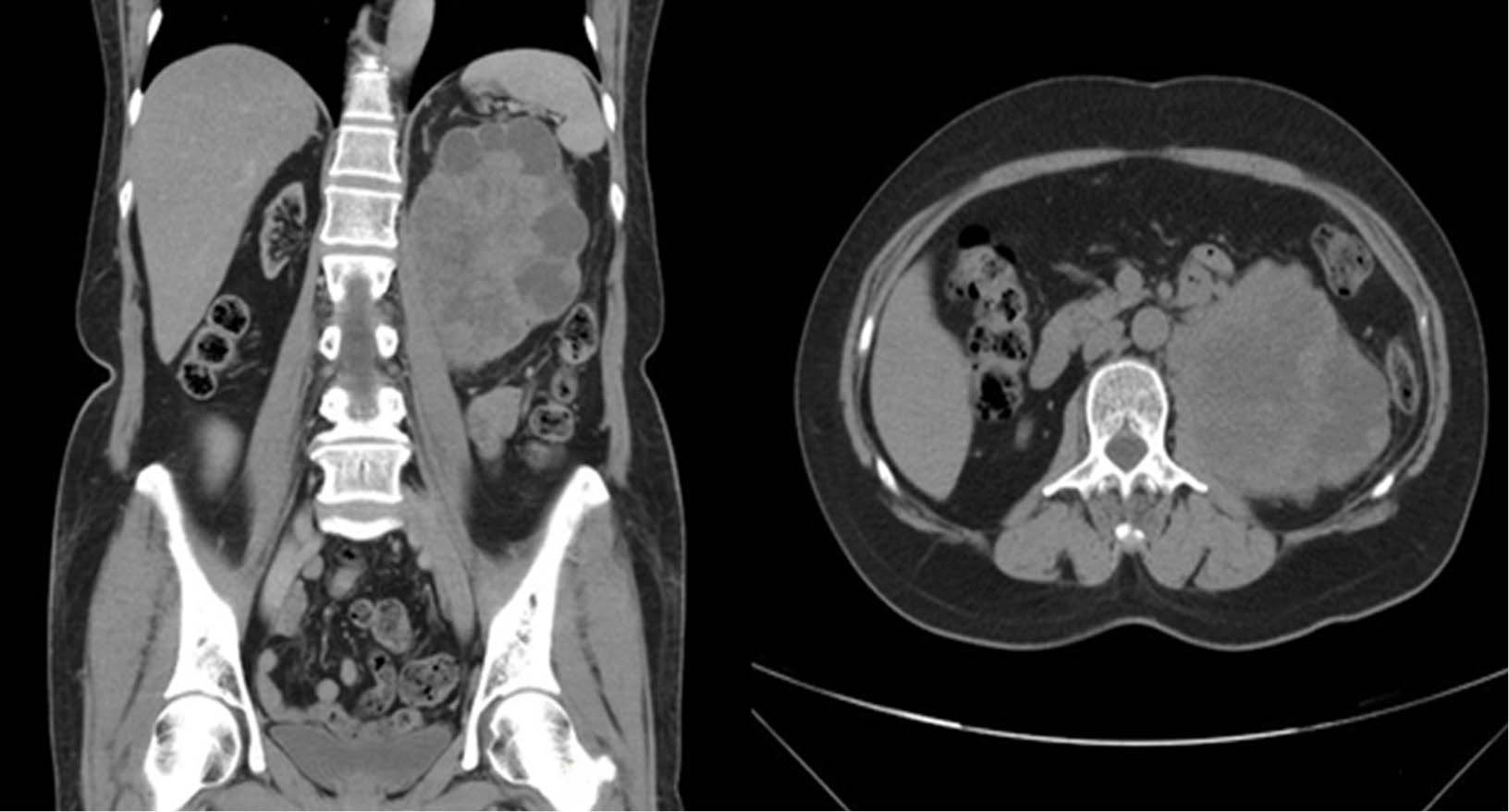
Extrapulmonary small cell carcinoma (EPSCC) is a rare neoplasm comprising 2.5% to 5% of small cell carcinomas (SCCs). Bladder SCC is the most common site of genitourinary tract. Primary renal SCC is extremely rare. We report a case of primary SCC of the kidney which is rarely reported in the urinary tract and presents an aggressive clinical picture. A 59-year-old female visited a urologic clinic with complaint of persistent left flank soreness 10 years after undergoing renal transplantation. Abdominal computed tomography showed a left renal pelvis tumor. After the patient received left nephroureterectomy with bladder cuff resection, her pathology results showed SCC. After surgery, she received adjuvant systemic chemotherapy, and her recovery has been uneventful as of 8 months. Primary renal SCC presents with an advanced tumor stage and a short median survival period, therefore early intervention and close follow-up are recommended.
Extrapulmonary small cell carcinoma (EPSCC) is a rare neoplasm comprising 2.5% to 5% of small cell carcinomas (SCCs). Bladder SCC is the most common site of genitourinary tract. Primary renal SCC is extremely rare. We report a case of primary SCC of the kidney which is rarely reported in the urinary tract and presents an aggressive clinical picture. A 59-year-old female visited a urologic clinic with complaint of persistent left flank soreness 10 years after undergoing renal transplantation. Abdominal computed tomography showed a left renal pelvis tumor. After the patient received left nephroureterectomy with bladder cuff resection, her pathology results showed SCC. After surgery, she received adjuvant systemic chemotherapy, and her recovery has been uneventful as of 8 months. Primary renal SCC presents with an advanced tumor stage and a short median survival period, therefore early intervention and close follow-up are recommended.
2013, 25(5): 613-614.
doi: 10.3978/j.issn.1000-9604.2013.10.09
Abstract:

