2014 Vol.26(2)
Display Mode: |
2014, 26(2): 132-132.
doi: 10.3978/j.issn.1000-9604.2014.04.01
 Abstract
 FullText HTML
 PDF 75KB
Abstract:
2014, 26(2): 133-134.
doi: 10.3978/j.issn.1000-9604.2014.04.04
Abstract:
2014, 26(2): 135-141.
doi: 10.3978/j.issn.1000-9604.2014.02.14
Abstract:
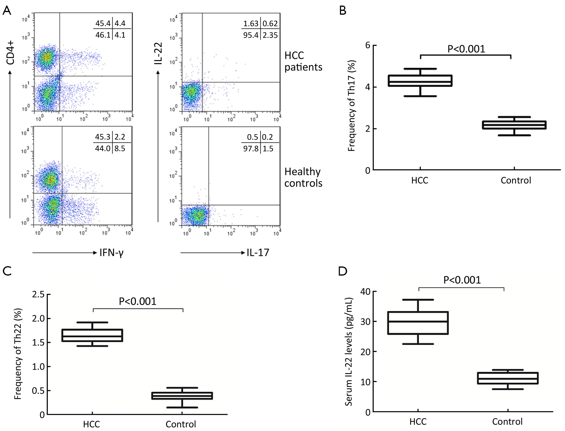
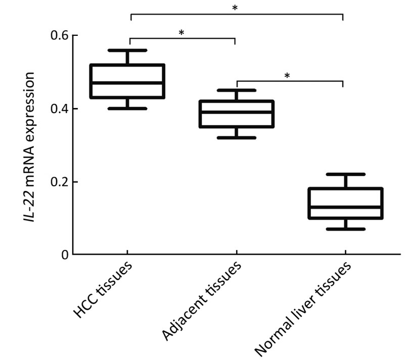
ObjectiveIL-22-producing CD4+ T helper cells (Th22 cells) have been identified as major inducers of tissue inflammation and immune responses. Currently, no previous study explored the role of Th22 cells in the pathogenesis of hepatocellular carcinoma (HCC). The study aimed to determine the biological function of Th22 cells and its effector IL-22 in HCC patients. MethodsForty-five HCC patients and 19 healthy controls were recruited and their peripheral blood was collected. The fresh HCC tissues, adjacent HCC tissues and ten normal liver tissues were also collected. Flow cytometry analysis was used to determine the frequencies of circulating Th22 cells and Th17 cells. Serum IL-22 levels were tested by enzyme-linked immunosorbent assay (ELISA). Immunohistochemical staining and real-time polymerase chain reaction (PCR) were used to detect IL-22 protein and mRNA in tissues specimens, respectively. ResultsCirculating Th22 cells, Th17 cells and serum IL-22 levels were significantly elevated in HCC patients compared with those of healthy controls (P<0.001). Th22 cells were showed to be positively correlated with IL-22 in HCC patients (P<0.05), but not in healthy controls. No significant differences were found in HCC patients with HBeAg positivity or negativity in term of Th22 cells and serum IL-22 levels. The expression of IL-22 protein and mRNA was highest in HCC tissues, followed by adjacent HCC tissues and normal liver tissues. Furthermore, Th22 cells, serum IL-22 levels and IL-22 mRNA were elevated at stage III-IV compared with stage I-II of HCC (P<0.05). ConclusionsElevation of circulating Th22 cells and IL-22 may be implicated in the pathogenesis of HCC, and potentially be cellular targets for therapeutic intervention.


2014, 26(2): 142-147.
doi: 10.3978/j.issn.1000-9604.2014.02.13
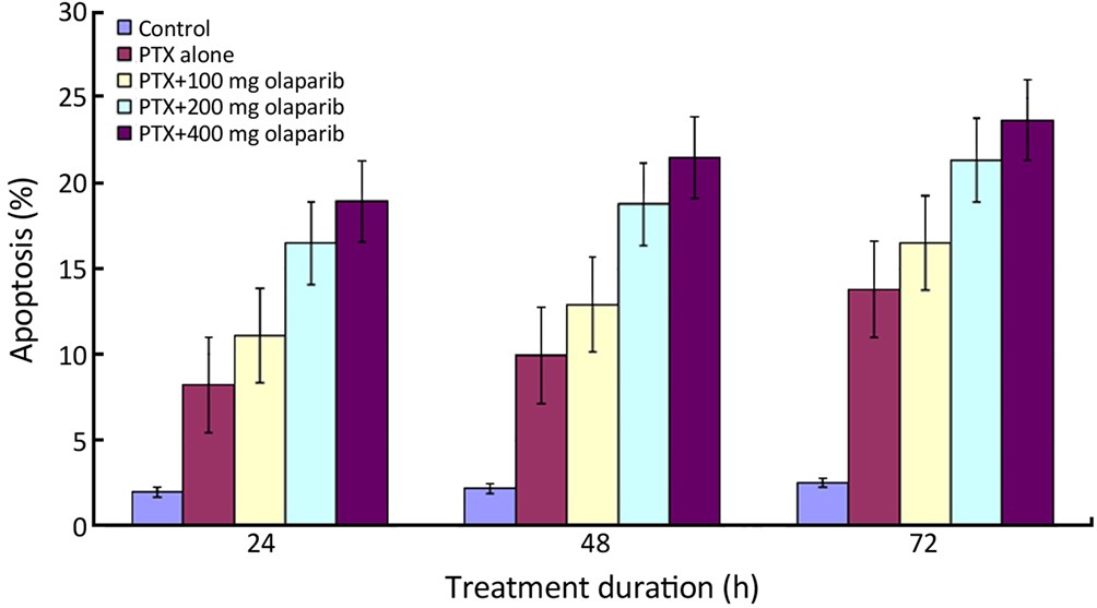
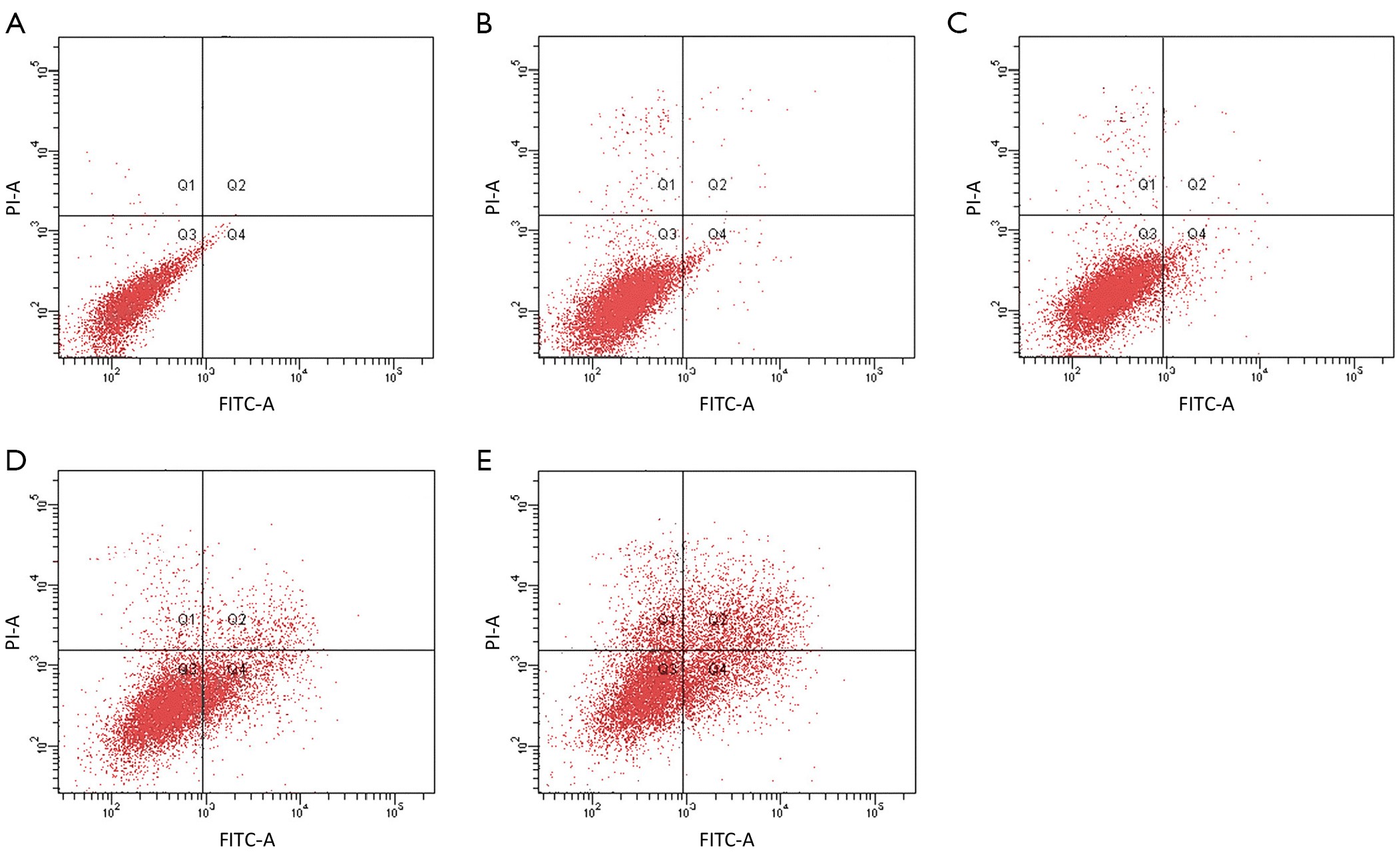
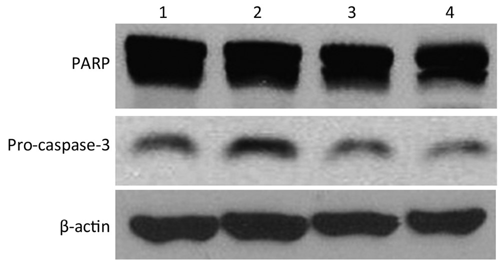
Abstract:
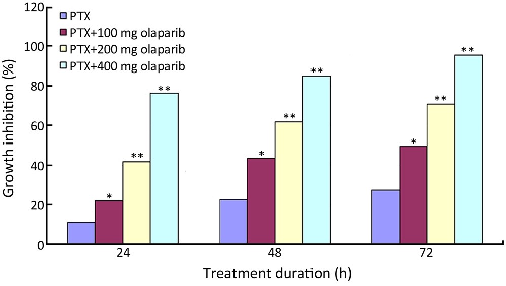
ObjectiveApoptosis is a reliable marker of chemotherapeutic efficacy. Olaparib and paclitaxel inhibit proliferation and induce apoptosis in a variety of cancers. We investigated the effects of paclitaxel combined with olaparib on apoptosis in breast cancer Bcap37 cells. MethodsProliferation and apoptosis were detected by MTT assay and PI staining. Degradation of pro-caspase-3 and poly(ADP-ribose) polymerase (PARP) was analyzed by Western blotting. ResultsCompared with paclitaxel alone, paclitaxel combined with 100 mg olaparib significantly reduced survival in Bcap37 cells at all tested treatment durations (P<0.05); inhibition increased with increasing olaparib dose and treatment time (P<0.01). Combined treatment yielded significantly higher rates of apoptosis (P<0.05), which also increased with time (P<0.01). Fluorescence micrographs showed that early and late apoptotic cells increased with treatment time. Pro-caspase-3 and PARP degradation was induced by paclitaxel and enhanced by olaparib in a dose-dependent manner. Thus, combined treatment was substantially more effective than treatment with paclitaxel alone. ConclusionsOur findings suggest that paclitaxel and olaparib inhibit breast cancer Bcap37 cell proliferation and induce apoptosis. Combined treatment further reduced cell growth and enhanced apoptosis, suggesting that this combination therapy may be a promising treatment for breast cancer.
2014, 26(2): 148-158.
doi: 10.3978/j.issn.1000-9604.2014.03.01
Abstract:
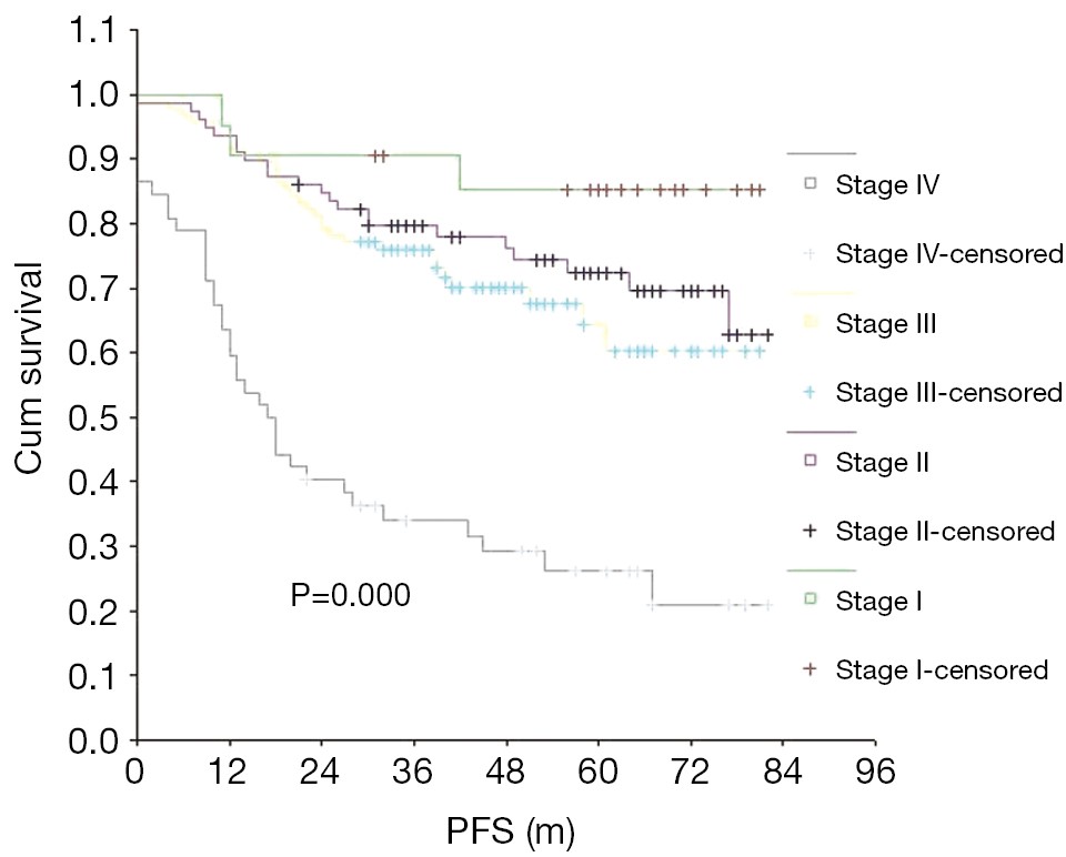
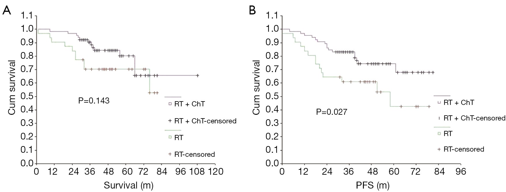
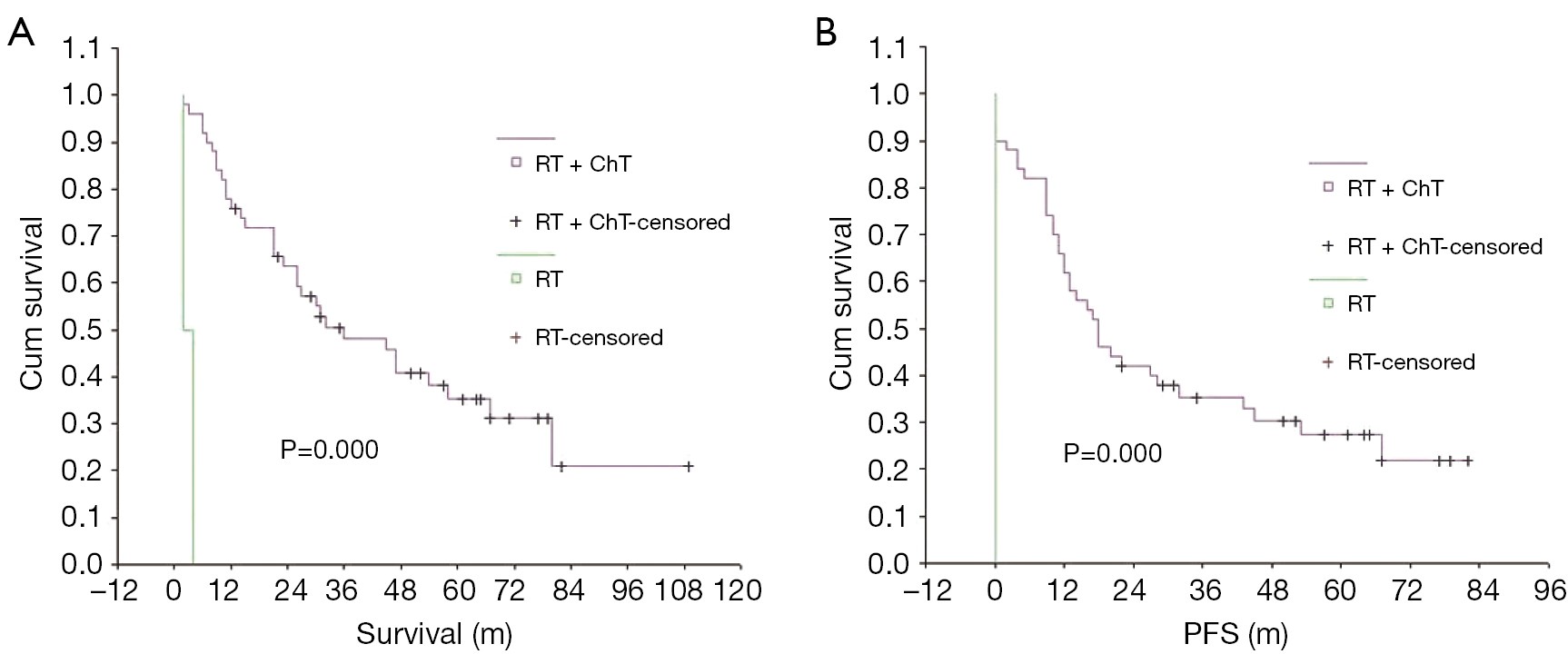
BackgroundNasopharyngeal carcinoma (NPC) is a common malignancy in Southeast Asia, however, a full consensus has not yet been reached as to the value of comprehensive treatment for NPC. This study was designed to evaluate the epidemiological characteristics of NPC and their prognostic value, as well as the long-term efficacy of NPC treatment. Patients and methodsA total of 248 patients, with different stages of NPC, were included in this study. ResultsThe 5-year overall survival (OS) rates for patients in stages I, II, III and IV were 90.48%, 76.71%, 76.89% and 33.87%, respectively (P=0.000), while the respective 5-year progression-free survival (PFS) rates were 85.15%, 72.36%, 63.88% and 26.26% (P=0.000). The respective 5-year OS rates, according to stage, for the group that received radiotherapy combined with chemotherapy and for the group that received radiotherapy only were as follows: stages I and II, 81.67% and 79.59% (P=0.753); stage III, 79.91% and 70.38% (P=0.143); stage IV, 35.22% and 0% (P=0.000). The respective 5-year PFS rates in these groups were as follows: stages I and II, 75.83% and 74.98% (P=0.814); stage III, 74.08% and 42.25% (P=0.027); stage IV, 27.31% and 0% (P=0.000). ConclusionsClinical staging appears to be the most important prognostic factor for NPC. As the stage number increases, both the 5-year OS and PFS significantly decrease. Adding chemotherapy to radiotherapy was not advantageous for patients with stage I or II NPC, however the addition of chemotherapy to radiotherapy significantly improved OS and PFS in patients with stage IV NPC. The addition of chemotherapy improved PFS, but not OS in patients with stage III NPC.


2014, 26(2): 159-165.
doi: 10.3978/j.issn.1000-9604.2014.03.03
Abstract:
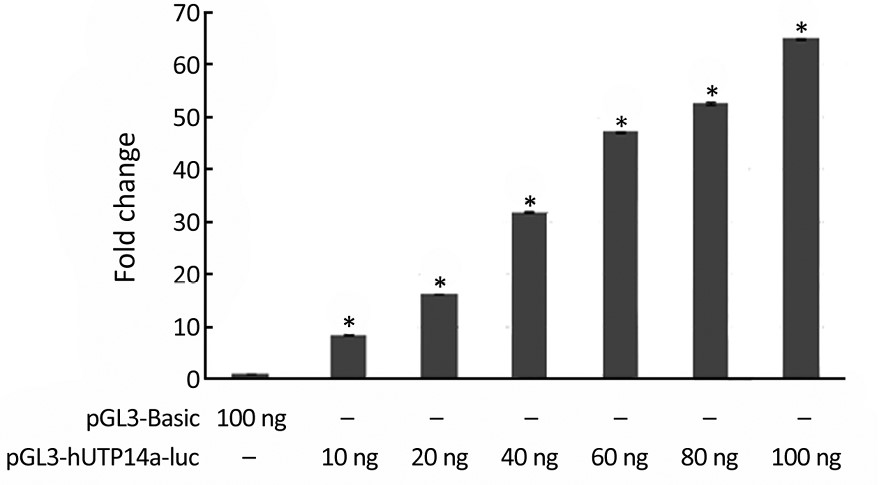
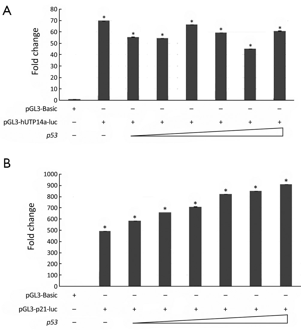
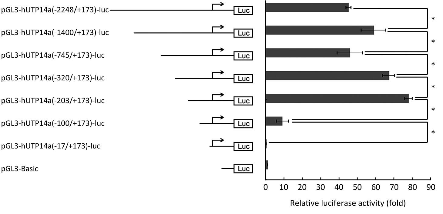
ObjectiveWe previously found that hUTP14a binds P53 and promotes P53 degradation. However, if hUTP14a is a downstream gene of P53 remains to be determined. This study aimed to identify the promoter of hUTP14a and investigate if hUTP14a is regulated by P53. MethodsThe hUTP14a promoter region was cloned into pGL3-Basic-luciferase reporter plasmid to get pGL3-hUTP14a-luc. The reporter plasmid was transfected into 293T cells and luciferase activity was evaluated by the Dual-Luciferase Reporter Assay System. Putative transcription factors were identified through searching MatInspector Professional and Algorismica i Genetica databases. Either pGL3-hUTP14a-luc or p21 promoter reporter plasmid was co-transfected with increasing dose of p53 plasmid, and luciferase activity was evaluated. A series of deletion constructs of pGL3-hUTP14a-luc were constructed and minimal promoter region of hUTP14a was determined. Differences of the luciferase activities between different groups were assessed by statistical analysis. ResultsThe hUTP14a gene promoter reporter construct was correctly cloned and was demonstrated to possess promoter activity. The transcription of hUTP14a was not regulated by P53. The minimal promoter region of hUTP14a gene is located between -203 to -100 of the transcription initiation site. ConclusionUnlike other P53-interacting proteins such as MDM2, Pirh2 and Cop I which promote P53 degradation and whose transcriptions are regulated by P53, does not hUTP14a transcription form a regulation feedback loop with P53.

2014, 26(2): 166-173.
doi: 10.3978/j.issn.1000-9604.2014.04.08
Abstract:
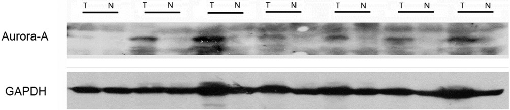
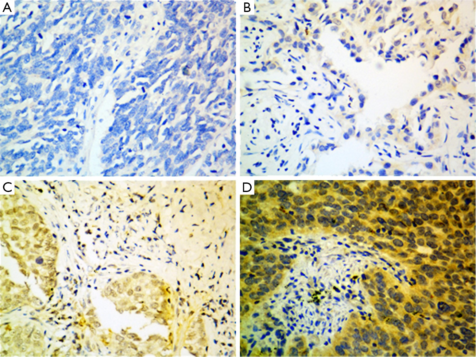
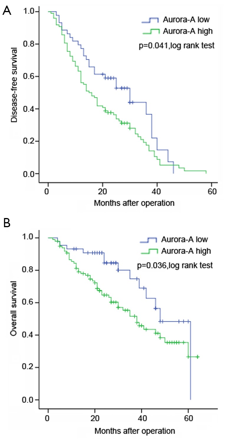
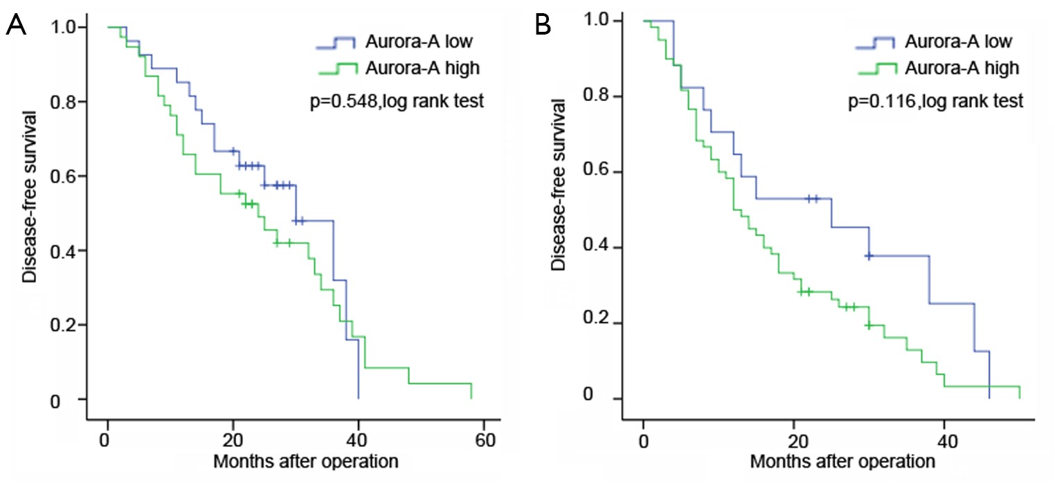
BackgroundThe Aurora-A (Aur-A) gene, a key regulator of mitosis, has been proved as an oncogene in a variety of cancers. The Aur-A overexpression has been proved correlated with aggressiveness of cancer cells. However, the frequency of Aur-A protein overexpression, as well as its association with clinicopathologic parameters and prognosis remain unclear in lung adenocarcinoma (ADC). This study tried to clarify the clinical significance of Aur-A in patients with resected lung ADC. Patients and methodsA total of 142 informative patients with surgically resected lung ADC and 20 normal lung tissues were enrolled. Western blot and immunohistochemistry (IHC) were utilized to assess protein expression of Aur-A. ResultThe expression of Aur-A was elevated in most of tumor tissues compared with the adjacent tissues by western blot. The IHC results showed that Aur-A protein was over-expressed in 98 of 142 (69.0%) tumor sections, while Aur-A was low-expressed in all normal lung sections. A positive correlation between Aur-A overexpression rate and ascending pathologic stages was observed (P<0.05). Kaplan-Meier analysis demonstrated that patients with Aur-A high expression had significantly inferior survival compared to those with Aur-A low expression. Both overall survival (OS) and disease-free survival (DFS) of positive overexpression patients were shorter than the negative group (P=0.036, P=0.041, respectively). Multivariate analysis confirmed that Aur-A expression, as an independent and significant factor for both DFS and OS, could predict a poor prognosis in patients with resected lung ADC (P=0.022, P=0.049, respectively). ConclusionsAur-A was overexpressed in lung ADC and overexpression of Aur-A might be a novel predictor for poor prognosis and potential therapeutic target in lung ADC.
2014, 26(2): 174-182.
doi: 10.3978/j.issn.1000-9604.2014.04.02
Abstract:
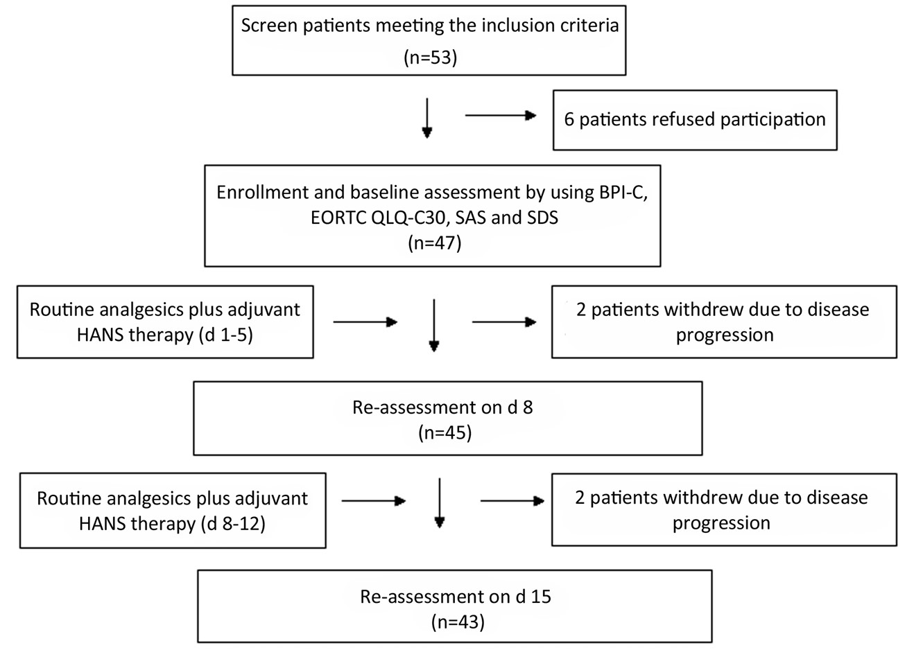
ObjectiveTo observe the adjuvant analgesic efficacy of Han’s Acupoint Nerve Stimulator (HANS) in opioid tolerant patients with cancer pain. MethodsA prospective non-controlled study was conducted. Opioid tolerant patients with cancer pain were enrolled and treated with both routinely analgesics and adjuvant HANS (2/100 Hz for 30 min/d, 5 d on and 2 d off for two weeks). Cancer pain, quality of life (QOL), anxiety and depression were assessed before enrollment and on d 8 and d 15 with the BPI-C, EORTC QLQ-C30, and self-rating anxiety scale (SAS)/self-rating depression scale (SDS), respectively; the therapeutic frequency of breakthrough pain (BP) and daily opioid dose were also recorded. ResultsTotally 47 patients meeting the inclusion criteria participated in this study; 43 patients completed the two-week treatment and assessment. The mean scores of patient’s “worst” and “least” pain intensity assessed with BPI-C decreased significantly on d 8 and d 15; the therapeutic frequency of BP also significantly decreased; but the average daily dose of opioids did not change significantly. For the nine symptoms in EORTC QLQ-C30 assessment, the mean scores of pain, fatigue, constipation and insomnia were significantly lower on d 8 and d 15 compared with baseline; the mean scores of the overall health status, nausea/vomiting and the incidence rates of both anxiety and depression also decreased significantly on d 15. ConclusionsTo opioid tolerant patients with cancer pain, adjuvant treatment with HANS could improve pain release and patients’ QOL by decreasing the severity of pain, fatigue, constipation, insomnia and other concomitant symptoms; it could also decrease the incidence rates of anxiety and depression.
2014, 26(2): 183-191.
doi: 10.3978/j.issn.1000-9604.2014.04.03
Abstract:
ObjectiveThis retrospective study was conducted to investigate the impact of more extended mediastinal lymphadenectomy on the outcome of lung cancer patients treated with R0 resection. MethodsDuring the investigation period, 325 lung cancer cases were enlisted and 278 cases entered the analysis. The patients were divided into Control group (n=116) and Research group (n=162) according to the different extents of mediastinal lymph node clearance at different time periods. Three major parameters were retrospectively assessed to compare the quality of surgical care: extent of lymph node clearance, resection volume, and postoperative recovery process and common complications. Comparison of the outcome between two groups was carried out. ResultsResearch group showed a significant quality improvement of lymphadenectomy, such as more mediastinal node stations investigated (more than 3 N2 stations investigated: Research group, 90.7% vs. Control group, 55.2%; P=0.001) and more nodes collection (total nodes 26.1±10.0 vs. 19.1±8.3, P=0.000; N2 nodes 15.5±7.2 vs. 9.8±5.6, P=0.000). However, overall survival (OS) and disease-free survival (DFS) were not significantly different either between two groups (5-year OS: Control group, 56.4±4.6% vs. Research group, 62.6±4.3%; P=0.271) or between subgroups from stage I to IIIa. TNM stage and histology were significant factors associated with OS and DFS in multivariate analysis; extent of mediastinal lymphadenectomy was not associated with OS or DFS. ConclusionsMore radical mediastinal lymphadenectomy may not lead to an improved oncological outcome for lung cancer treated with R0 resection.


2014, 26(2): 192-199.
doi: 10.3978/j.issn.1000-9604.2014.04.06
Abstract:
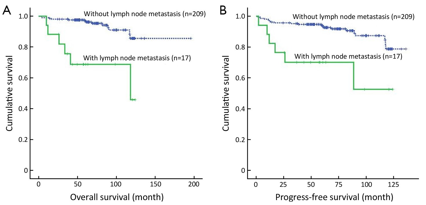
ObjectiveTo clarify the relationship between clinicopathological features and lymph node metastasis and to propose the potential indications of lymph node metastasis for prognosis in early gastric cancer (EGC) patients. MethodsWe retrospectively observed 226 EGC patients with lymph node resection, and analyzed the associations between lymph node metastasis and clinicopathological parameters using the chi-square test in univariate analysis and logistic regression analysis in multivariate analysis. Overall survival analysis was determined using the Kaplan-Meier and log-rank test. We conducted multivariate prognosis analysis using the Cox proportional hazards model. ResultsOf all the EGC patients, 7.5% (17/226) were histologically shown to have lymph node metastasis. The differentiation, lymphovascular invasion and depth of invasion were independent risk factors for lymph node metastasis in EGC. The 5- and 10-year survival rates were significantly lower in patients with lymph node metastasis than in those without and the patients also had shorter progress-free survival time. Lymph node metastasis and tumor size were independent prognostic factors for EGC. The status of the lymph nodes was a significant factor in predicting recurrence or metastasis after surgery. ConclusionsThe undifferentiated carcinoma and lymphovascular and/or submucosal invasion were associated with a higher incidence of lymph node metastasis in EGC patients, whom need to perform subsequent D2 lymphadenectomy or laparoscopic lymph node dissection and more rigorous follow-up or additional chemotherapy/radiation after D2 gastrectomy for poor prognosis and high recurrence/metastasis rate.
2014, 26(2): 200-207.
doi: 10.3978/j.issn.1000-9604.2014.04.07
Abstract:
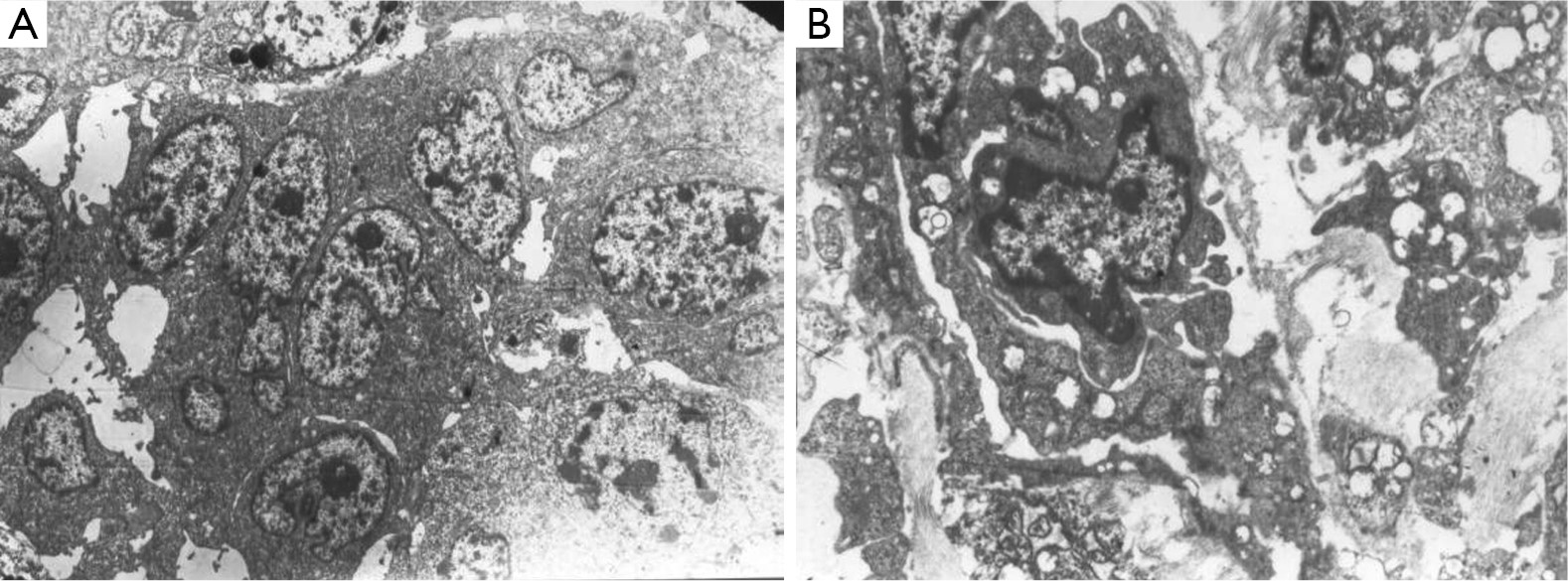
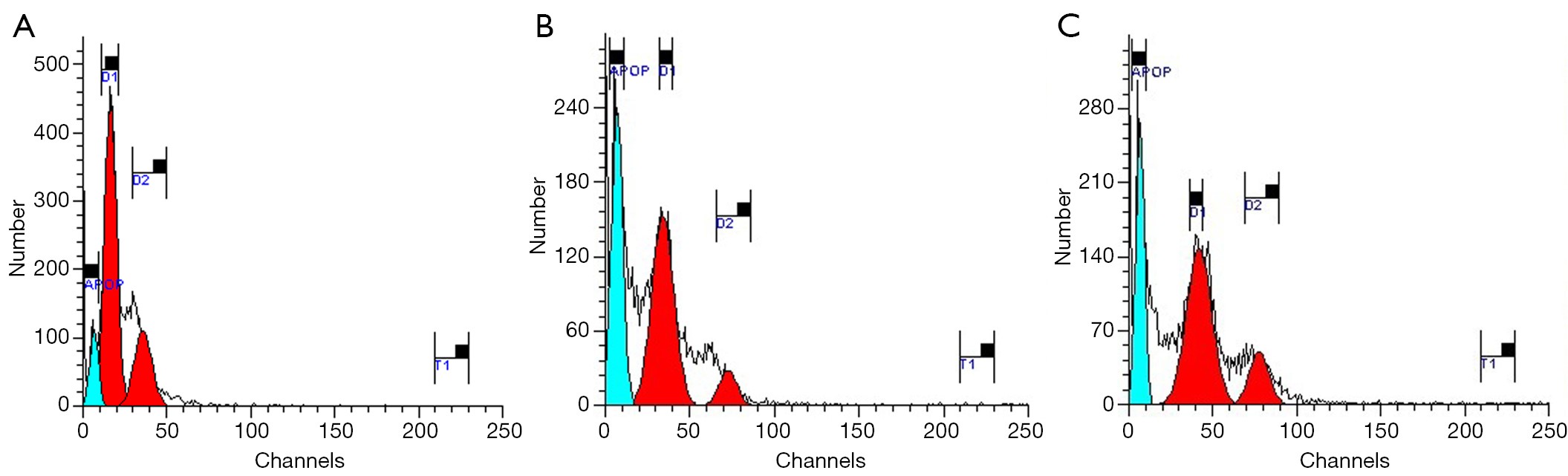
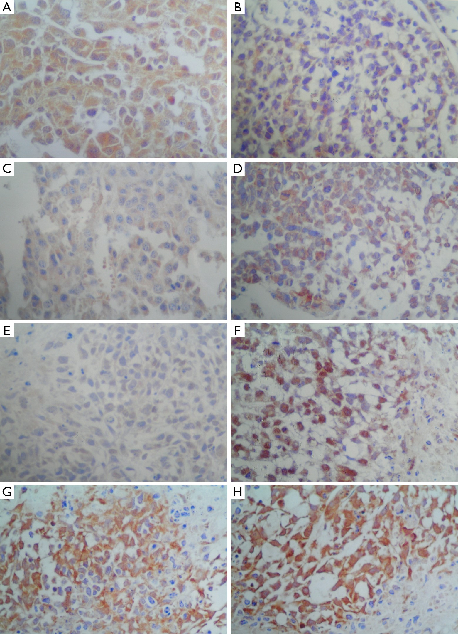
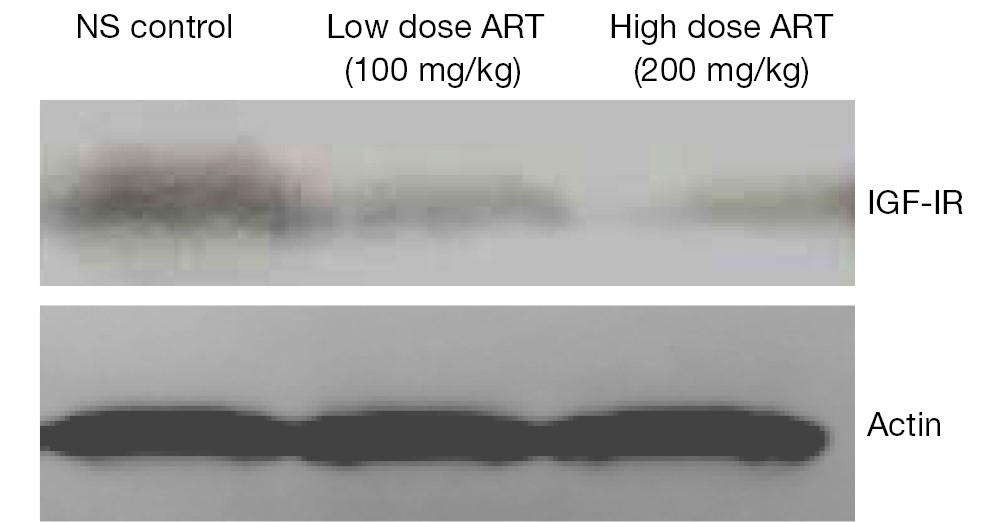
PurposeThe objective of this study was to investigate the anti-tumor effects and analyze the mechanism of artesunate (ART) action on breast cancer in vivo using tumor transplanted nude mice. MethodsThe human breast tumor cell line MCF-7 was transplanted into nude mice, and the animals were treated with various doses of ART alone or in combination with cyclophosphamide (CTX) or normal saline (NS). The tumor inhibitory effects were observed and compared, and the ultrastructural morphology of the transplanted tumor cells was observed by electron microscopy. The apoptosis rates and cell cycle status were detected by flow cytometry (FCM). The expression of apoptosis-related proteins p53, Bcl-2, Bax and Caspase-3 were detected by immunohistochemistry and IGF-IR was detected by western blot. The expression correlation for these proteins was also analyzed. ResultsThe tumor inhibition rates in the low dose ART group, high dose ART group, CTX group and combined drug therapy group were (24.39±10.20)%, (40.24±7.02)%, (57.01±5.84)% and (68.29±5.1)%, respectively. The cell cycle was arrested in phase G0/G1 after treatment with ART. The expression of Bcl-2 was significantly reduced, and the expression levels of Bax and Caspase-3 were significantly increased in the ART group compared to the negative control saline group. There was no significant difference detected in p53 expression. The Bcl-2 level was negatively related to Bax and Caspase-3. The western blotting results showed IGF-IR downregulation. ConclusionsART inhibits the growth of MCF-7 breast tumor cell xenografts in nude mice. The anti-tumor mechanism of ART for human breast carcinoma in nude mice might be correlated with the alteration of apoptosis related protein expression, which may further induce apoptosis and inhibit cell proliferation.
2014, 26(2): 208-210.
doi: 10.3978/j.issn.1000-9604.2014.04.05
Abstract:
Due to the complexity of the splenic hilar vessels, their anatomical variation and the narrow and deep space, as well as the bleeding-prone splenic parenchyma and the difficulty to manage splenic or vascular bleeding at the splenic hilum,the procedure remains challenging and technically demanding procedure for the performance of laparoscopic pancreas- and spleen-preserving splenic hilar lymph nodes dissection. Based on our experiences, we gradually explored a set of procedural operation steps called “Huang’s three-step maneuver”. In this paper, we not only provide the concrete operation steps for the surgeon, but we also provide our recommended technique of pulling and exposure for assistants. This new maneuver simplifies the complicated procedure and improves the efficiency of laparoscopic spleen-preserving splenic hilar lymphadenectomy, making it easier to master and allowing for its widespread adoption.
Due to the complexity of the splenic hilar vessels, their anatomical variation and the narrow and deep space, as well as the bleeding-prone splenic parenchyma and the difficulty to manage splenic or vascular bleeding at the splenic hilum,the procedure remains challenging and technically demanding procedure for the performance of laparoscopic pancreas- and spleen-preserving splenic hilar lymph nodes dissection. Based on our experiences, we gradually explored a set of procedural operation steps called “Huang’s three-step maneuver”. In this paper, we not only provide the concrete operation steps for the surgeon, but we also provide our recommended technique of pulling and exposure for assistants. This new maneuver simplifies the complicated procedure and improves the efficiency of laparoscopic spleen-preserving splenic hilar lymphadenectomy, making it easier to master and allowing for its widespread adoption.

2014, 26(2): 211-214.
doi: 10.3978/j.issn.1000-9604.2014.02.12
Abstract:
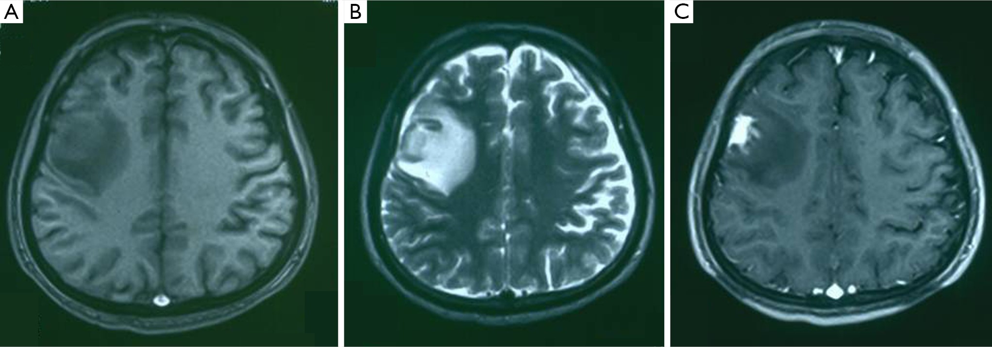
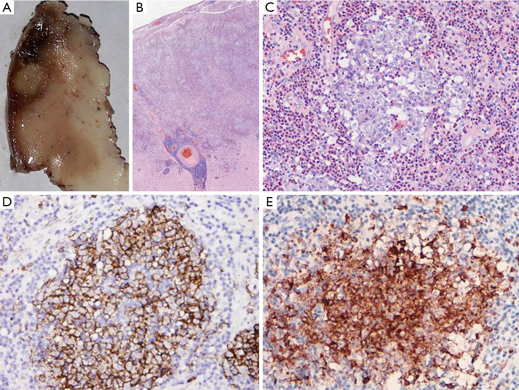
The brain parenchymal Langerhans cell histiocytosis (LCH) without systemic disease or lytic skull lesions is extremely rare. We report a 23-year-old male presenting with new onset 1 hour seizure with loss of consciousness 20 days prior to admission, and recurrent seizure 2 weeks later. Brain magnetic resonance imaging (MRI) showed an irregularly mass with enhancement involving the right frontal lobe. Microscopically, the lesion was characterized by sheets of Langerhans cells in addition to reactive inflammatory elements. Immunohistochemically, Langerhans cells were positive for Langerin, CD1a and S-100 protein. The patient received no chemotherapy or radiotherapy after surgery. After 24 months of follow-up, no recurrence or other systemic lesions were observed. Although there is no standard treatment for solitary cerebral LCH, the prognosis generally appears to be good.
The brain parenchymal Langerhans cell histiocytosis (LCH) without systemic disease or lytic skull lesions is extremely rare. We report a 23-year-old male presenting with new onset 1 hour seizure with loss of consciousness 20 days prior to admission, and recurrent seizure 2 weeks later. Brain magnetic resonance imaging (MRI) showed an irregularly mass with enhancement involving the right frontal lobe. Microscopically, the lesion was characterized by sheets of Langerhans cells in addition to reactive inflammatory elements. Immunohistochemically, Langerhans cells were positive for Langerin, CD1a and S-100 protein. The patient received no chemotherapy or radiotherapy after surgery. After 24 months of follow-up, no recurrence or other systemic lesions were observed. Although there is no standard treatment for solitary cerebral LCH, the prognosis generally appears to be good.
2014, 26(2): 215-218.
doi: 10.3978/j.issn.1000-9604.2014.02.15
Abstract:
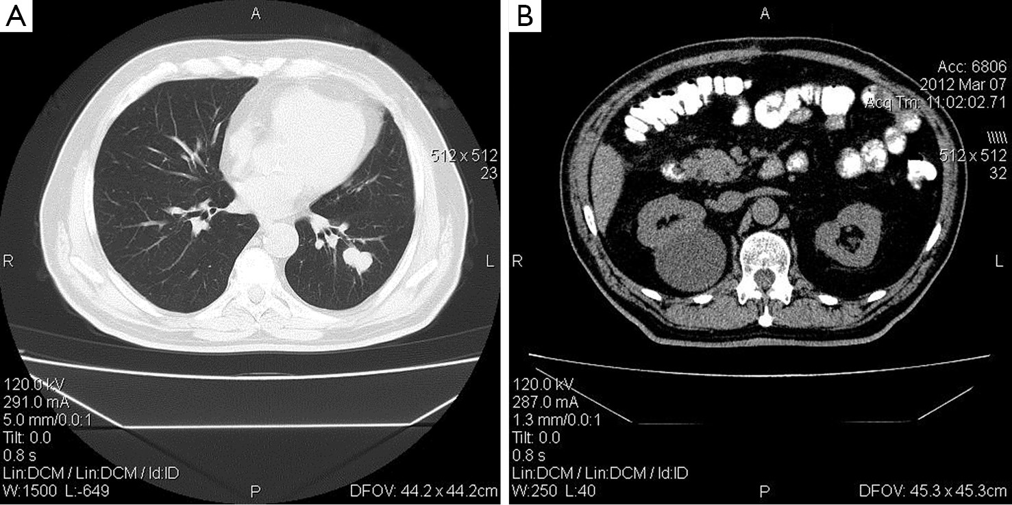
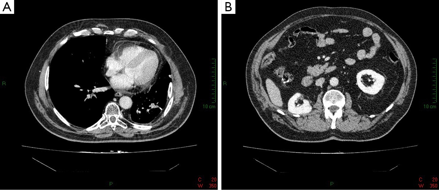
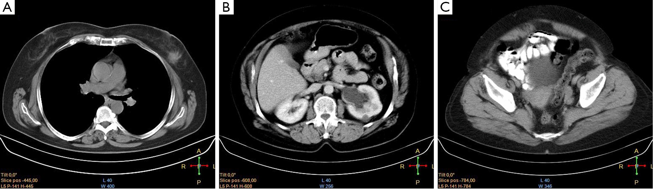
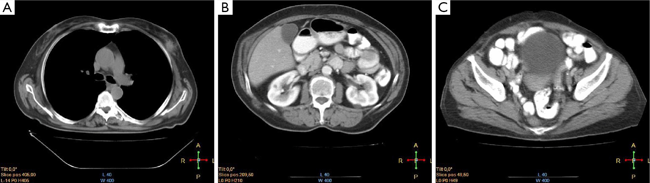
The diagnosis of multiple primary malignancies (MPMs) in a patient has been reported rather frequently during the past decade. Here we present two cases with three synchronous primary malignant tumors. The first patient is a 66-year-old male with synchronous colorectal cancer, renal cell carcinoma (RCC) and non-small cell lung cancer (NSCLC). The second patient is a 64-year-old female with breast cancer, transitional cell carcinoma of the ureter and endometrial cancer. MPMs seem to be diagnosed in a higher incidence than that predicted only by the influence of hazard and, whenever found, they raise questions regarding not only possible common etiologic factors or same pathogenetic mechanisms but also they cause a lot of troubles to both clinicians and patients because the therapeutic options usually become limited.
The diagnosis of multiple primary malignancies (MPMs) in a patient has been reported rather frequently during the past decade. Here we present two cases with three synchronous primary malignant tumors. The first patient is a 66-year-old male with synchronous colorectal cancer, renal cell carcinoma (RCC) and non-small cell lung cancer (NSCLC). The second patient is a 64-year-old female with breast cancer, transitional cell carcinoma of the ureter and endometrial cancer. MPMs seem to be diagnosed in a higher incidence than that predicted only by the influence of hazard and, whenever found, they raise questions regarding not only possible common etiologic factors or same pathogenetic mechanisms but also they cause a lot of troubles to both clinicians and patients because the therapeutic options usually become limited.
2014, 26(2): 219-221.
doi: 10.3978/j.issn.1000-9604.2014.03.02
Abstract:
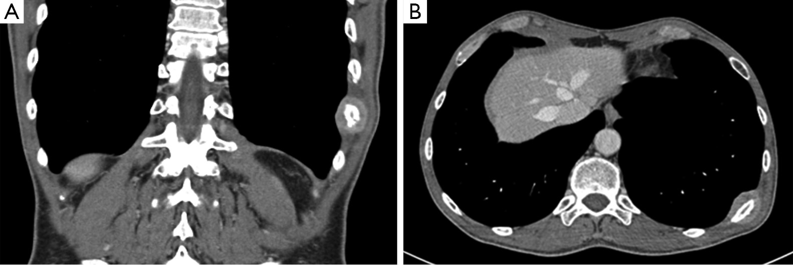
We report a case of a 49-year-old man who developed solitary rib metastasis of nasopharyngeal cancer. Patient had been treated for primary carcinoma with radiation therapy and concomitant chemotherapy. The bone metastasis presented as bulky, solid, painful mass in the posterior arch of 10th rib, within nine months the end of treatment. Biopsy of the solitary lesion presented the same histological characteristics as those of primary lesion. Although there are reported in literature series of nasopharyngeal cancer metastasizing to bone, we did not find previously published report of a nasopharyngeal carcinoma metastasizing only to a rib.
We report a case of a 49-year-old man who developed solitary rib metastasis of nasopharyngeal cancer. Patient had been treated for primary carcinoma with radiation therapy and concomitant chemotherapy. The bone metastasis presented as bulky, solid, painful mass in the posterior arch of 10th rib, within nine months the end of treatment. Biopsy of the solitary lesion presented the same histological characteristics as those of primary lesion. Although there are reported in literature series of nasopharyngeal cancer metastasizing to bone, we did not find previously published report of a nasopharyngeal carcinoma metastasizing only to a rib.