2014 Vol.26(3)
Display Mode: |
2014, 26(3): 222-223.
doi: 10.3978/j.issn.1000-9604.2014.06.12
 Abstract
 FullText HTML
 PDF 77KB
Abstract:
2014, 26(3): 224-225.
doi: 10.3978/j.issn.1000-9604.2014.06.13
Abstract:
2014, 26(3): 226-230.
doi: 10.3978/j.issn.1000-9604.2014.06.14
Abstract:
2014, 26(3): 231-233.
doi: 10.3978/j.issn.1000-9604.2014.06.15
Abstract:
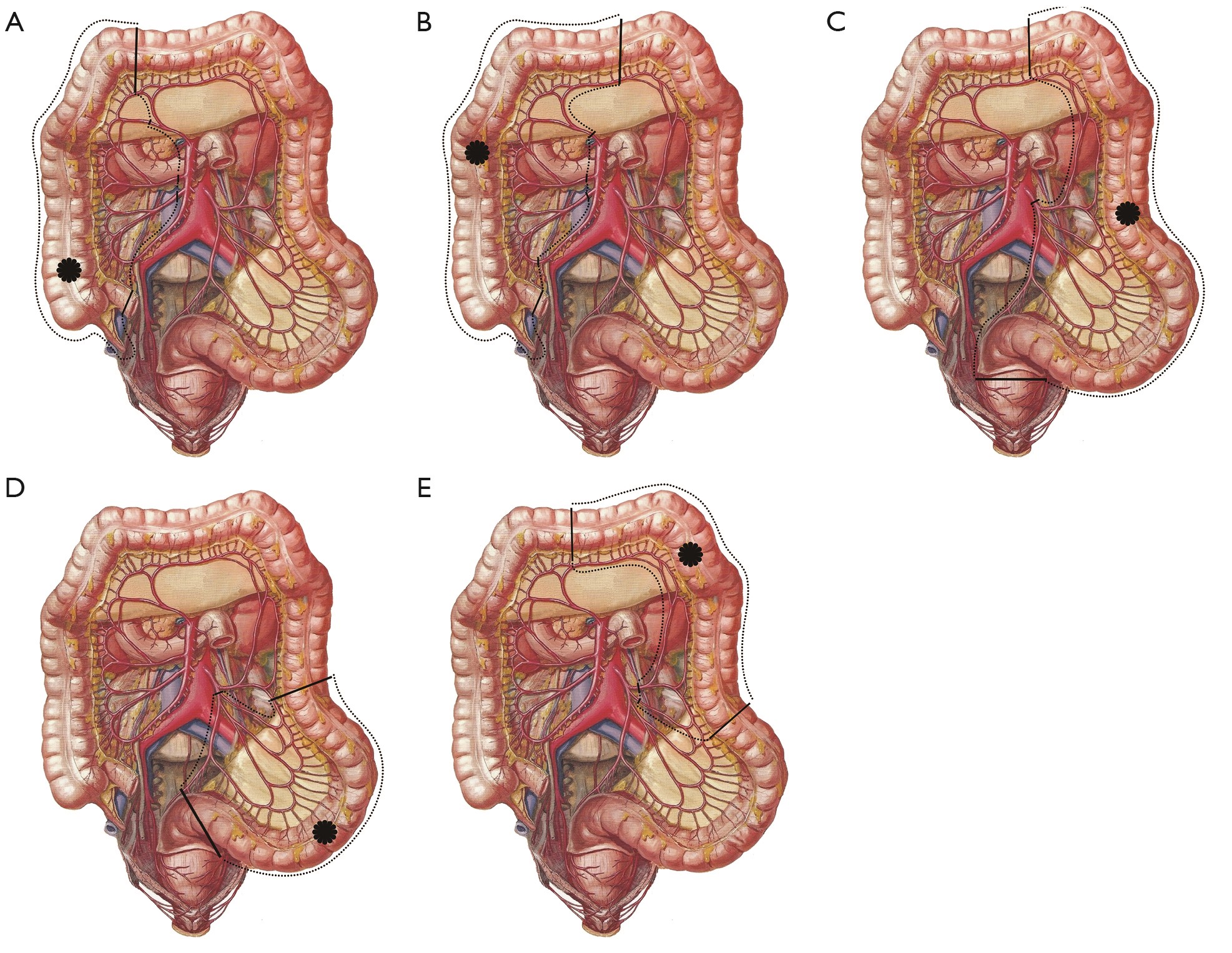
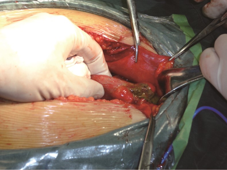
Qualified radical gastrectomy with lymph node dissection is very important to the prognosis of patients with gastric cancer. Now D2 lymph node dissection is standard procedure for gastric cancer surgery, and spleen hilar lymph node dissection is mandatory for gastric cancer in upper body. Because the anatomy of vessels in this area is very complicated, D2 lymph node dissection is technical challenging not only for open gastrectomy but also for laparoscopic one. Adapting a new technique is important to all surgeons, but we surgeons should always consider a patient’s safety as the most important factor during surgery and that efforts should be based on scientific rationale with oncologic principles. I hope that the recent report by Huang et al. about laparoscopic spleen preserving hilar lymph node dissection would be helpful to young surgeons who will perform laparoscpic total gastrectomy for gastric cancer.
Qualified radical gastrectomy with lymph node dissection is very important to the prognosis of patients with gastric cancer. Now D2 lymph node dissection is standard procedure for gastric cancer surgery, and spleen hilar lymph node dissection is mandatory for gastric cancer in upper body. Because the anatomy of vessels in this area is very complicated, D2 lymph node dissection is technical challenging not only for open gastrectomy but also for laparoscopic one. Adapting a new technique is important to all surgeons, but we surgeons should always consider a patient’s safety as the most important factor during surgery and that efforts should be based on scientific rationale with oncologic principles. I hope that the recent report by Huang et al. about laparoscopic spleen preserving hilar lymph node dissection would be helpful to young surgeons who will perform laparoscpic total gastrectomy for gastric cancer.
2014, 26(3): 234-236.
doi: 10.3978/j.issn.1000-9604.2014.06.19
Abstract:
2014, 26(3): 237-242.
doi: 10.3978/j.issn.1000-9604.2014.06.17
Abstract:
Based on Siewert classification, adenocarcinomas of the esophagogastric junction (AEGs) have different behaviors of perigastric-mediastinal nodal metastasis. Siewert type I AEGs have higher incidence of mediastinal nodal metastasis than those of type II or III, especially at middle-upper mediastinum. With regard to the necessity of mediastinal lymphadenectomy, theoretically, transthoracic esophagogastrectomy with complete mediastinal lymphadenectomy is suggested for Siewert type I AEGs, while transhiatal total gastrectomy with lower mediastinal and D2 perigastric lymphadenectomy is a standard surgery for type II-III AEGs. Nevertheless, the mediastinal nodal metastasis is an independent factor of poor prognosis for any type of AEG.
Based on Siewert classification, adenocarcinomas of the esophagogastric junction (AEGs) have different behaviors of perigastric-mediastinal nodal metastasis. Siewert type I AEGs have higher incidence of mediastinal nodal metastasis than those of type II or III, especially at middle-upper mediastinum. With regard to the necessity of mediastinal lymphadenectomy, theoretically, transthoracic esophagogastrectomy with complete mediastinal lymphadenectomy is suggested for Siewert type I AEGs, while transhiatal total gastrectomy with lower mediastinal and D2 perigastric lymphadenectomy is a standard surgery for type II-III AEGs. Nevertheless, the mediastinal nodal metastasis is an independent factor of poor prognosis for any type of AEG.
2014, 26(3): 243-244.
doi: 10.3978/j.issn.1000-9604.2014.06.16
Abstract:
2014, 26(3): 245-246.
doi: 10.3978/j.issn.1000-9604.2014.06.20
Abstract:
2014, 26(3): 247-254.
doi: 10.3978/j.issn.1000-9604.2014.05.02
Abstract:
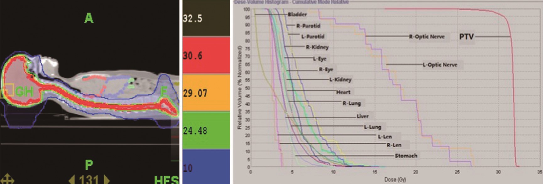
ObjectiveTo evaluate the short-term clinical outcomes of intracranial germinoma patients treated with craniospinal irradiation (CSI) using helical tomotherapy (HT) system in our center. MethodsTwenty-three patients who were treated with CSI in our center from January 2008 to July 2012 were collected, with an average age of 20. All of the patients’ CSI used the HT system. The total doses were 27-36 Gy/15-20 F (1.5-2 Gy per fraction), and total local doses were 46-60 Gy/30-50 F (5 fractions per week). All female patients for CSI were treated with left-right parallel-opposed field irradiation to protect their ovarian functions. Median follow-up time was 30.9 months (range, 5-67 months). The SPSS19.0 software was used, and the overall survival (OS) was calculated using the Kaplan-Meier method. ResultsAmong 17 patients with assessable tumors, 9 cases (52.9%) were CR, 7 cases (41.2%) were PR, and 1 case (5.9%) was SD. Hematological toxicity was the severest side-effect occurred in the procedure of CSI. The level 1-4 acute leukopenia were 8.7%, 30.4%, 34.8% and 21.7% and the level 1-4 acute thrombopenia were 8.7%, 30.4%, 21.7% and 8.7%, respectively. ConclusionsFor primary intracranial germinomas, HT can be used to implement CSI for simplifying radiotherapy procedures, improving radiotherapy accuracy, enhancing protection of peripheral organs at risk (ORA) and guaranteeing therapeutic effects. With the acceptable acute and long-term toxicity, CSI using HT in intracranial germinoma patients can be a safe and alternative mode.

2014, 26(3): 255-267.
doi: 10.3978/j.issn.1000-9604.2014.06.01
Abstract:
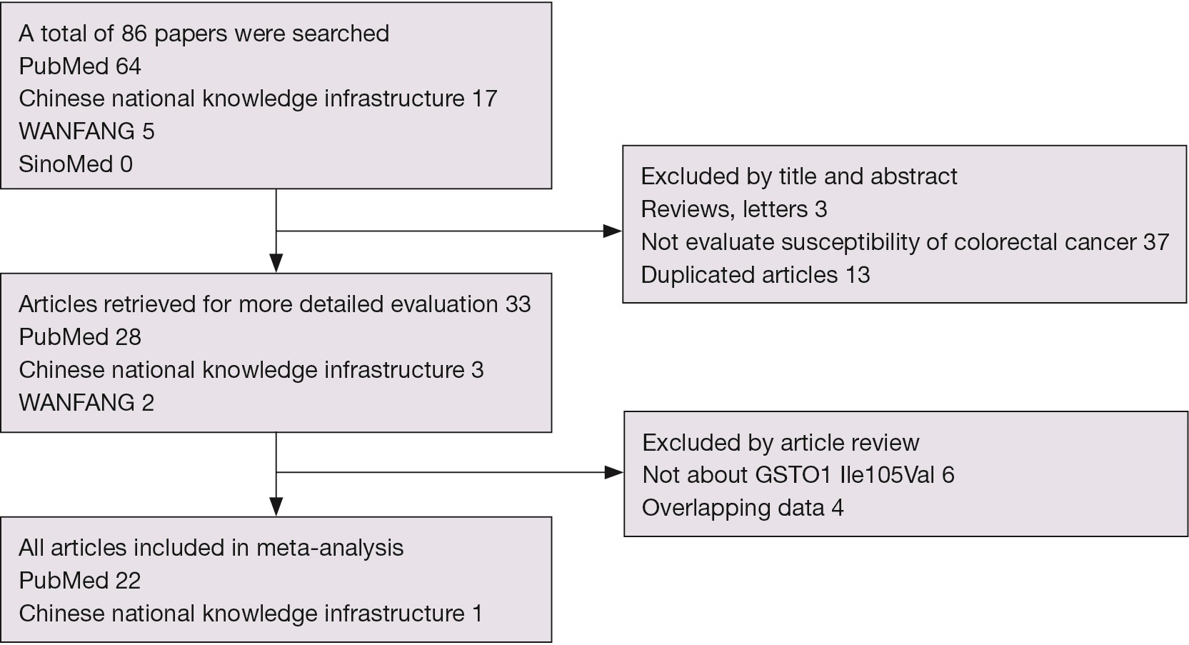
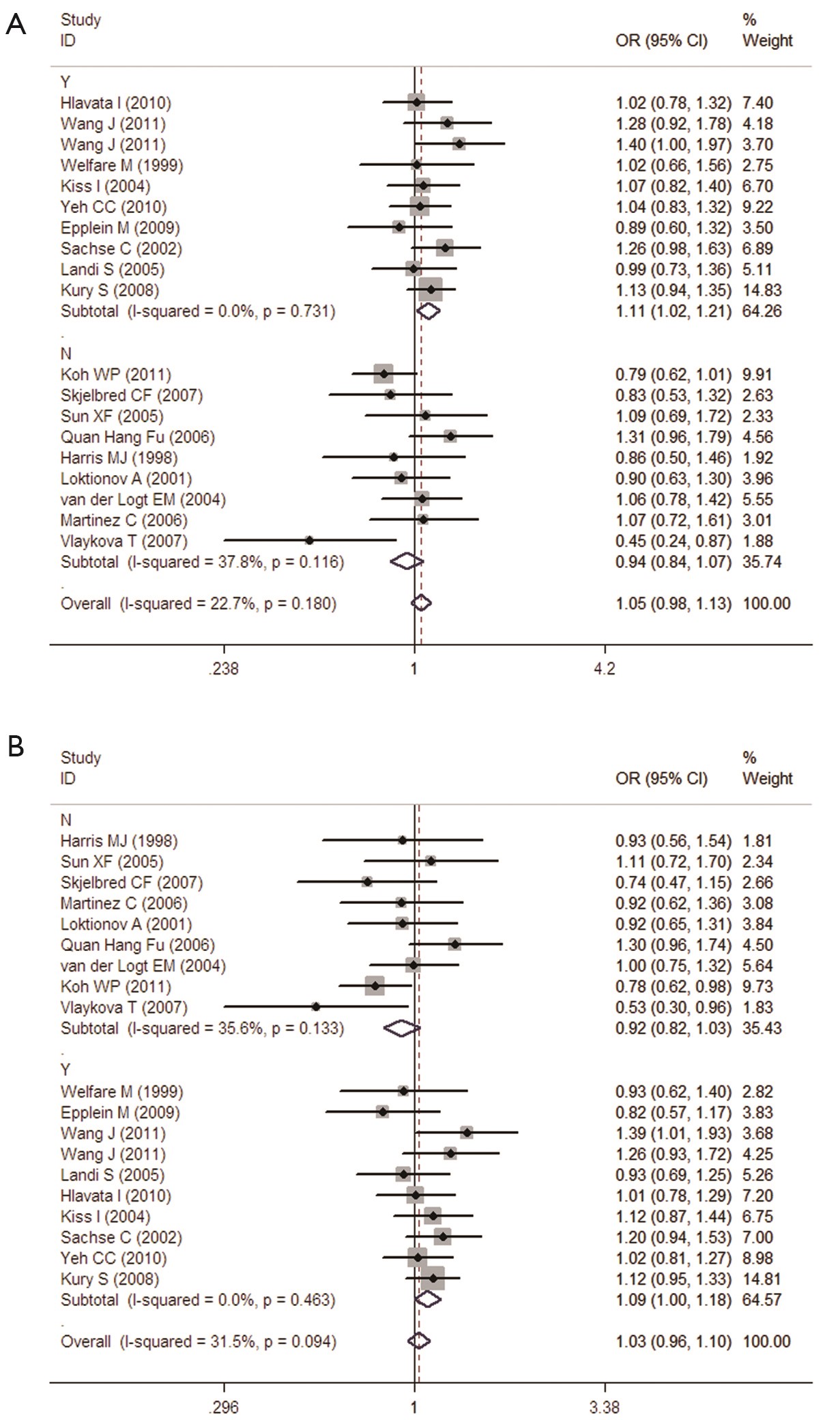
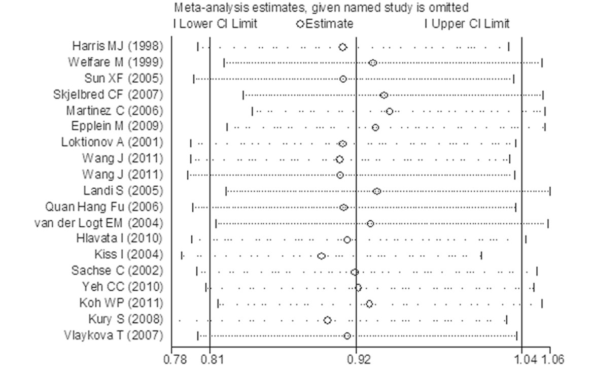
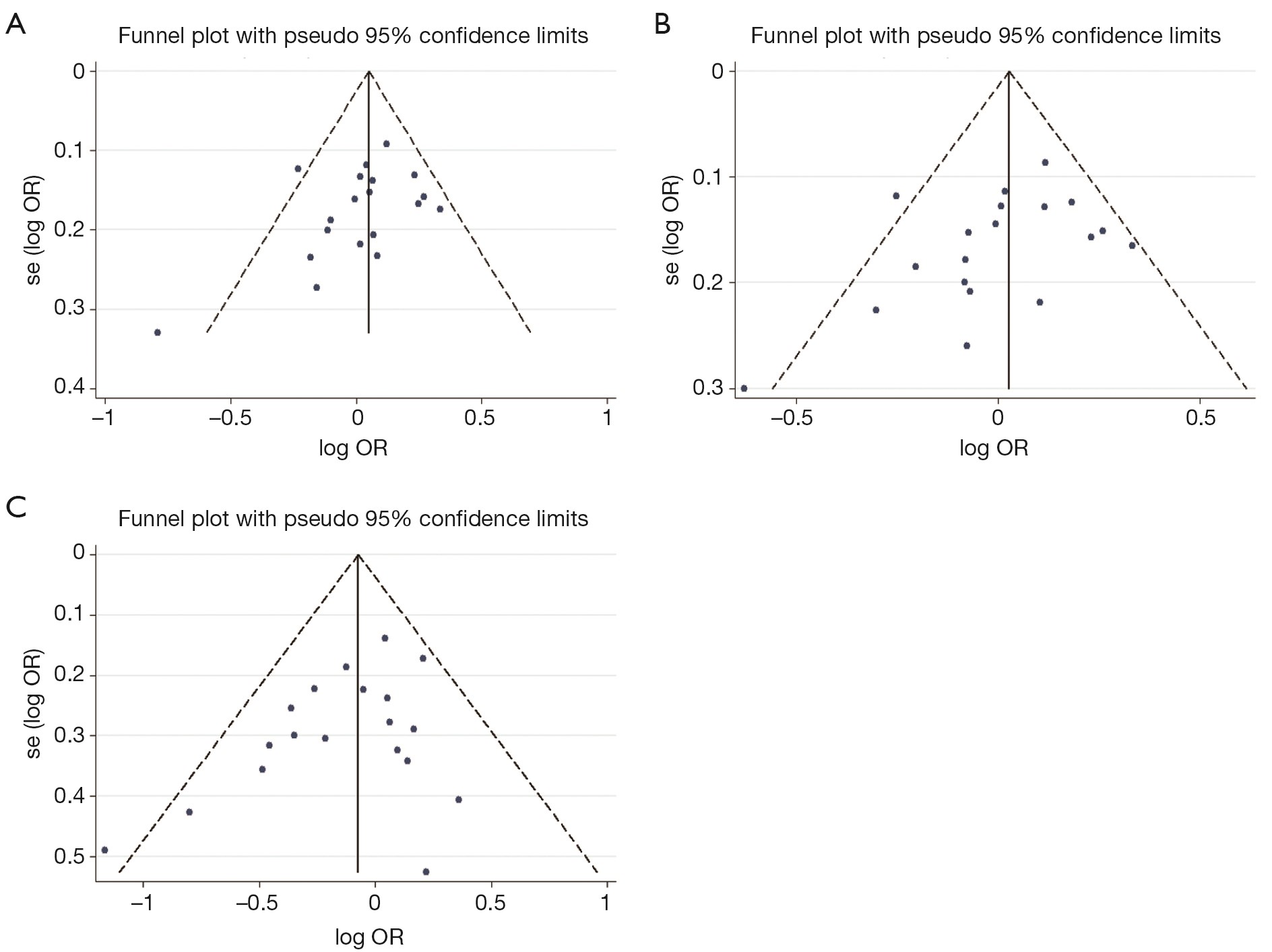
ObjectivesTo investigate the correlation between glutathione S-transferase P1 (GSTP1) Ile105Val polymorphism and colorectal cancer (CRC) risk. MethodsStudies were identified to investigate the association between GSTP1 Ile105Val polymorphism and CRC risk. Systematic computerized searches of the PubMed, Chinese National Knowledge Infrastructure, WANFANG and SinoMed were performed. Summary odds ratios (OR) and 95% confidence intervals (95% CI) were used to measure GSTP1 Ile105Val polymorphisms and CRC risk. ResultsA total of 23 retrospective studies were included in the meta-analysis. During all studies including 6,981 cases and 8,977 controls, sample sizes ranged from 146 to 2,144. Overall, the pooled results revealed that Ile105Val polymorphism was not associated with CRC risk and confused results were found in subgroup analyses. Further meta-analyses were conducted after excluding low-quality studies. GSTP1 Ile105Val is associated with increased risk of CRC limited in studies with matched control. There was no significant heterogeneity in all genetic comparisons, but heterogeneity existed in subgroup analyses of heterozygous and dominant comparisons. The meta-regression analyses indicated that matched controls were the significant factor influencing between-study heterogeneity in all possible influential factors including published year, ethnicity, source of control, sample size, Hardy-Weinberg equilibrium (HWE) in control and matched controls. Sensitivity analysis revealed the pooled ORs were not changed before and after removal of each single study in all genetic comparisons, indicating the robustness of the results. ConclusionsGSTP1 Ile105Val might be associated with increased risk of CRC. However, more high-quality case-control studies should be performed to confirm the authenticity of our conclusion.

2014, 26(3): 268-276.
doi: 10.3978/j.issn.1000-9604.2014.05.03
Abstract:
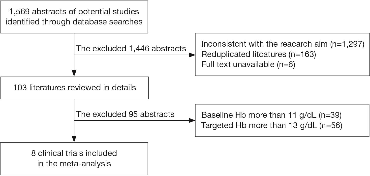
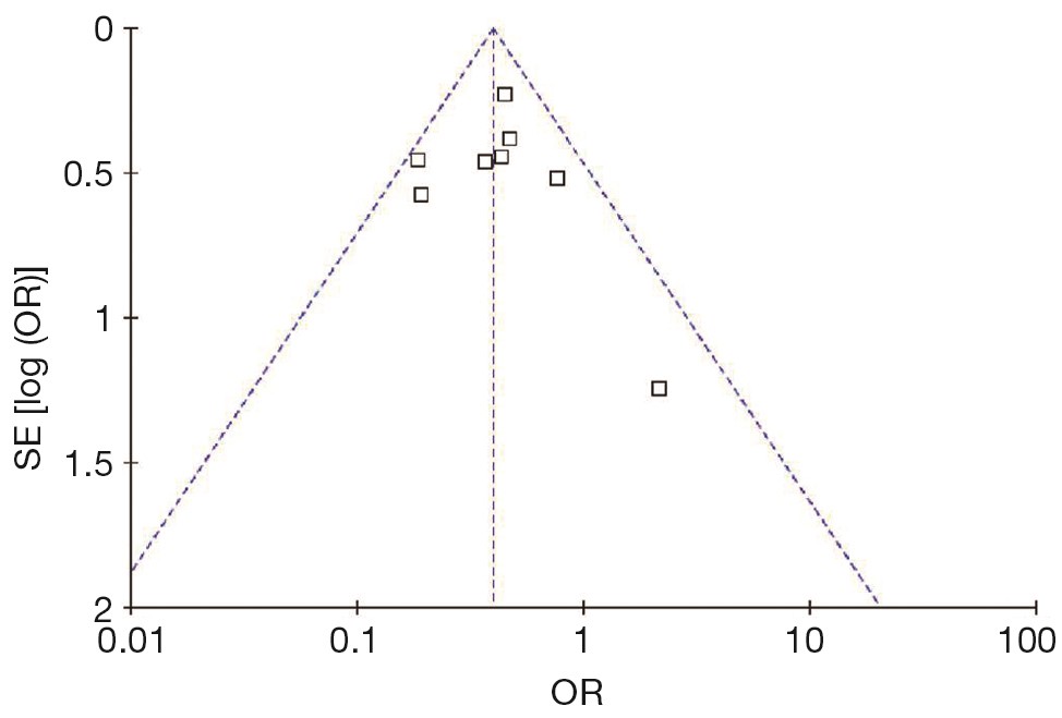
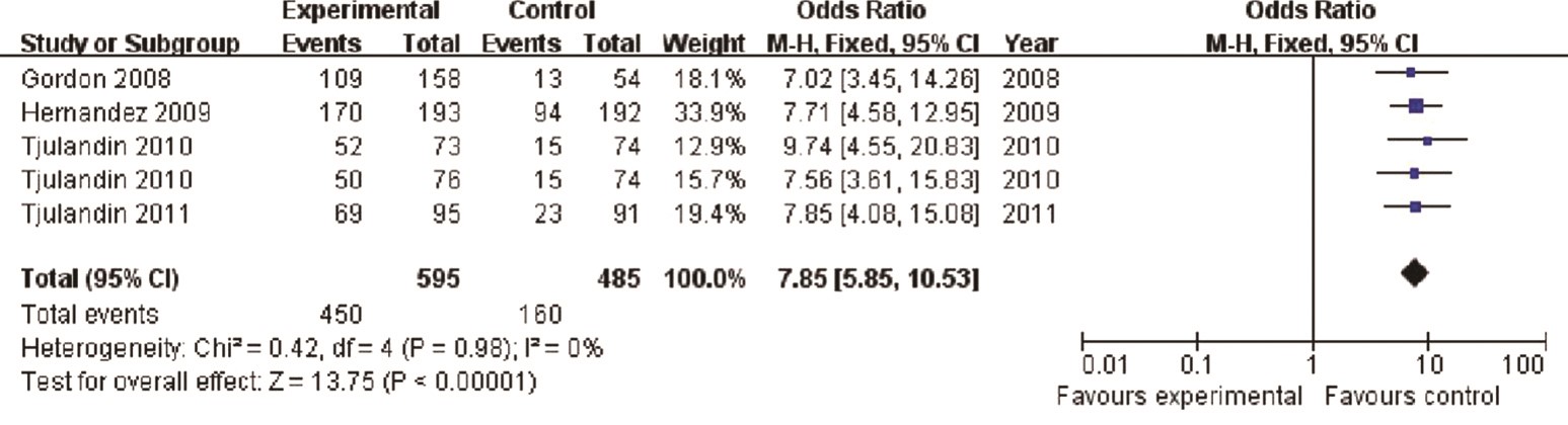
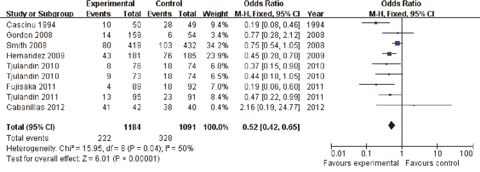
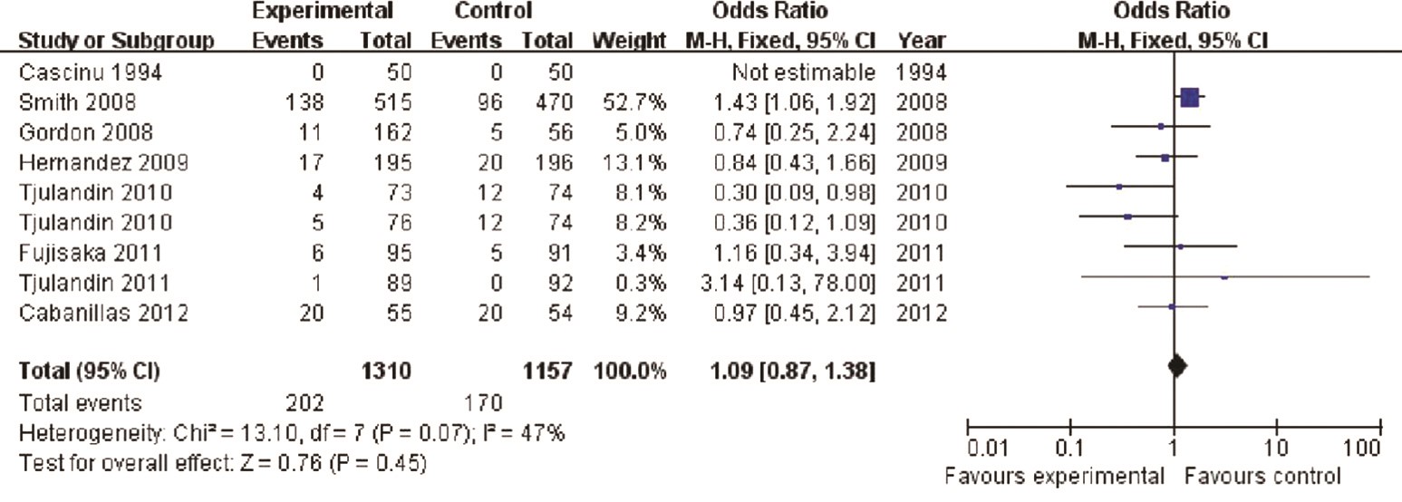
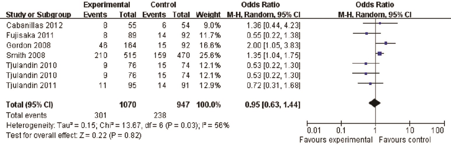
BackgroundErythropoiesis-stimulating agents (ESAs) are widely used in the management of anemia in cancer patients. Despite their apparent effectiveness, recent studies have suggested that ESAs could result in serious adverse events and even higher mortality. The aim of the current study was to evaluate the benefits and risks of ESAs in the management of cancer patients with anemia using a meta-analysis. MethodsThe initial literature search covered Medline, PubMed, Embase, and the Cochrane Center Register of Controlled Trials, and identified 1,569 articles. The final meta-analysis included eight randomized controlled trials (n=2,387) in cancer patients with <11 g/dL hemoglobin (Hb) at the baseline and target Hb (for stopping ESA treatment) at no more than 13 g/dL. The assessment measures included Hb response, blood transfusion rate and adverse events that included venous thromboemblism (VTE), hypertension, and on-study mortality. The results are expressed as pooled odds ratio (OR). Publication bias was assessed using funnel plot analysis. ResultsESAs significantly increased the Hb concentration [OR 7.85, 95% confidence interval (CI): 5.85 to 10.53, P<0.001] and reduced the red blood cell (RBC) transfusion rate (OR 0.52, 95% CI: 0.42 to 0.65, P<0.001). ESAs did not increase the accumulated adverse events (OR 0.95, P=0.82), or the on-study mortality (OR 1.09, P=0.47). ConclusionsESAs are not associated with increased frequency of severe adverse events in anemic cancer patients when the target Hb value is no more than 13 g/dL.
2014, 26(3): 277-284.
doi: 10.3978/j.issn.1000-9604.2014.06.05
Abstract:
ObjectiveTo compare internal with external drainage of the pancreatic duct during pancreaticoduodenectomy with regard to the incidence of postoperative pancreatic fistula (POPF) and other complications. MethodsWe retrospectively analyzed 316 patients who underwent pancreaticoduodenectomy with a placed drainage tube (external, n=128; internal, n=188) in the pancreatic duct from 1 January 1999 to 31 December 2011 in Tianjin Third Central Hospital of China. The incidence of POPF and some other complications were compared. ResultsThere was no difference in the incidence rates of POPF between those given an internal or external drainage tube (P=0.788), but POPF was more severe in the former (P=0.014). Intraperitoneal bleeding rate was also higher in the patients with internal drainage (P=0.040), but operative time and postoperative hospitalization were longer in those with external drainage (P=0.002 and P=0.007, respectively). There was no difference between the groups with regard to the incidence rates of gastrointestinal bleeding, delayed gastric emptying, pulmonary infection, or incision infection and in-hospital mortality. ConclusionsExternal drainage of the pancreatic duct during pancreaticoduodenectomy can decrease the severity of POPF, but operative time and postoperative hospitalization will be extended.
2014, 26(3): 285-292.
doi: 10.3978/j.issn.1000-9604.2014.06.09
Abstract:
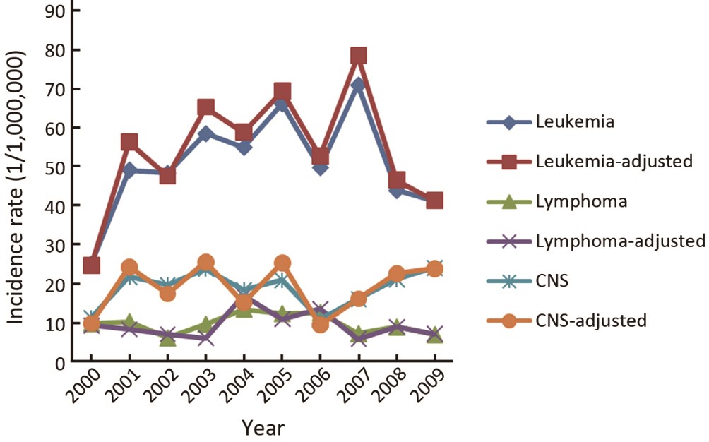
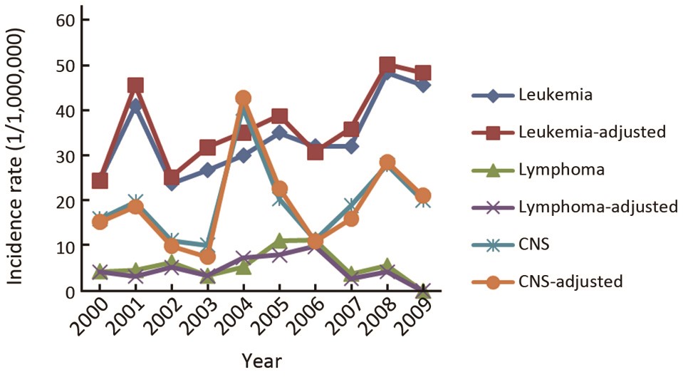
ObjectiveTo investigate the characteristics and incidence trends of childhood cancer in Beijing, China, from 2000 to 2009. MethodsA total of 1,274 cases with childhood cancer in Beijing from 2000 to 2009 were included in the study. All rates were age-standardized using the direct method to the world standard population and expressed per million person-years. Incidence trends were characterized by calculating annual percent change (APC) using Joinpoint Regression Program. ResultsThe crude incidence rate was 106.47 per million [age-standardized rate (ASR) 113.34] between 2000 and 2009 in Beijing with the most common diagnoses, leukemia (N=505, 39.64%, ASR 45.20), followed by central nervous system (CNS) tumors (N=228, 17.90%, ASR 19.28) and lymphoma (N=91, 7.14%, ASR 6.97). The incidence for all childhood cancers combined has increased during the study period, with an APC of 5.84% [95% confidence interval (95% CI): 1.0-10.9] after adjusted by world population. The ASR of all combined cancers in boys showed a slight, but no significant increase, with an APC of 5.33% (95% CI: –0.6-11.6); for girls, the trends increased significantly, with an APC of 6.54% (95% CI: 1.5-11.8). ConclusionsThe incidence rate of childhood cancer in Beijing was higher than the average level of China and lower than that of western countries. The incidence trends of childhood cancer, especially leukemia among girls showed a significantly increase from 2000 to 2009. While among boys, no substantially change was seen during the observed time period. Some sex-specific trends by subcategories and trends of major cancers in different age groups by cancer site merit further investigation.
2014, 26(3): 293-298.
doi: 10.3978/j.issn.1000-9604.2014.06.02
Abstract:
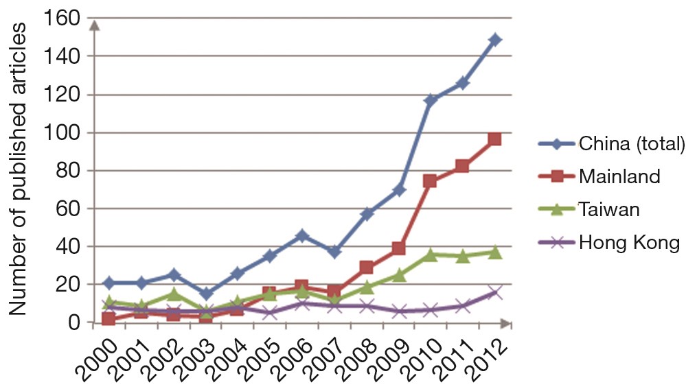
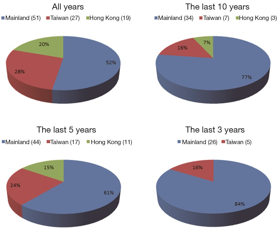
AimsTo investigate the research status of radiation oncology in China through survey of literature in international radiation oncology journals and retrospectively compare the outputs of radiation oncology articles of the three major regions of China—Mainland (ML), Taiwan (TW) and Hong Kong (HK). MethodsRadiation oncology journals were selected from “oncology” and “radiology, nuclear & medical image” category from Science Citation Index Expand (SCIE). Articles from the ML, TW and HK were retrieved from MEDLINE. The number of total articles, clinical trials, case reports, impact factors (IF), institutions and articles published in each journals were conducted for quantity and quality comparisons. ResultsA total 818 articles from 13 radiation oncology journals were searched, of which 427 are from ML, 259 from TW, and 132 from HK. Ninety-seven clinical trials and 5 case reports are reported in China. Accumulated IF of articles from ML (1,417.11) was much higher than that of TW (1,003.093) and HK (544.711), while the average IF of articles from ML is the lowest. ConclusionsThe total number of articles from China especially ML increased significantly in the last decade. The number of articles published from the ML has exceeded those from TW and HK. However, the quality of articles from TW and HK is better than that from ML.

2014, 26(3): 299-308.
doi: 10.3978/j.issn.1000-9604.2014.06.08
Abstract:
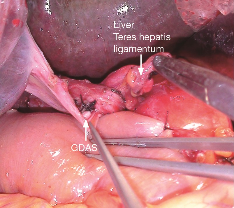
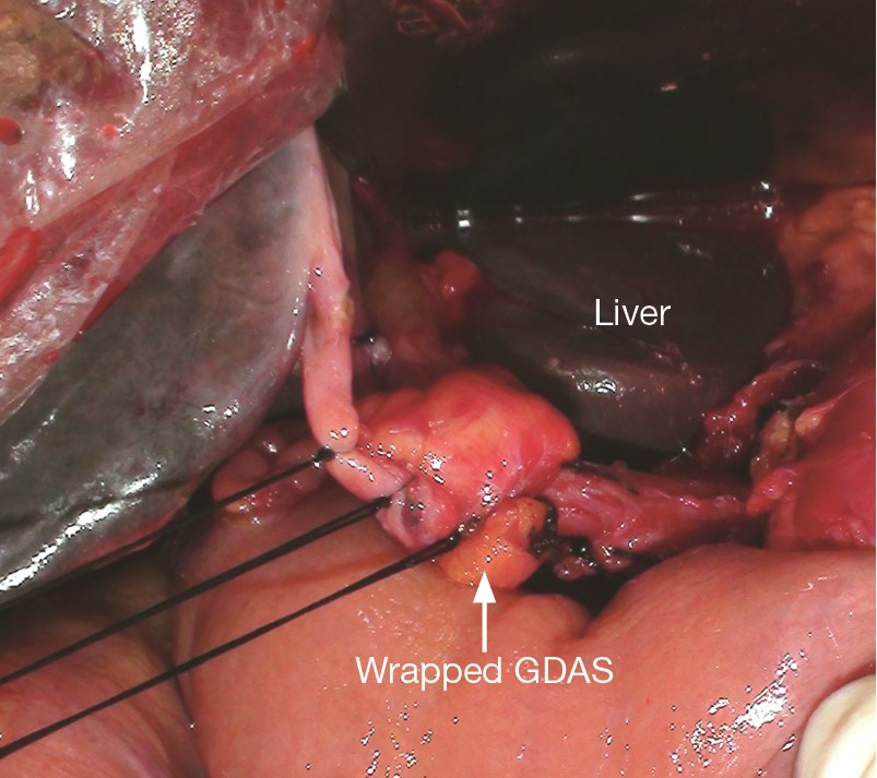
ObjectiveAfter pancreaticoduodenectomy (PD), the postoperative gastroduodenal artery stump (GDAS) hemorrhage is one of the most serious complications. The purpose of this study is to determine whether wrapping the GDAS during PD could decrease the postoperative GDAS hemorrhage incidence. MethodsA retrospective review involving 280 patients who underwent PD from 2005 to 2012 was performed. Wrapping the GDAS during PD was defined as “Wrapping the GDAS using the teres hepatis ligamentum during PD”. A total of 140 patients accepted the “wrapping” procedure (wrapping group). The other 140 patients didn’t apply the procedure (non-wrapping group). Age, sex, preoperative data, estimated intraoperative blood loss, postoperative complications, pathologic parameters and hospitalization time were compared between two groups. ResultsThere were no significant differences in patient characteristics between two groups. After wrapping, the incidence of postoperative GDAS bleeding decreased significantly (1/140 vs. 9/140, P=0.01). The rates of the other complications (such as intra-abdominal infection pancreatic fistula, billiary fistula, gastrointestinal bleeding, et al.) showed no significant differences. ConclusionsWrapping the GDAS during PD significantly reduced the postoperative GDAS hemorrhage incidence. And the “wrapping” had no obvious influence on other complications.
2014, 26(3): 309-314.
doi: 10.3978/j.issn.1000-9604.2014.06.10
Abstract:
ObjectiveTo explore the risk factors of intra-abdominal bacterial infection (IAI) after liver transplantation (LT) in patients with hepatocellular carcinoma (HCC). MethodsA series of 82 HCC patients who received LT surgeries in our department between March 2004 and April 2010 was recruited in this study. Then we collected and analyzed the clinical data retrospectively. Statistical analysis system (SPSS) software was adopted to perform statistical analysis. Chi-square test, t-test and Wilcoxon rank sum test were used to analyze the clinical data and compute the significance of the incidences of early-stage IAI after LT for HCC patients. Binary logistic regression was performed to screen out the risk factors, and multiple logistic regression analyses were performed to compute the independent risk factors. ResultsA series of 13 patients (13/82, 15.9%) had postoperative IAI. The independent risk factors of postoperative intra-abdominal bacterial infections after LT for HCC patients were preoperative anemia [Hemoglobin (HGB) <90 g/L] and postoperative abdominal hemorrhage (72 hours >400 mL), with the odds ratios at 8.121 (95% CI, 1.417 to 46.550, P=0.019) and 5.911 (95% CI, 1.112 to 31.432, P=0.037). ConclusionsPostoperative IAI after LT in patients with HCC was a common complication. Preoperative moderate to severe anemia, as well as postoperative intra-abdominal hemorrhage more than 400 mL within the first 72 hours might independently indicate high risk of IAI for these patients.
2014, 26(3): 315-322.
doi: 10.3978/j.issn.1000-9604.2014.06.18
Abstract:
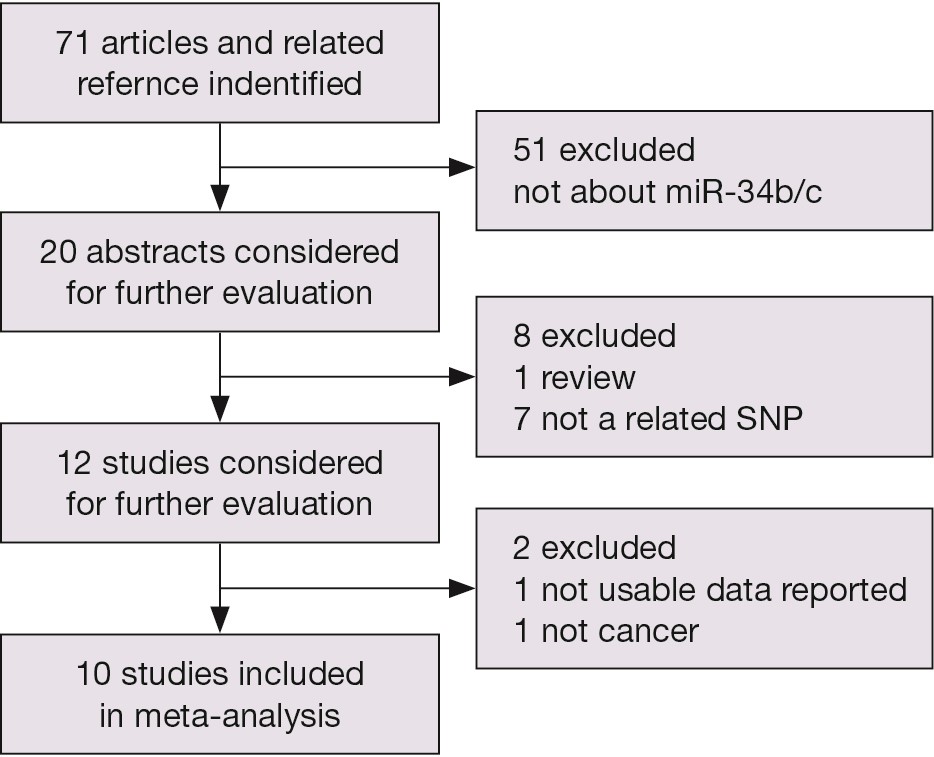
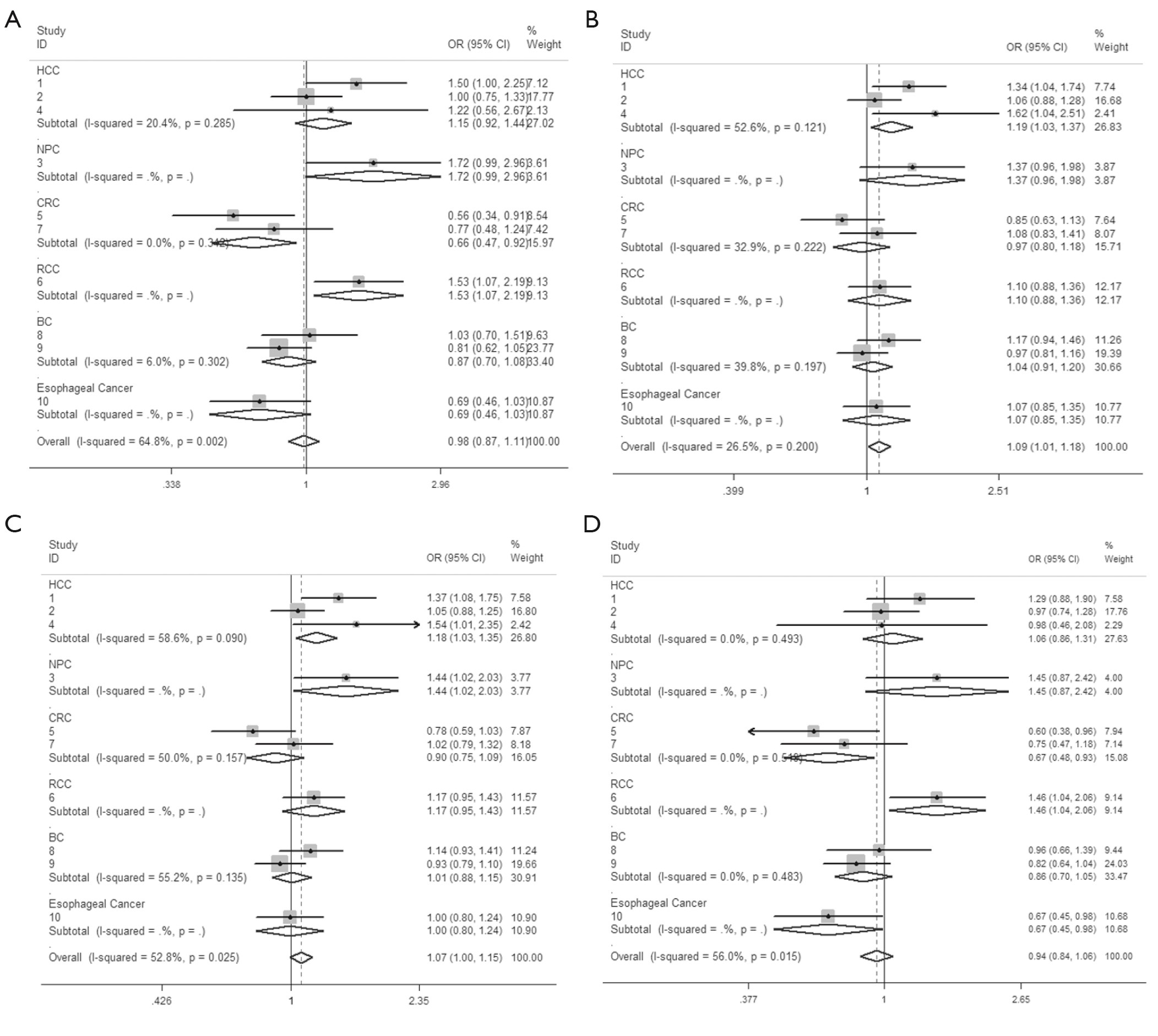
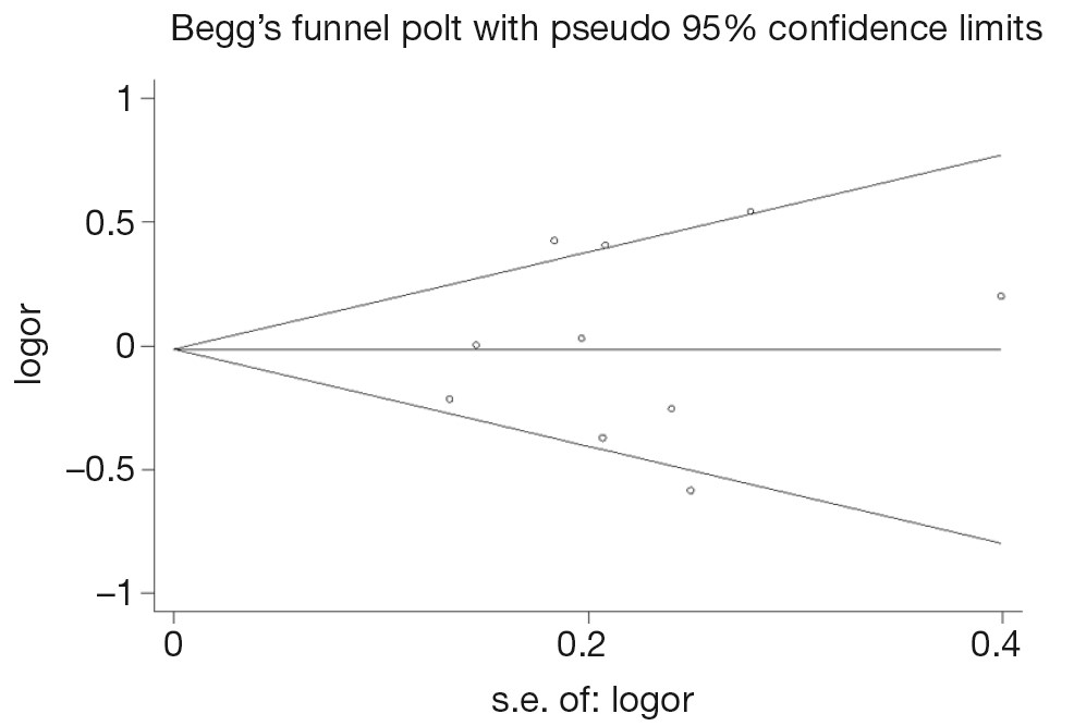
ObjectiveEmerging evidence shows that microRNAs (miRNAs) function as tumor suppressors or oncogenes in human carcinogenesis. A single nucleotide polymorphism (SNP) located in the pri-miRNA promoter may affect the processing and expression of mature miRNA. However, previous studies showed conflicting results regarding the association of hsa-miR-34b/c rs4938723 T > C promoter polymorphism with cancer. Therefore, we conducted a meta-analysis to determine the association of polymorphism with cancer risk. MethodsA computerized search of PubMed, Web of Science, and Chinese National Knowledge Infrastructure (CNKI) for publications on hsa-miR-34b/c rs4938723 T > C promoter polymorphism and cancer risk was performed and the genotype data were analyzed in a meta-analysis. Odds ratios (ORs) with 95% confidence intervals (CIs) were estimated to assess the association. Test of heterogeneity, cumulative meta-analysis, sensitivity analysis and assessment of bias were performed in our meta-analysis by STATA software 12.0. ResultsThere was no significant association between hsa-miR-34b/c rs4938723 polymorphism and overall cancer risk in the comparison models. Moreover, subgroup analysis revealed that the variant CT (OR =1.19, 95% CI: 1.03-1.37) and CC/CT (OR =1.18, 95% CI: 1.03-2.35) genotypes were associated with an increased risk of hepatocellular carcinoma (HCC) compared with wild-type TT genotype. However, a decreased risk of colorectal cancer (CRC) was found in the genetic model of CC/TT (OR =0.66, 95% CI: 0.47-0.92) and CC/CTTT (OR =0.67, 95% CI: 0.49-0.93). ConclusionsThe results suggest that hsa-miR-34b/c rs4938723 polymorphism may play an opposite role in different types of cancer based on current studies, which is the main origin of heterogeneity in this meta-analysis. Further large-scale studies and functional studies between this polymorphism and cancer risk are warranted.

2014, 26(3): 323-330.
doi: 10.3978/j.issn.1000-9604.2014.06.07
Abstract:
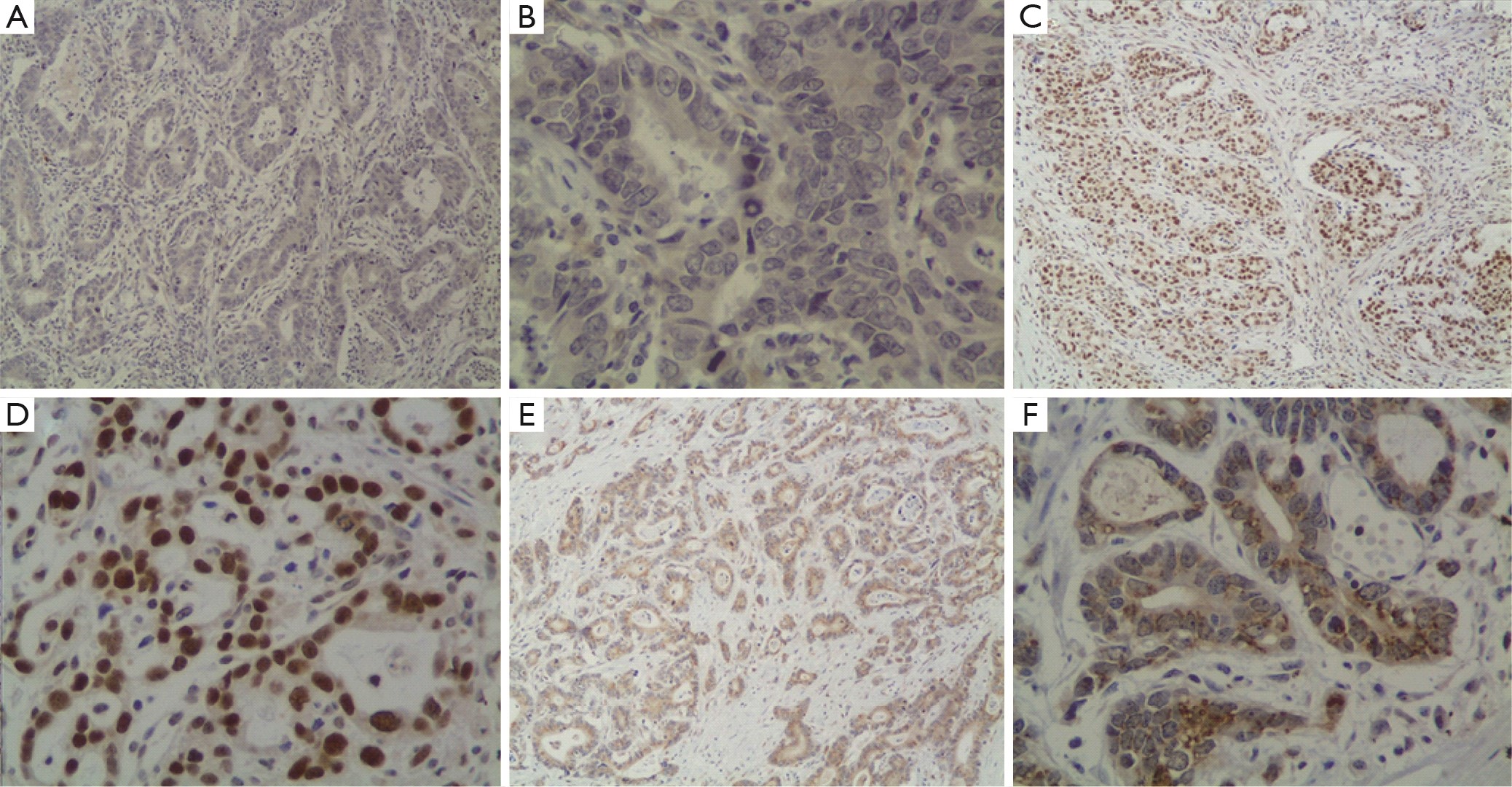
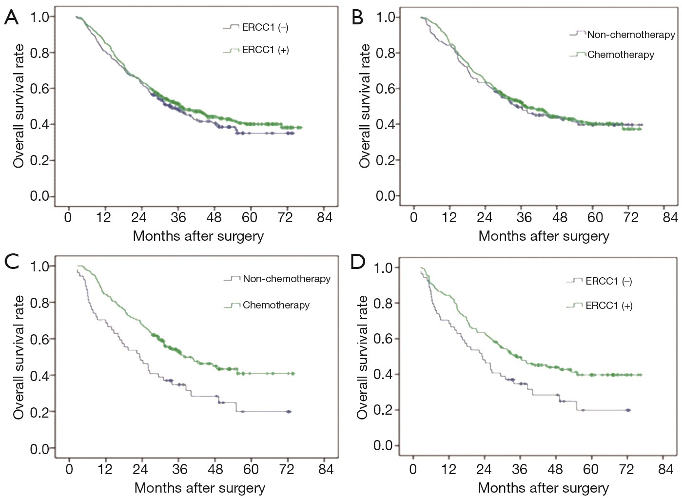
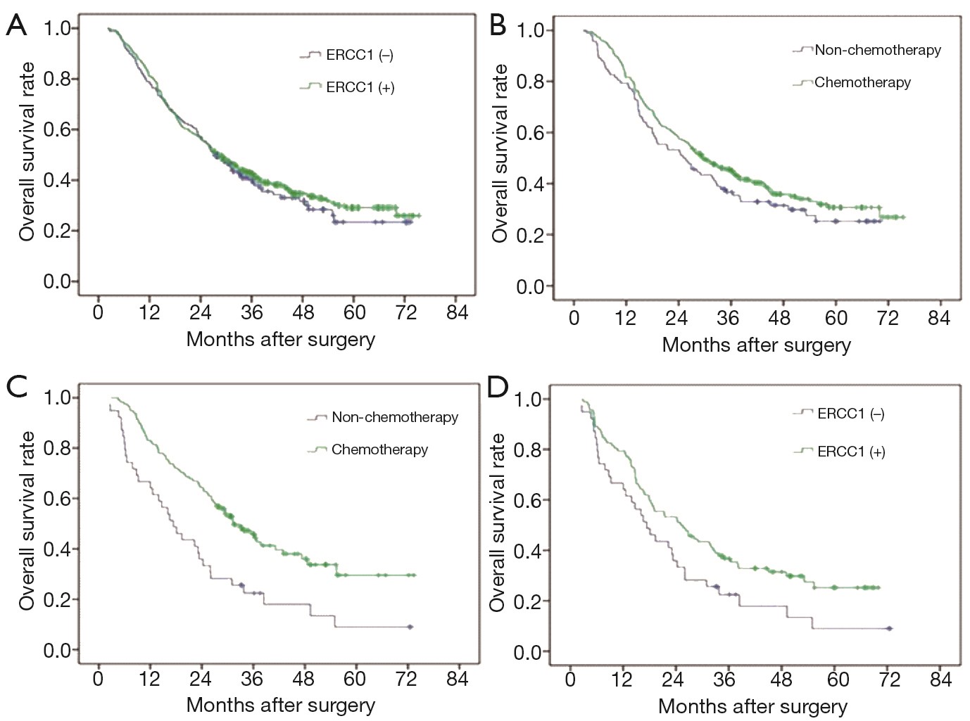
AimThis study explored the correlation between the expression of excision repair cross-complementation group 1 (ERCC1) and the prognosis of gastric cancer patients. MethodsFrom January 2005 to December 2008, 605 patients who underwent radical surgery in The First Affiliated Hospital of Nanjing Medical University were enrolled. We conducted the follow-up every 6 months and its contents included a comprehensive medical history, tumor markers and abdominal ultrasound or CT and other imaging findings. Deadline was April 30, 2013 and follow-up time between 51 to 91 months. Survival time is calculated from the date of diagnosis to death or last follow-up date. Immunohistochemistry (IHC) was used to assess the expression of ERCC1 in resected samples. The relationship between ERCC1 expression and survival of patients was investigated. The comparison of count data were analyzed by Chi-square test. Median survival time (MST) and the 5-year survival rate were calculated by life table analysis. The Kaplan-Meier curves were used for survival analysis. ResultsERCC1 expression was positive in 412 patients (68.1%). There is no significant difference between ERCC1-positive group and ERCC1-negative group in terms of the MST and 5-year survival rate (P=0.455). The MST and 5-year survival rate have no significant difference (P=0.162) between group with chemotherapy and group with no chemotherapy in patients with ERCC1-positive expression. However, the MST and 5-year survival rate in patients with ERCC1-negative expression benefited more from with chemotherapy (P=0.019). The ERCC1-positive patients survived longer than those ERCC1-negative patients (P=0.183) in subgroup with no adjuvant chemotherapy. In the subgroup analysis, ERCC1 expression had no significant relationship with overall survival in patients with stage II or III gastric cancer (P>0.05). ConclusionsERCC1 might be a good prognostic factor for the patients of gastric cancer after radical resection. Patients with ERCC1-negative expression could benefit more from adjuvant chemotherapy.
2014, 26(3): 331-340.
doi: 10.3978/j.issn.1000-9604.2014.06.11
Abstract:
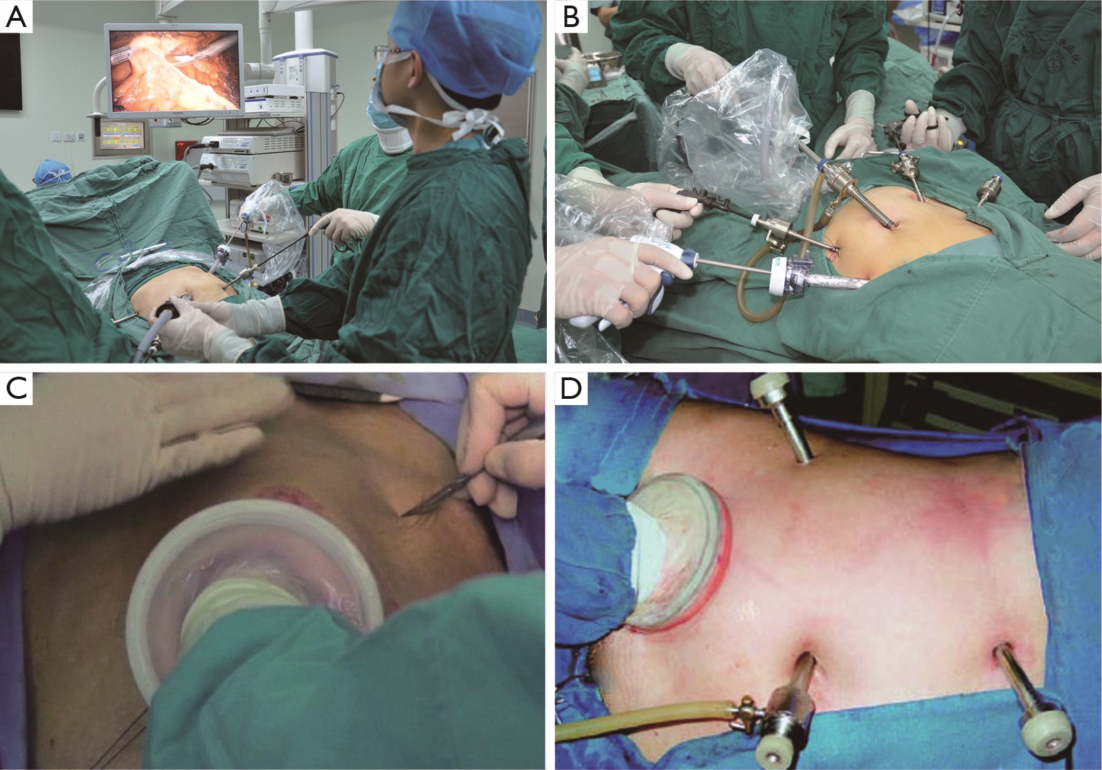
Among the colorectal cancers, the incidence of colon cancer has obviously increased. As a result, the actual incidence of colon cancer has exceeded that of rectal cancer, which dramatically changed the long-existing epidemiological profile. The acute complications of colon cancer include bleeding, obstruction, and perforation, which were among the common acute abdominal surgical conditions. The rapid and accurate diagnosis of these acute complications was very important, and laparoscopic techniques can be applied in abdominal surgery for management of the complications.
Among the colorectal cancers, the incidence of colon cancer has obviously increased. As a result, the actual incidence of colon cancer has exceeded that of rectal cancer, which dramatically changed the long-existing epidemiological profile. The acute complications of colon cancer include bleeding, obstruction, and perforation, which were among the common acute abdominal surgical conditions. The rapid and accurate diagnosis of these acute complications was very important, and laparoscopic techniques can be applied in abdominal surgery for management of the complications.
2014, 26(3): 341-344.
doi: 10.3978/j.issn.1000-9604.2014.05.01
Abstract:
The side effects of tamoxifen are generally mild, including the effect on lipoprotein metabolism. However, there are few cases of severe tamoxifen induced hypertriglyceridemia. Hypertriglyceridemia is a marked risk factor for acute pancreatitis and approximately 2% to 5% of cases of acute pancreatitis are related to drugs. We report on tamoxifen-induced hypertriglyceridemia and acute pancreatitis in a 40 years old woman with type 2 diabetes mellitus occurred by dexamethasone. She was treated with insulin infusion and fenofibrate, and goserelin acetate was started instead of tamoxifen after discharge from the hospital. Also, probable pathogenic hypotheses about the correlation between tamoxifen and dexamethasone induced type 2 diabetes mellitus on severe acute pancreatitis are provided. Clinicians should take care of risks of severe acute pancreatitis on using tamoxifen, especially for patients with dexamethasone induced diabetes mellitus. These individuals should undergo pre-post tamoxifen lipid screening and careful history taking of drugs, including dexamethasone.
The side effects of tamoxifen are generally mild, including the effect on lipoprotein metabolism. However, there are few cases of severe tamoxifen induced hypertriglyceridemia. Hypertriglyceridemia is a marked risk factor for acute pancreatitis and approximately 2% to 5% of cases of acute pancreatitis are related to drugs. We report on tamoxifen-induced hypertriglyceridemia and acute pancreatitis in a 40 years old woman with type 2 diabetes mellitus occurred by dexamethasone. She was treated with insulin infusion and fenofibrate, and goserelin acetate was started instead of tamoxifen after discharge from the hospital. Also, probable pathogenic hypotheses about the correlation between tamoxifen and dexamethasone induced type 2 diabetes mellitus on severe acute pancreatitis are provided. Clinicians should take care of risks of severe acute pancreatitis on using tamoxifen, especially for patients with dexamethasone induced diabetes mellitus. These individuals should undergo pre-post tamoxifen lipid screening and careful history taking of drugs, including dexamethasone.
2014, 26(3): 345-350.
doi: 10.3978/j.issn.1000-9604.2014.06.06
Abstract:
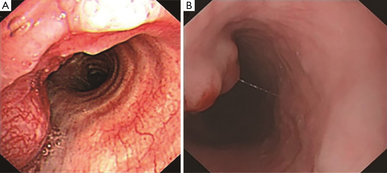
The incidence of multiple primary cancers involving trachea is rare. We present a case of synchronous double primary cancer of trachea and esophagus in a 70-year-old woman, with a special symptom of ventricular tachycardia and no history of smoking and alcohol drinking. Biopsies from multiple foci demonstrated the patient had primary small cell cancer of trachea and squamous cell carcinoma in situ of esophagus. The patient was successfully treated with four cycles of chemotherapy consisting of etoposide and carboplatin (EC) followed by thoracic radiotherapy (60 Gy in 30 fractions, in 6 weeks), and was evaluated to have complete response of tumor. To our knowledge, there is no synchronous cancer of trachea and esophagus has been reported in English literature, and our experience showed sequential EC chemotherapy and radiotherapy provided an effective treatment to control both cancers.
The incidence of multiple primary cancers involving trachea is rare. We present a case of synchronous double primary cancer of trachea and esophagus in a 70-year-old woman, with a special symptom of ventricular tachycardia and no history of smoking and alcohol drinking. Biopsies from multiple foci demonstrated the patient had primary small cell cancer of trachea and squamous cell carcinoma in situ of esophagus. The patient was successfully treated with four cycles of chemotherapy consisting of etoposide and carboplatin (EC) followed by thoracic radiotherapy (60 Gy in 30 fractions, in 6 weeks), and was evaluated to have complete response of tumor. To our knowledge, there is no synchronous cancer of trachea and esophagus has been reported in English literature, and our experience showed sequential EC chemotherapy and radiotherapy provided an effective treatment to control both cancers.
2014, 26(3): 351-354.
doi: 10.3978/j.issn.1000-9604.2014.06.03
Abstract:
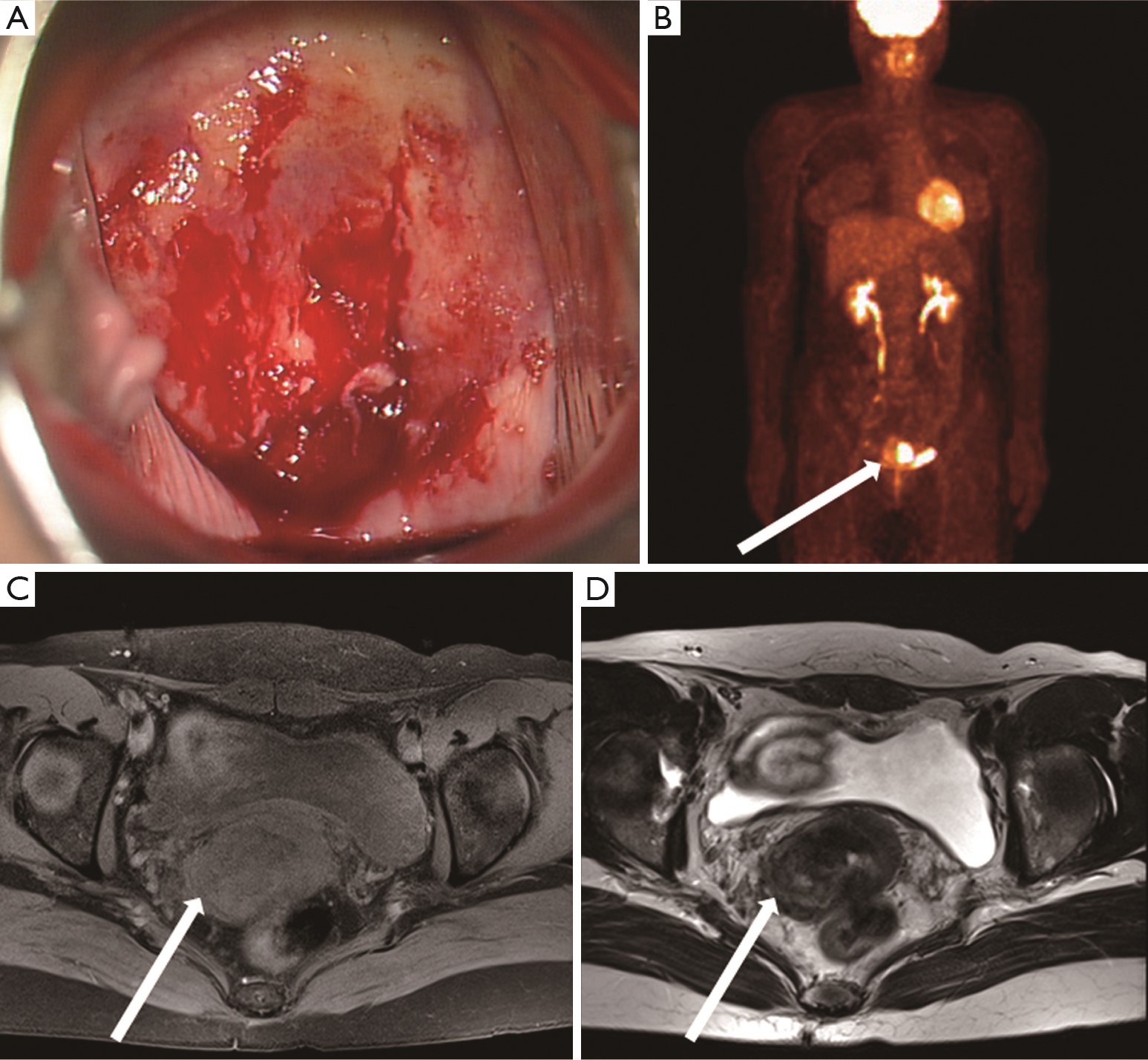
Historically, the lack of melanocytes in the vaginal and cervical mucus membranes has deterred the findings of primary melanomas. Mainly due to its rarity, difficulty to diagnose, and poor prognosis, there has been no absolute agreement on comprehensive treatment so far. In this case report, we present a case of a 46-year-old woman with primary malignant melanoma of uterine cervix. She underwent neo-adjuvant chemotherapy initially followed by a radical hysterectomy. After adjuvant concurrent chemo-radiation, the patient has been followed up for 24 months. So far, she has not shown any symptoms or signs of recurrence. Further studies with more cases based on variable combinations of treatment regimen have been on the way.
Historically, the lack of melanocytes in the vaginal and cervical mucus membranes has deterred the findings of primary melanomas. Mainly due to its rarity, difficulty to diagnose, and poor prognosis, there has been no absolute agreement on comprehensive treatment so far. In this case report, we present a case of a 46-year-old woman with primary malignant melanoma of uterine cervix. She underwent neo-adjuvant chemotherapy initially followed by a radical hysterectomy. After adjuvant concurrent chemo-radiation, the patient has been followed up for 24 months. So far, she has not shown any symptoms or signs of recurrence. Further studies with more cases based on variable combinations of treatment regimen have been on the way.
2014, 26(3): 355-359.
doi: 10.3978/j.issn.1000-9604.2014.06.04
Abstract:
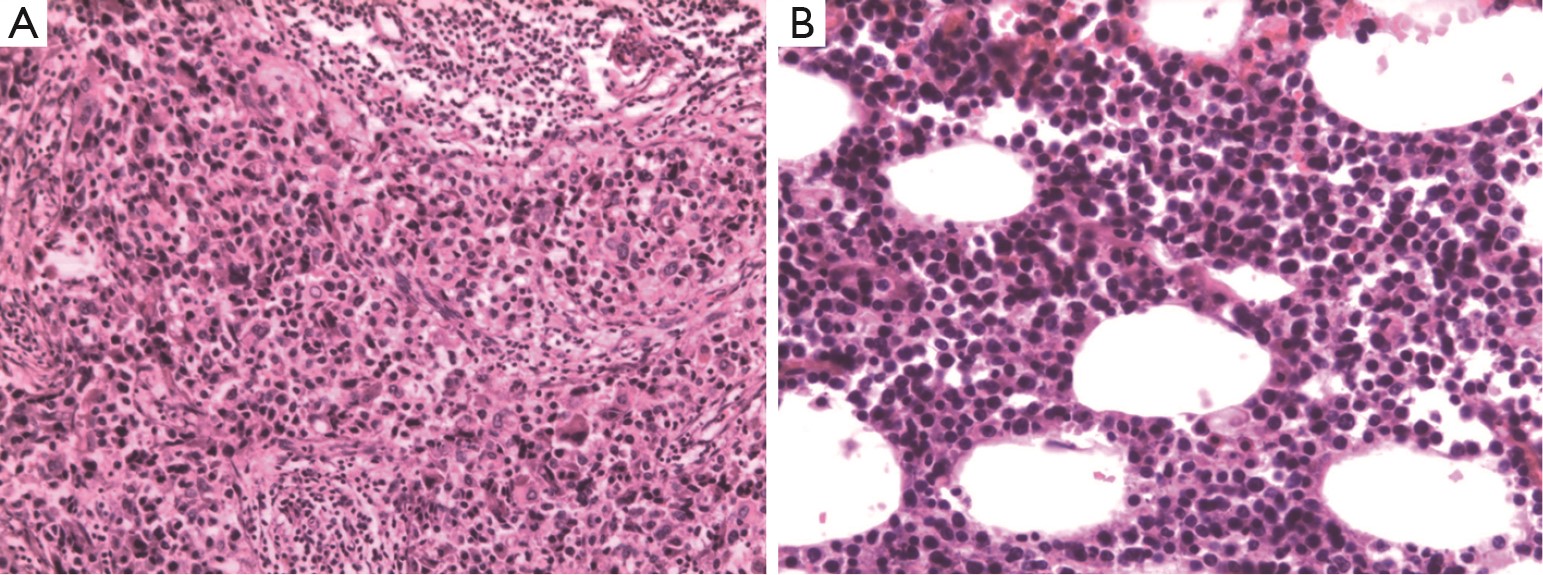
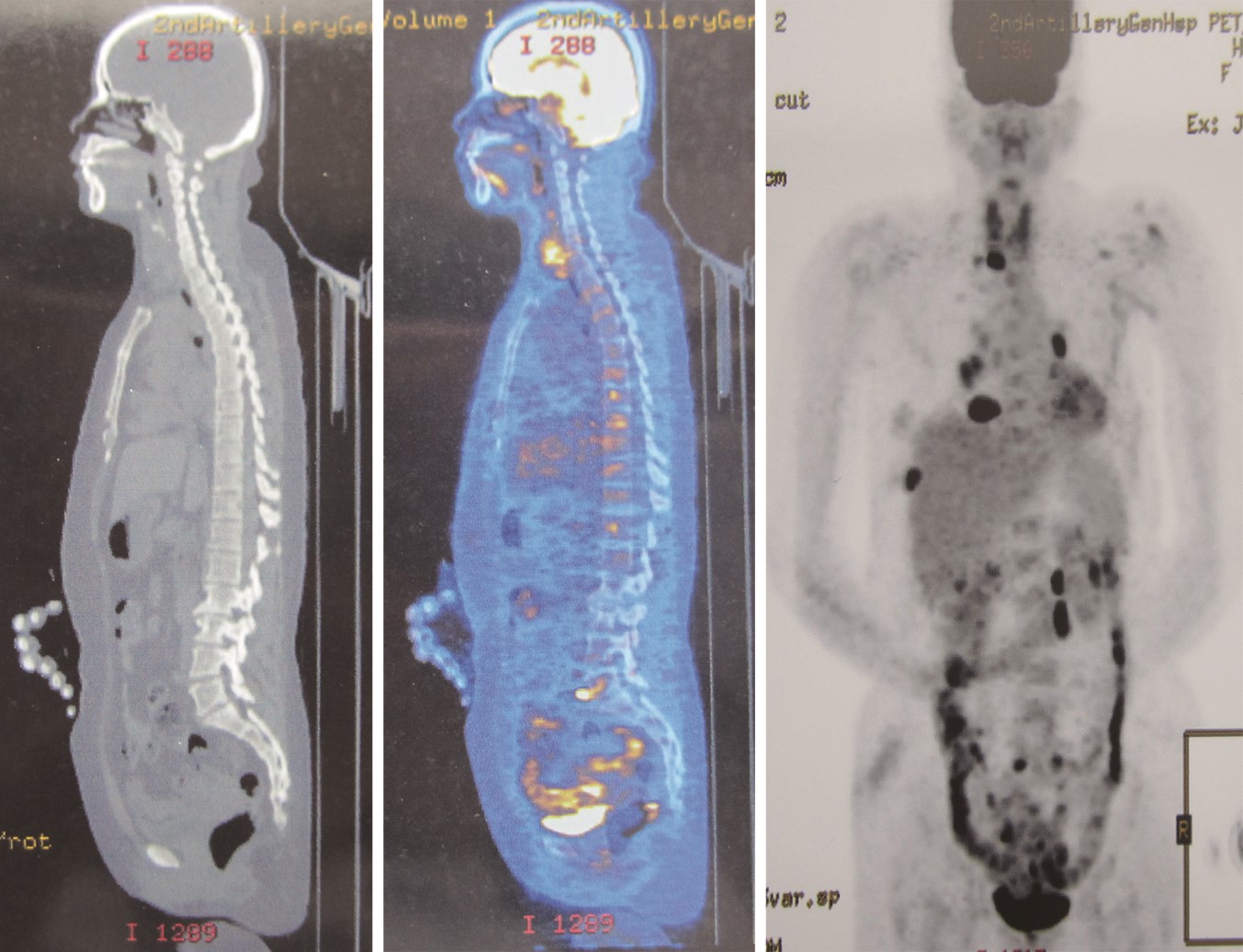
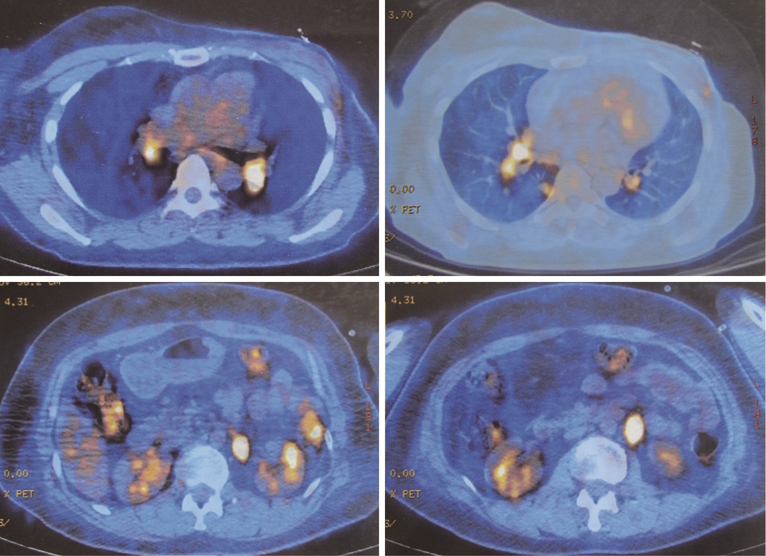
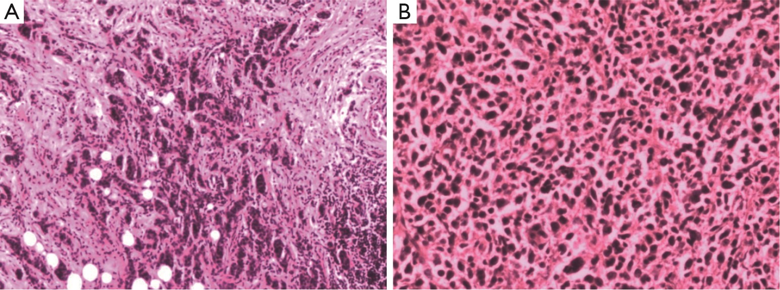
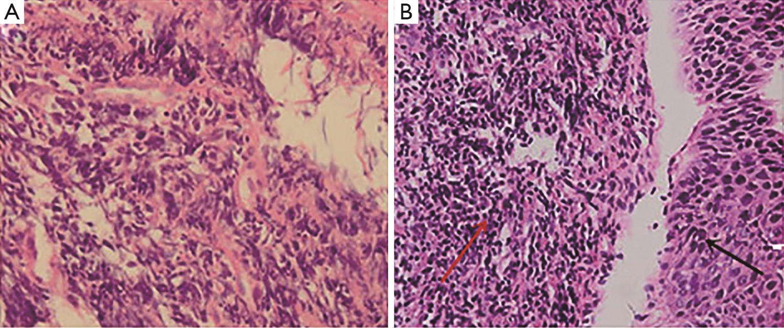
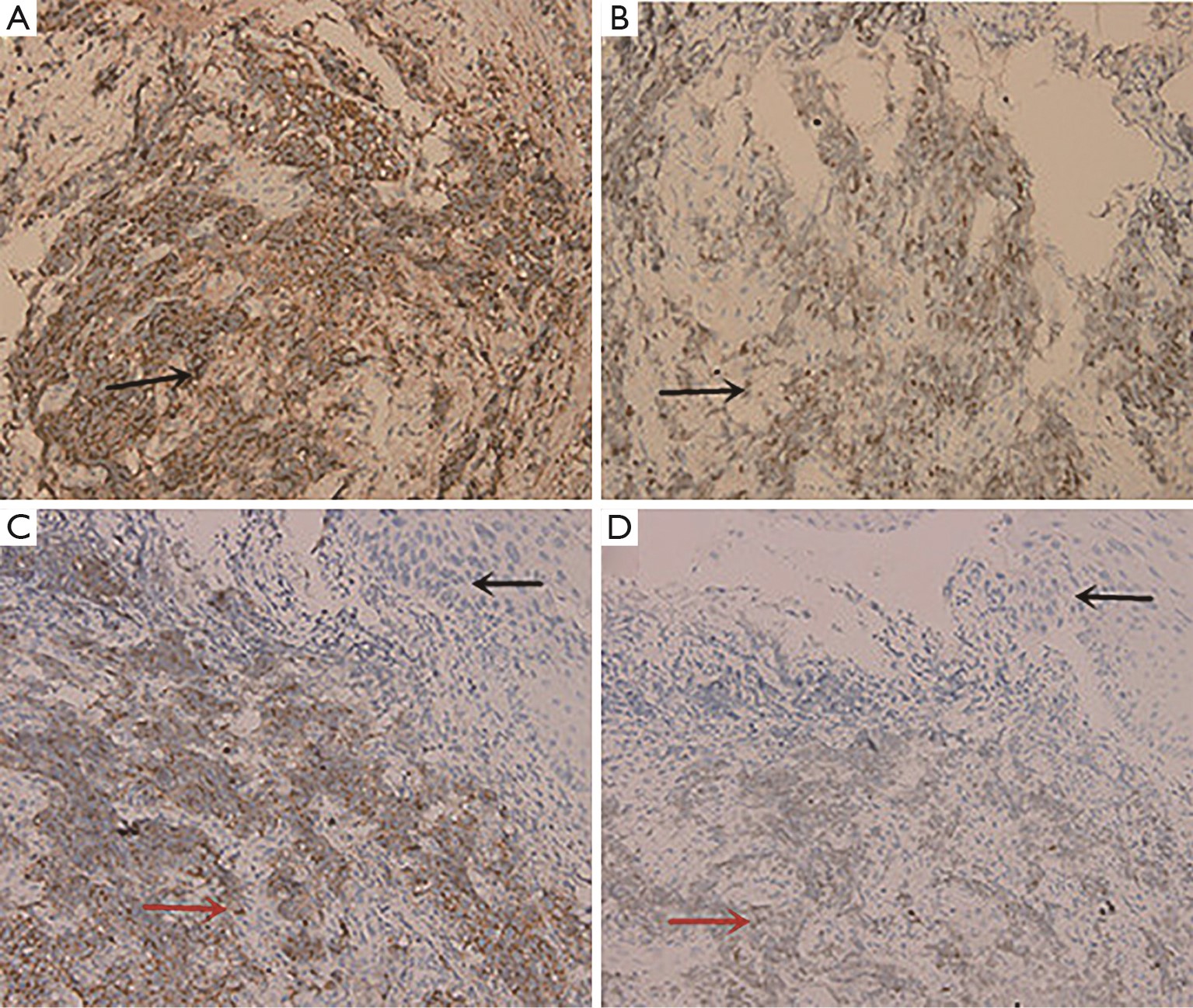
Synchronous breast cancer and breast lymphoma are rare. It is of high rate of misdiagnosis in clinical practice. Here we present two cases with this presentation. They are both middle-aged women, with stage I invasive ductal carcinoma of the breast. One patient happened to have primary breast lymphoma (PBL); the other was secondary breast lymphoma (SBL). Their pathology and immunohistochemistry (IHC) findings supported the diagnosis of multiple primary carcinoma. Both patients had a surgery. Then they both received CHOP regime chemotherapy and subsequent endocrine therapy.
Synchronous breast cancer and breast lymphoma are rare. It is of high rate of misdiagnosis in clinical practice. Here we present two cases with this presentation. They are both middle-aged women, with stage I invasive ductal carcinoma of the breast. One patient happened to have primary breast lymphoma (PBL); the other was secondary breast lymphoma (SBL). Their pathology and immunohistochemistry (IHC) findings supported the diagnosis of multiple primary carcinoma. Both patients had a surgery. Then they both received CHOP regime chemotherapy and subsequent endocrine therapy.