2015 Vol.27(3)
Display Mode: |
2015, 27(3): 221-230.
doi: 10.3978/j.issn.1000-9604.2015.04.04
 Abstract
 FullText HTML
 PDF 179KB
Abstract:
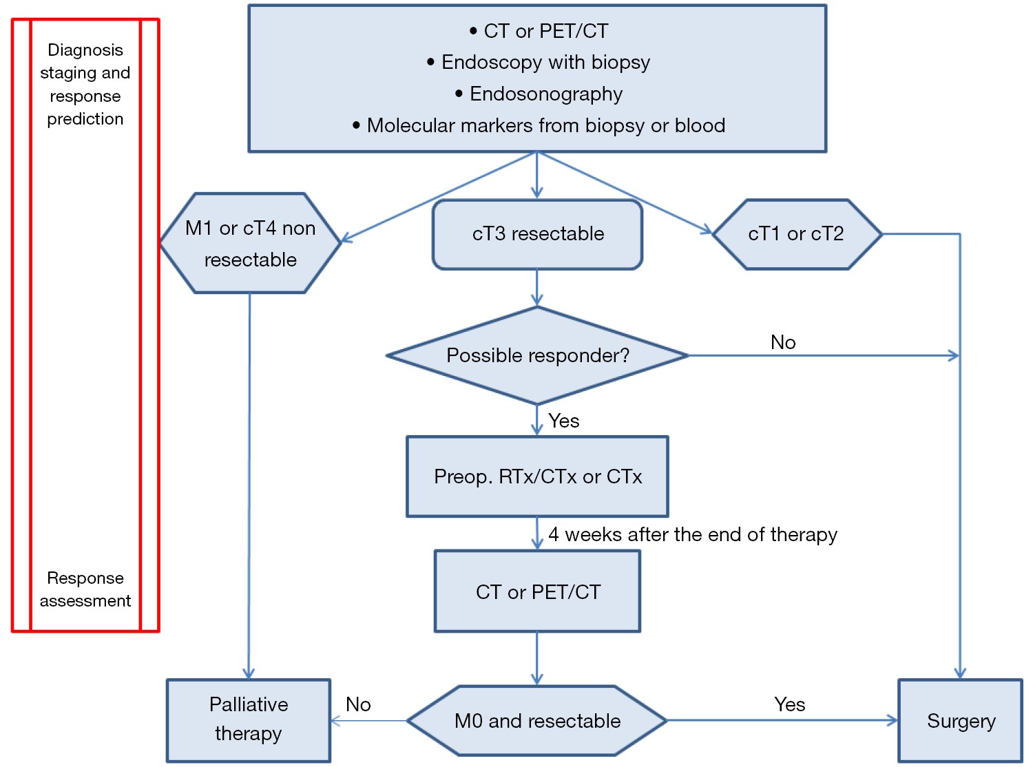
Patients with advanced esophageal cancer (T3-4, N) have a poor prognosis. Chemoradiation or chemotherapy before esophagectomy with adequate lymphadenectomy is the standard treatment for patients with resectable advanced esophageal carcinoma. However, only patients with major histopathologic response (regression to less than 10% of the primary tumor) after preoperative treatment will have a prognostic benefit of preoperative chemoradiation. Using current therapy regimens about 40% to 50% of the patients show major histopathological response. The remaining cohort does not benefit from this neoadjuvant approach but might benefit from earlier surgical resection. Therefore, it is an aim to develop tools for response prediction before starting the treatment and for early response assessment identifying responders. The current review discusses the different imaging techniques and the most recent studies about molecular markers for early response prediction. The results show that [18F]-fluorodeoxyglucose-positron emission tomography (FDG-PET) has a good sensitivity but the specificity is not robust enough for routine clinical use. Newer positron emission tomography detector technology, the combination of FDG-PET with computed tomography, additional evaluation criteria and standardization of evaluation may improve the predictive value. There exist a great number of retrospective studies using molecular markers for prediction of response. Until now the clinical use is missing. But the results of first prospective studies are promising. A future perspective may be the combination of imaging technics and special molecular markers for individualized therapy. Another aspect is the response assessment after finishing neoadjuvant treatment protocol. The different clinical methods are discussed. The results show that until now no non-invasive method is valid enough to assess complete histopathological response.
Patients with advanced esophageal cancer (T3-4, N) have a poor prognosis. Chemoradiation or chemotherapy before esophagectomy with adequate lymphadenectomy is the standard treatment for patients with resectable advanced esophageal carcinoma. However, only patients with major histopathologic response (regression to less than 10% of the primary tumor) after preoperative treatment will have a prognostic benefit of preoperative chemoradiation. Using current therapy regimens about 40% to 50% of the patients show major histopathological response. The remaining cohort does not benefit from this neoadjuvant approach but might benefit from earlier surgical resection. Therefore, it is an aim to develop tools for response prediction before starting the treatment and for early response assessment identifying responders. The current review discusses the different imaging techniques and the most recent studies about molecular markers for early response prediction. The results show that [18F]-fluorodeoxyglucose-positron emission tomography (FDG-PET) has a good sensitivity but the specificity is not robust enough for routine clinical use. Newer positron emission tomography detector technology, the combination of FDG-PET with computed tomography, additional evaluation criteria and standardization of evaluation may improve the predictive value. There exist a great number of retrospective studies using molecular markers for prediction of response. Until now the clinical use is missing. But the results of first prospective studies are promising. A future perspective may be the combination of imaging technics and special molecular markers for individualized therapy. Another aspect is the response assessment after finishing neoadjuvant treatment protocol. The different clinical methods are discussed. The results show that until now no non-invasive method is valid enough to assess complete histopathological response.
2015, 27(3): 231-238.
doi: 10.3978/j.issn.1000-9604.2015.05.06
Abstract:
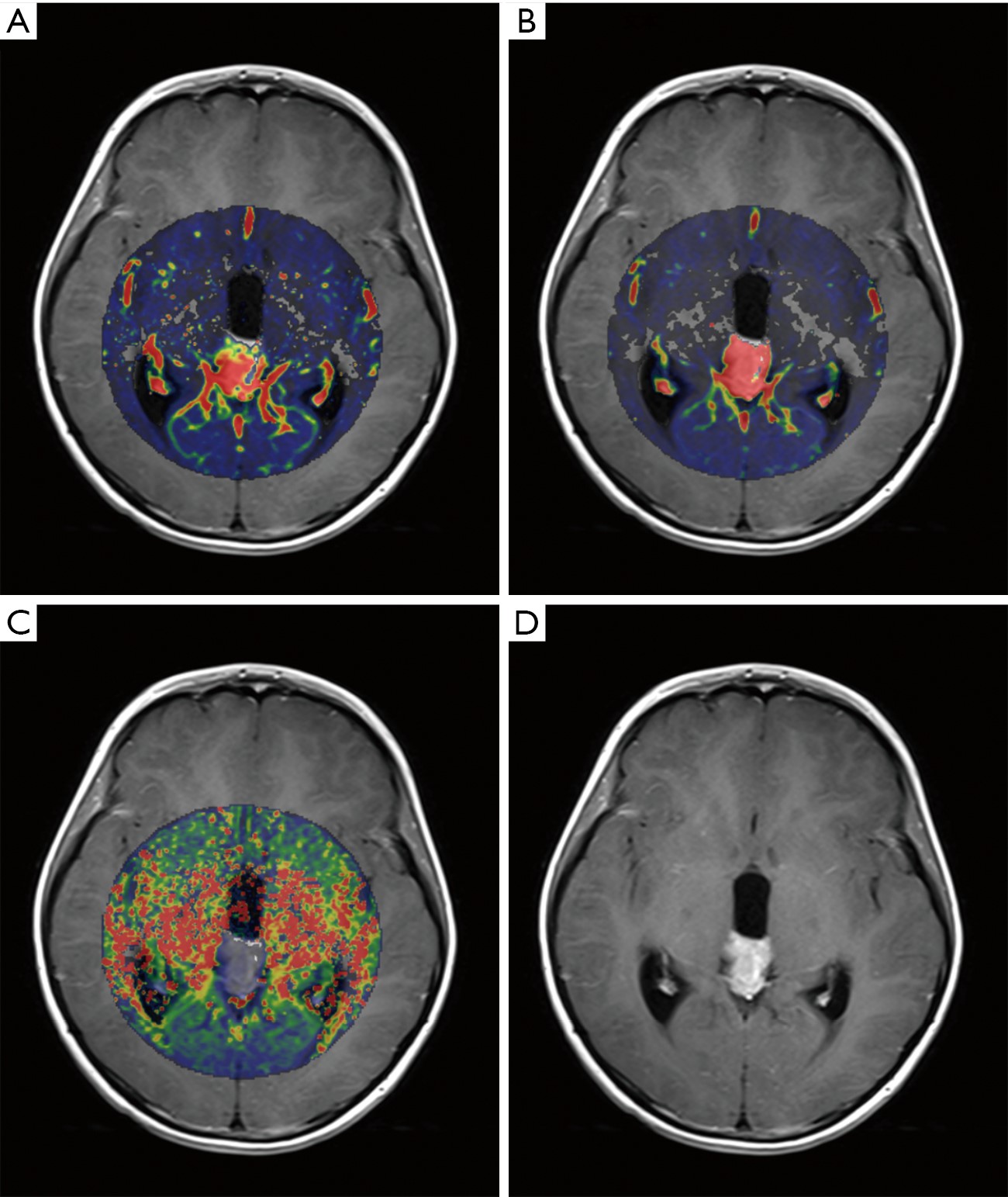
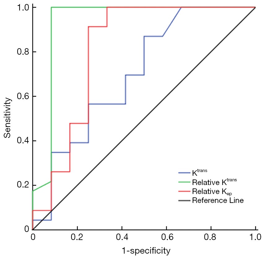
ObjectiveTo evaluate the feasibility of dynamic contrast-enhanced magnetic resonance imaging (DCE-MRI) for predicting tumor response to radiotherapy in patients with suspected primary central nervous system (CNS) germ cell tumors (GCTs). MethodsDCE-MRI parameters of 35 patients with suspected primary CNS GCTs were obtained prior to diagnostic radiation, using the Tofts and Kermode model. Radiosensitivity was determined in tumors diagnosed 2 weeks after radiation by observing changes in tumor size and markers as a response to MRI. Taking radiosensitivity as the gold standard, the cut-off value of DCE-MRI parameters was measured by receiver operating characteristic (ROC) curve. Diagnostic accuracy of DCE-MRI parameters for predicting radiosensitivity was evaluated by ROC curve. ResultsA significant elevation in transfer constant (Ktrans) and extravascular extracellular space (Ve) (P=0.000), as well as a significant reduction in rate constant (Kep) (P=0.000) was observed in tumors. Ktrans, relative Ktrans, and relative Kep of the responsive group were significantly higher than non-responsive groups. No significant difference was found in Kep, Ve, and relative Ve between the two groups. Relative Ktrans showed the best diagnostic value in predicting radiosensitivity with a sensitivity of 100%, specificity of 91.7%, positive predictive value (PPV) of 95.8%, and negative predictive value (NPV) of 100%. ConclusionsRelative Ktrans appeared promising in predicting tumor response to radiation therapy (RT). It is implied that DCE-MRI pre-treatment is a requisite step in diagnostic procedures and a novel and reliable approach to guide clinical choice of RT.

2015, 27(3): 239-246.
doi: 10.3978/j.issn.1000-9604.2015.06.08
Abstract:
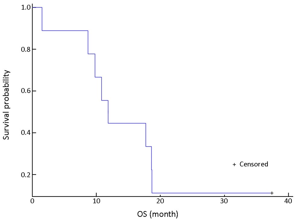
ObjectiveTo determine the maximum tolerated dose (MTD), dose-limiting toxicity (DLT) and efficacy of sorafenib in combination with FOLFOX4 (oxaliplatin/leucovorin (LV)/5-fluorouracil) as first-line treatment for advanced gastric cancer, we performed a phase I dose-finding study in nine evaluable patients with unresectable locally advanced or metastatic gastric cancer or gastroesophageal junction adenocarcinoma. MethodsAccording to modified Fibonacci method, the design of this study was to guide elevation of the sorafenib dosage to the next level (from 200 mg twice daily to 400 mg twice daily and then, if tolerated, 600 mg twice daily). If the patient achieved complete response (CR), partial response (PR) or stable disease (SD) after eight cycles of treatment, combination chemotherapy was scheduled to be discontinued and sorafenib monotherapy continued at the original dose until either disease progression or unacceptable toxicity. ResultsIn sorafenib 200 mg twice daily group, DLT was observed in 1 of 6 patients, and in 400 mg twice daily group, it was observed in 2 of 3 patients. Seven of 9 (77.8%) evaluable patients achieved PR, with a median overall survival (OS) of 11.8 [95% confidence interval (CI): 8.9-14.7] months. Common adverse effects include hand-foot syndrome, leukopenia, neutropenia, anorexia, and nausea. ConclusionsTwice-daily dosing of sorafenib 200 mg in combination with FOLFOX4 was proven effective and safe for the treatment of advanced gastric cancer, and could be an appropriate dosage for subsequent phase II clinical studies.
2015, 27(3): 247-257.
doi: 10.3978/j.issn.1000-9604.2015.04.07
Abstract:
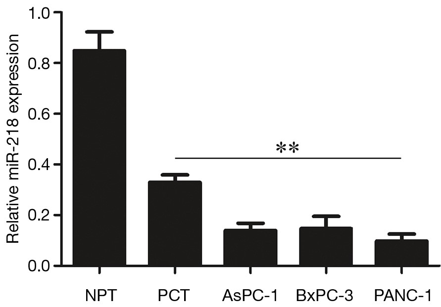
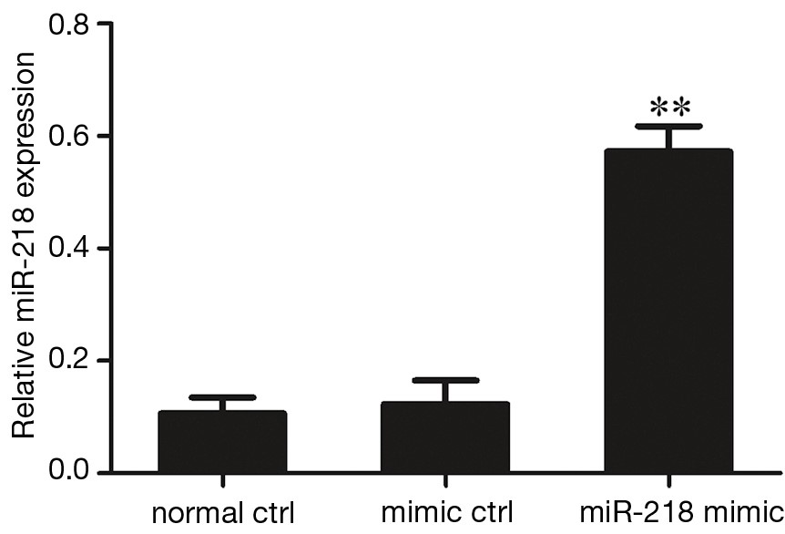
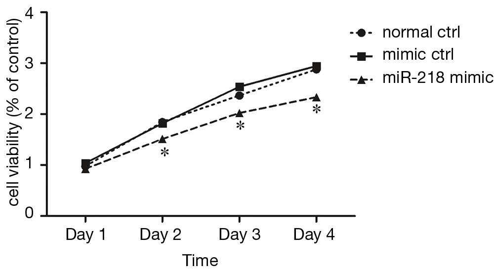
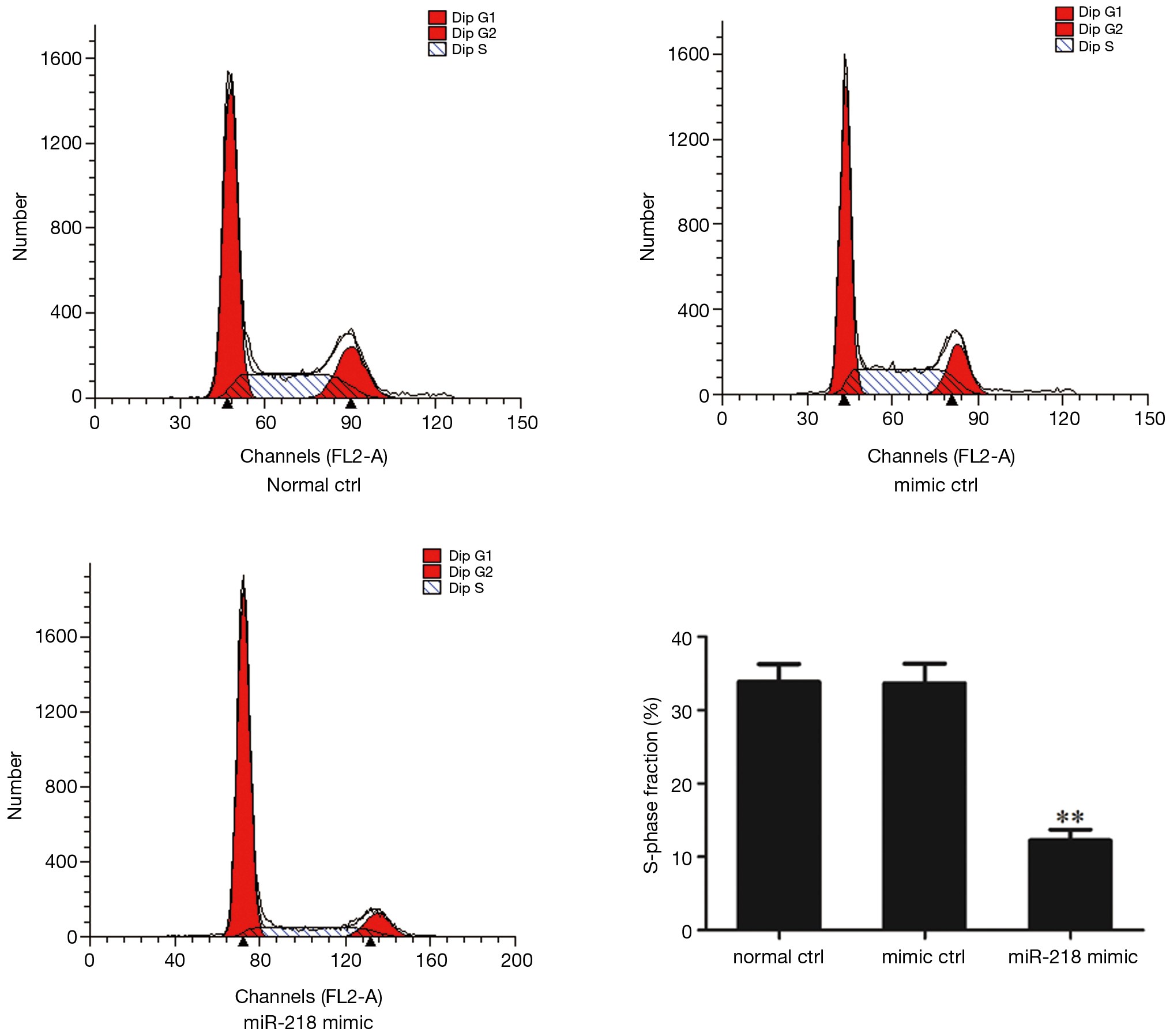
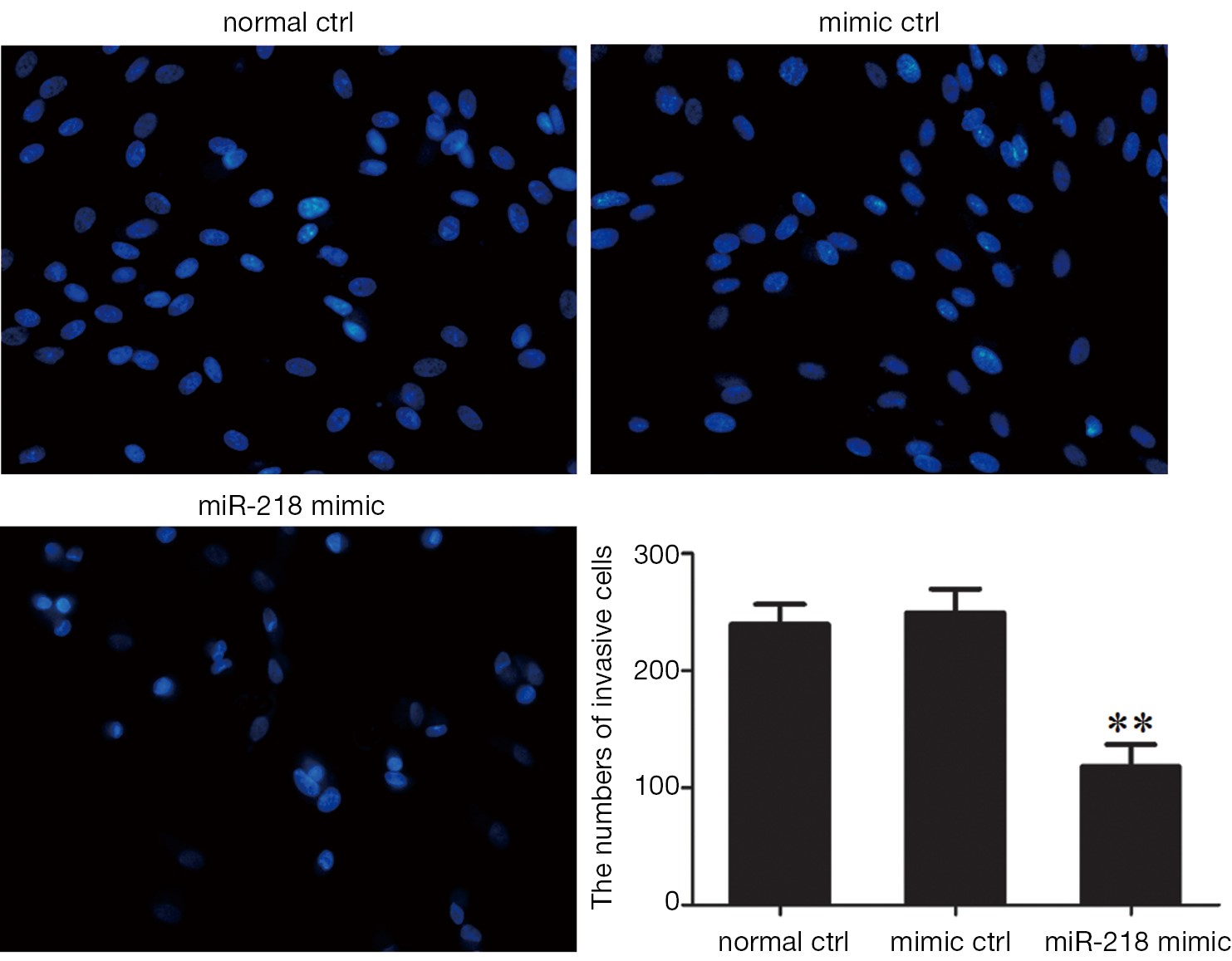
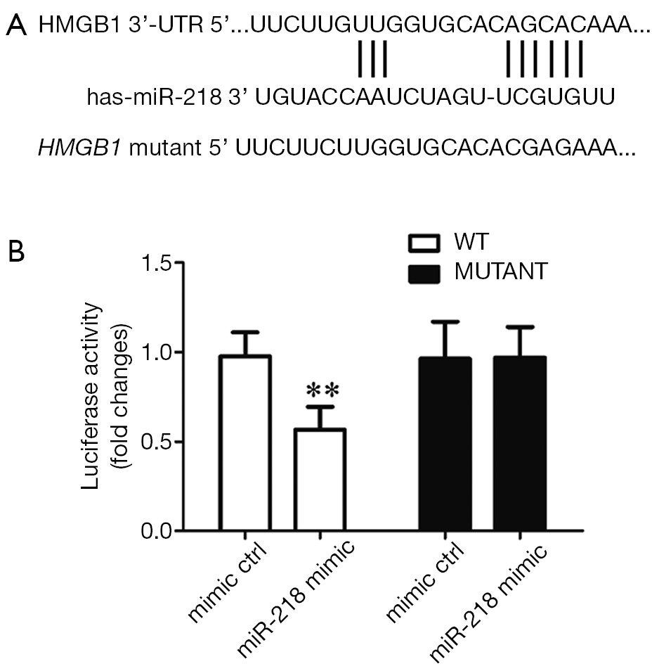
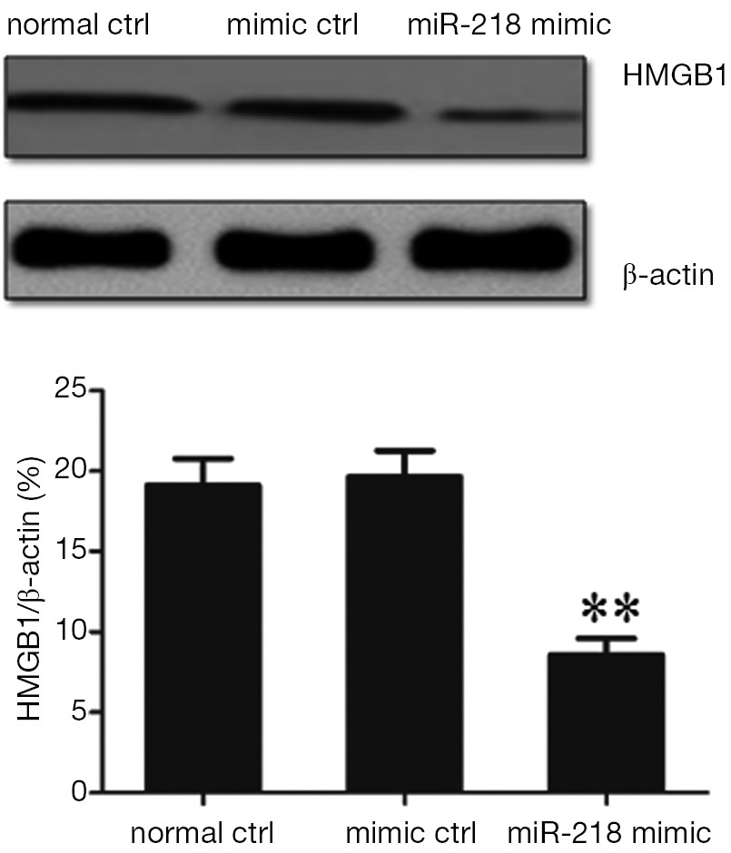
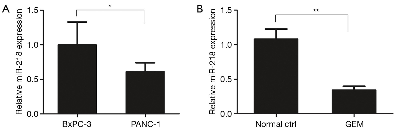
ObjectiveTo detect the expression profiles of microRNA-218 (miR-218) in human pancreatic cancer tissue (PCT) and cells and their effects on the biological features of human pancreatic cancer cell line PANC-1 and observe the effect of miR-218 on the expression of the target gene high mobility group box 1 (HMGB1), with an attempt to provide new treatment methods and strategies for pancreatic cancer. MethodsThe expressions of miR-218 in PCT and normal pancreas tissue as well as in various pancreatic cancer cell lines including AsPC-1, BxPC-3, and PANC-1 were determined with quantitative real-time reverse transcription polymerase chain reaction (qRT-PCR). The change of miR-218 expression in PANC-1 cells was detected using qRT-PCT after the transfection of miR-218 mimic for 48 h. Cell Counting Kit-8 (CCK-8) was applied for detecting the effect of miR-218 on the activity of PANC-1 cells. The effects of miR-218 on the proliferation and apoptosis of PANC-1 cells were analyzed using the flow cytometry. The effect of miR-218 on the migration of PANC-1 cells was detected using the Trans-well migration assay. The HMGB1 was found to be a target gene of miR-218 by luciferase reporter assay, and the effect of miR-218 on the expression of HMGB1 protein in cells were determined using Western blotting. ResultsAs shown by qRT-PCR, the expressions of miR-218 in PCT and in pancreatic cancer cell line significantly decreased when compared with the normal pancreatic tissue (NPT) (P<0.01). Compared with the control group, the miR-218 expression significantly increased in the PANC-1 group after the transfection of miR-218 mimic for 48 h (P<0.01). Growth curve showed that the cell viability significantly dropped after the overexpression of miR-218 in the PANC-1 cells for two days (P<0.05). Flow cytometry showed that the S-phase fraction significantly dropped after the overexpression of miR-218 (P<0.01) and the percentage of apoptotic cells significantly increased (P<0.01). As shown by the Trans-well migration assay, the enhanced miR-218 expression was associated with a significantly lower number of cells that passed through a Transwell chamber (P<0.01). Luciferase reporter assay showed that, compared with the control group, the relative luciferase activity significantly decreased in the miR-218 mimic group (P<0.01). As shown by the Western blotting, compared with the control group, the HMGB1 protein expression significantly decreased in the PANC-1 group after the transfection of miR-218 mimic for 48 h (P<0.01). ConclusionsThe miR-218 expression decreases in human PCT and cell lines. miR-218 can negatively regulate the HMGB1 protein expression and inhibit the proliferation and invasion of pancreatic cancer cells. A treatment strategy by enhancing the miR-218 expression may benefit the patients with pancreatic cancer.

2015, 27(3): 258-266.
doi: 10.3978/j.issn.1000-9604.2015.06.04
Abstract:
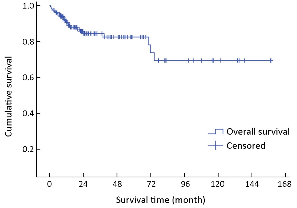
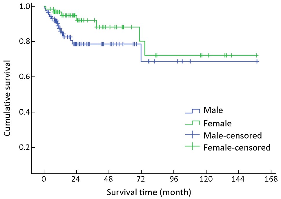
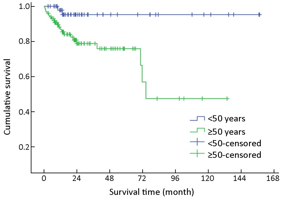
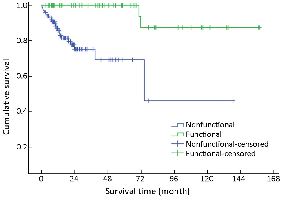
ObjectiveTo investigate the clinicopathological features, survival and prognostic factors for gastroenteropancreatic neuroendocrine neoplasms (GEP-NENs) in a Chinese population. MethodsWe investigated 154 consecutive patients (88 males, 66 females; median age 56 years, age range 9-86 years) diagnosed with GEP-NENs between 2001 and 2013 at The Affiliated Hospital of Qingdao University. Demographic, clinical and pathological variables and survival data were retrieved. ResultsThe pancreas was the most common site of involvement (63/154, 40.9%). Tumor size varied from 0.3 to 16.0 cm (median, 1.2 cm). The patients were followed up for a median period of 22 months (range, 1-157 months). The estimated 3- and 5-year overall survival (OS) rates for all patients were 84.0% and 81.9%, respectively. Multivariate analysis showed that larger tumor size, lymphatic metastases and distant metastases were significant predictors for poor survival outcome. ConclusionsOur data provide further information on the clinicopathological features of GEP-NENs in China. Additionally, we identified tumor size, lymphatic metastases and distant metastases as independent prognostic factors for long-term survival.


2015, 27(3): 267-278.
doi: 10.3978/j.issn.1000-9604.2015.04.06
Abstract:
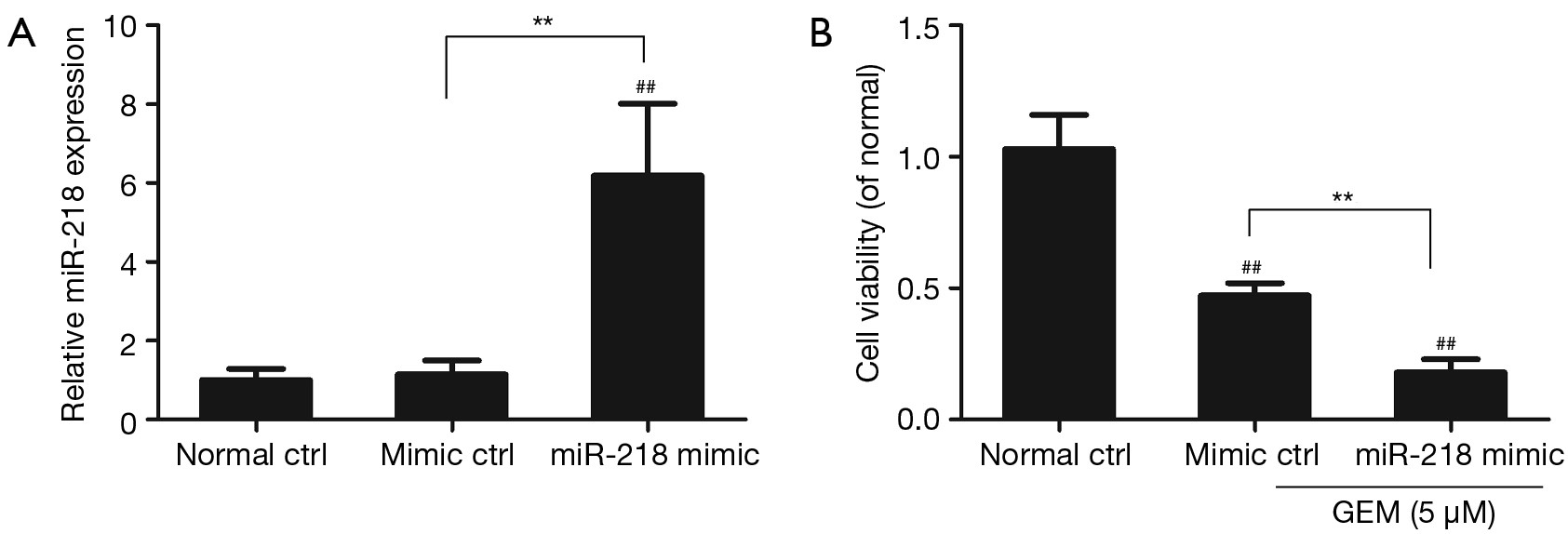
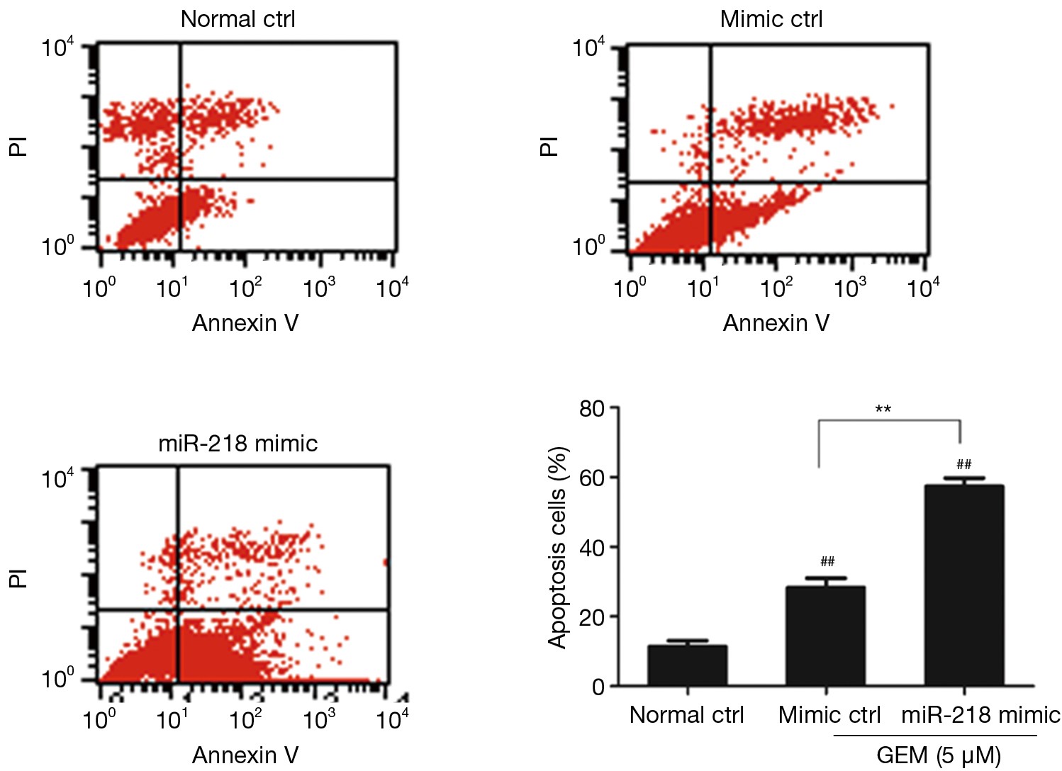
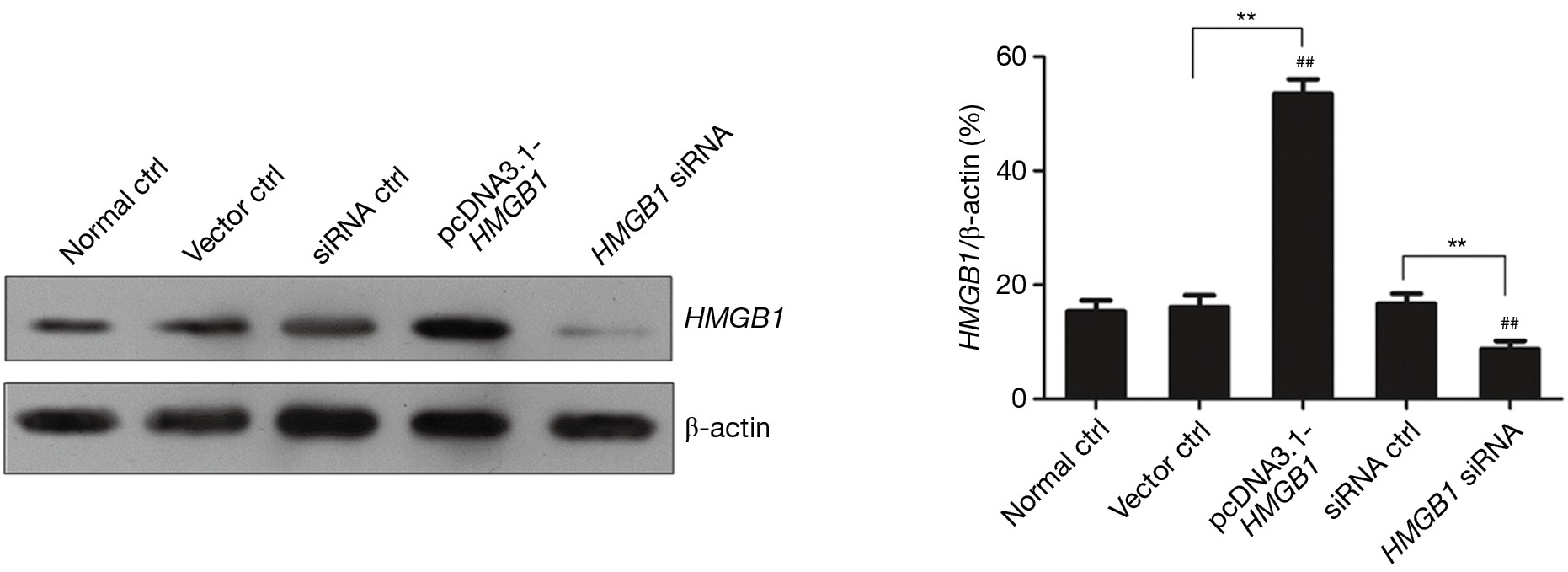
ObjectiveThe purpose of this study was to examine the effect of gemcitabine (GEM) on microRNA-218 (miR-218) expression in human pancreatic cancer cells. MethodsQuantitative reverse transcription polymerase chain reaction (qRT-PCR) was performed to examine the differences in miR-218 expression between the GEM-sensitive BxPC-3 pancreatic cancer cells and GEM-resistant PANC-1 cells. The effect of GEM on the expression of miR-218 in PANC-1 cells was also investigated. PANC-1 cells were transfected either with HMGB1 siRNA to knock down the expression of HMGB1 or with the recombinant HMGB1 expression vector (pcDNA3.1-HMGB1) to overexpress HMGB1. The effect of ectopic expression of HMGB1 on the apoptosis of miR-218-transfected and GEM-treated PANC-1 cells was examined by flow cytometric analysis. ResultsThe miR-218 expression level was lower in GEM-resistant PANC-1 cells compared to GEM-sensitive BxPC-3 cells (P<0.05). The percentage of apoptotic PANC-1 cells was significantly increased in the miR-218 mimic + GEM group compared to the mimic ctrl + GEM group and the normal control group (P<0.01). The HMGB1 expression level was markedly decreased in PANC-1 cells transfected with HMGB1 siRNA but was significantly increased in PANC-1 cells transfected with the recombinant HMGB1 expression vector, pcDNA3.1-HMGB1 (P<0.01). The proportion of apoptotic PANC-1 cells was significantly lower in the miR-218 mimic + GEM + pcDNA3.1-HMGB1 group compared to the miR-218 mimic + GEM + HMGB1 siRNA group (P<0.01). ConclusionsThe expression level of miR-218 was downregulated in the GEM-resistant cell line. miR-218 promoted the sensitivity of PANC-1 cells to GEM, which was achieved mainly through regulating the expression of HMGB1 in PANC-1 cells.


2015, 27(3): 279-287.
doi: 10.3978/j.issn.1000-9604.2015.01.04
Abstract:
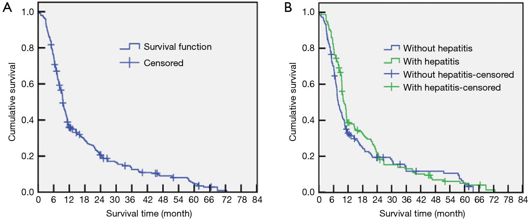
ObjectiveTo compare the clinical characteristics and prognosis between hepatitis virus-related hepatocellular carcinoma (viral HCC) and non-B, non-C HCC (NBC-HCC) among Uyghur patients in Xinjiang province, China. MethodsBetween 01/01/2000 and 31/12/2012, 319 Uyghur HCC patients were treated at the Cancer Centre of The First Affiliated Hospital of Xinjiang Medical University. The data for the patients were obtained from a retrospective review of the patients’ medical records. A total of 18 patients were excluded from the study because of incomplete information. The patients were classified into two groups: viral HCC and NBC-HCC. The clinical characteristics and prognostic factors were statistically analysed. ResultsFor all 301 patients, gender (P=0.000), area of residence (P=0.002), diabetes mellitus (P=0.009), BMI (P=0.000), cirrhosis (P=0.000), tumour stage (P=0.004), Child-Pugh class (P=0.000), the TBIL level (P=0.000), and the alpha-fetoprotein (AFP) level (P=0.000) were significantly different between the NBC-HCC and viral HCC groups. The NBC-HCC patients tended to be diagnosed at advanced stages; however, the NBC-HCC patients exhibited lower Child-Pugh scores than the viral HCC patients. In all patients examined, the 0.5-, 1-, 3- and 5-year overall survival (OS) rates were 35.6%, 20.3%, 12.6% and 4.5%, respectively. No significant difference in OS was observed between the two groups (P=0.124). Cox multivariate analysis revealed that age (RR =1.539, P=0.001), TNM stage (RR =12.708, P=0.000), portal vein tumour thrombus (PVTT) (RR =2.003, P=0.000), Child-Pugh class (RR =1.715, P=0.000), and TACE + radiotherapy/RFA (RR =0.567, P=0.000) were significant independent prognostic factors for HCC patients. ConclusionsThe clinical characteristics differ between Uyghur patients with NBC-HCC and viral HCC. HCC in the Xinjiang region displays specific regional characteristics. Age, TNM stage, PVTT, Child-Pugh class and TACE + radiotherapy/RFA are significant risk factors that influence patient survival.
2015, 27(3): 288-293.
doi: 10.3978/j.issn.1000-9604.2015.04.11
Abstract:
BackgroundThere is strong evidence that delayed diagnosis of breast cancer is associated with poor survival. The objectives were to determine the frequency of breast cancer patients with delayed presentation, the reasons of delay and its association with different socio-demographic variables in our settings. MethodsWe interviewed 315 histologically confirmed breast cancer patients. Delay was defined as more than 3 months from appearance of symptoms to the consultation from doctor. Questions were asked from each patient which could reflect their understanding about the disease and which could be the likely reasons for their delayed presentation. ResultsA total of 39.01% (n=123) of patients presented late and out of those, 40.7% wasted time using alternative medicines; 25.2% were not having enough resources; 17.1% presented late due to painless lump; 10.6% felt shyness and 6.5% presented late due to other reasons. Higher age, negative family history, <8 school years of education and low to middle socio-economic status were significantly associated with delayed presentation (P<0.05). Education and socioeconomic status were two independent variables related to the delayed presentation after adjustment for others [odds ratios (OR) of 2.26, 2.29 and 95% confidence intervals (CI) was 1.25-4.10, 1.06-4.94 respectively]. ConclusionsSignificant percentage of women with breast cancer in North Pakistan is experiencing presentation delay due to their misconceptions about the disease. Coordinated efforts with public health department are needed to educate the focused groups and mitigating the barriers identified in the study. Long term impact will be reduced overall burden of the disease in the region.
2015, 27(3): 294-300.
doi: 10.3978/j.issn.1000-9604.2015.05.03
Abstract:
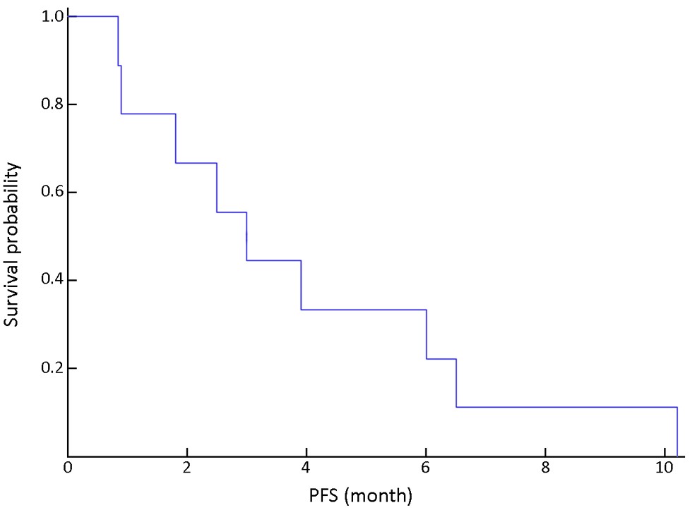
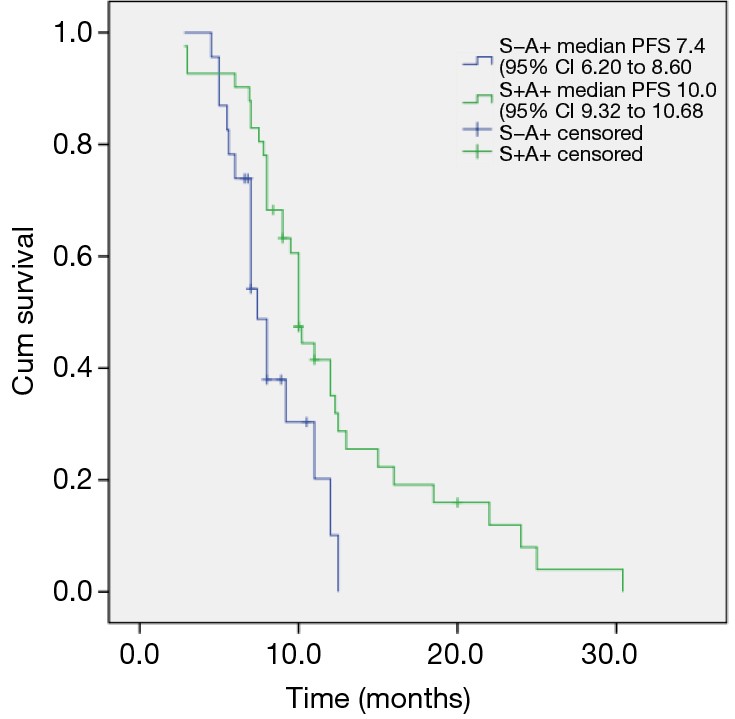
BackgroundEpidermal growth factor receptor (EGFR) mutation is the key predictor of EGFR tyrosine kinase inhibitors (TKIs) efficacy in non-small cell lung cancer (NSCLC). We conducted this study to verify the feasibility of EGFR mutation analysis in cytological specimens and investigate the responsiveness to gefitinib treatment in patients carrying EGFR mutations. MethodsA total of 210 cytological specimens were collected for EGFR mutation detection by both direct sequencing and amplification refractory mutation system (ARMS). We analyzed EGFR mutation status by both methods and evaluated the responsiveness to gefitinib treatment in patients harboring EGFR mutations by overall response rate (ORR), disease control rate (DCR) and progression free survival (PFS). ResultsOf all patients, EGFR mutation rate was 28.6% (60/210) by direct sequencing and 45.2% (95/210) by ARMS (P<0.001) respectively. Among the EGFR wild type patients tested by direct sequencing, 26.7% of them were positive by ARMS. For the 72 EGFR mutation positive patients treated with gefitinib, the ORR, DCR and median PFS were 69.4%, 90.2% and 9.3 months respectively. The patients whose EGFR mutation status was negative by direct sequencing but positive by ARMS had lower ORR (48.0% vs. 80.9%, P=0.004) and shorter median PFS (7.4 vs. 10.5 months, P=0.009) as compared with that of EGFR mutation positive patients by both detection methods. ConclusionsOur study verified the feasibility of EGFR analysis in cytological specimens in advanced NSCLC. ARMS is more sensitive than direct sequencing in EGFR mutation detection. EGFR Mutation status tested on cytological samples is applicable for predicting the response to gefitinib. Abundance of EGFR mutations might have an influence on TKIs efficacy.
2015, 27(3): 301-308.
doi: 10.3978/j.issn.1000-9604.2014.11.03
Abstract:
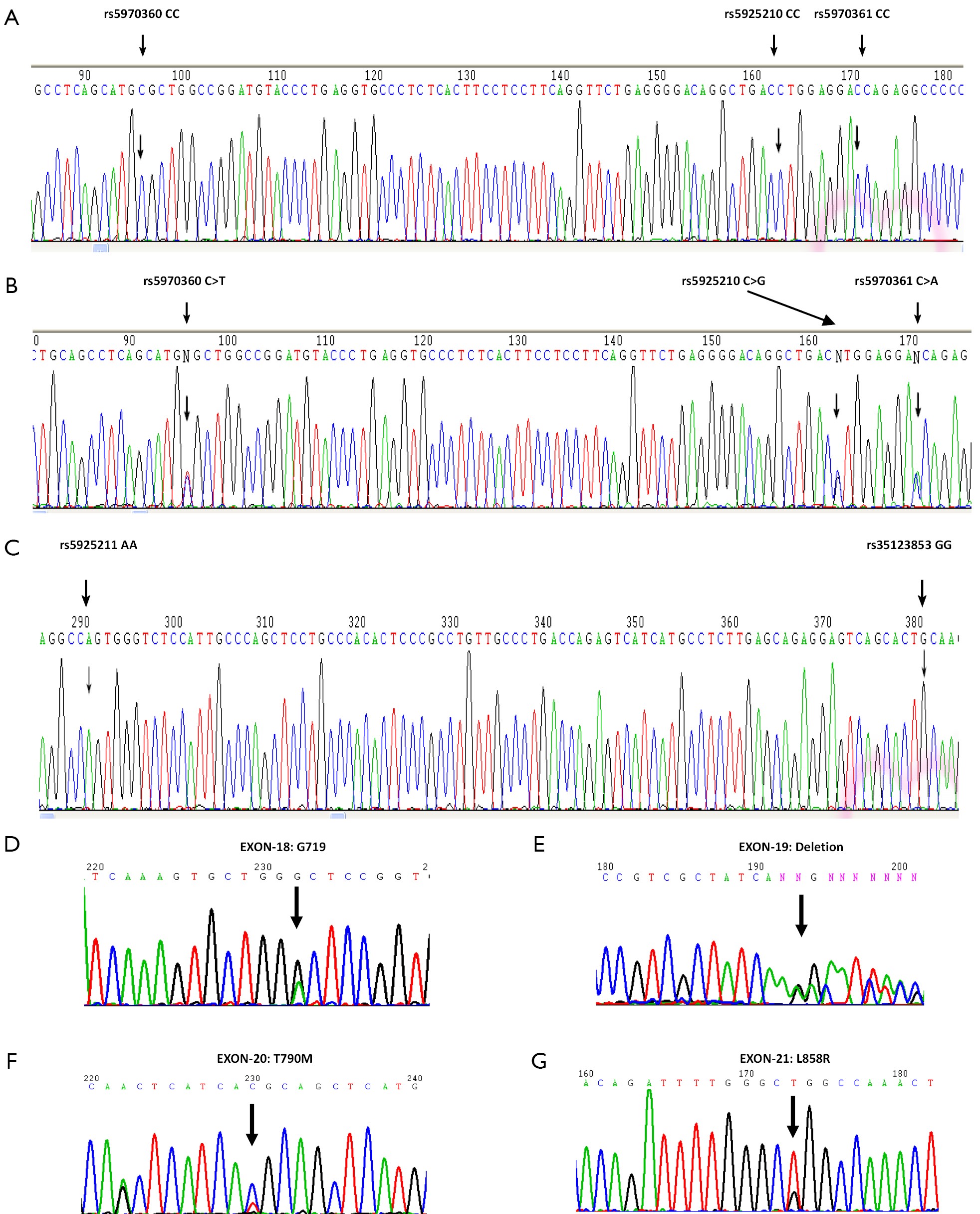
BackgroundAvailable study revealed advanced tumors have a higher expression rate of MAGE-A3 gene which has a lot of single nucleotide polymorphism (SNP) loci with polymorphisms. This study aimed to analyze the allele frequency of SNP loci in MAGE-A3 gene and investigate the relationship between MAGE-A3 gene polymorphisms and clinical factors. MethodsTumor samples of a cohort of 191 NSCLC patients were collected. EGFR mRNA expression were detected by qRT-PCR. SNPs in whole length of MAGE-A3 gene were detected by direct sequencing. Frequencies of the SNPs were correlated to gene expression, mutation status of EGFR and clinical factors. ResultsSequencing analysis confirmed that allele frequencies of genotypes on SNP loci rs5970360, rs5925210, rs5970361, rs5925211 and rs35123853 were CC (0.681)/CT (0.319), CC (0.660)/CG (0.340), CC (0.681)/CA (0.319), AA (0.984)/AT (0.016) and GG (1.000)/GA (0.000), respectively, which were different from the frequencies and genotypes of MAGE-A3 in SNP database. Chi-square tests showed the EGFR mRNA expression level had significant correlation with the genotypes of SNP loci rs5970360 and rs5925210. But all frequencies of each MAGE-A3 SNPs were not found significantly different between EGFR mutant and wild type patients. MAGE-A3 gene polymorphisms had no significant effects on survival of NSCLC patients. ConclusionsChinese patients with NSCLC had different SNP patterns of MAGE-A3 in comparison with those in international SNP database. These MAGE-A3 SNP loci might have not prognostic significance. MAGE-A3 SNP loci rs5970360 and rs5925210 might be predictive for EGFR mRNA expression levels and helpful to the selection of patients for epidermal growth factor receptor (EGFR) targeted immunotherapy.

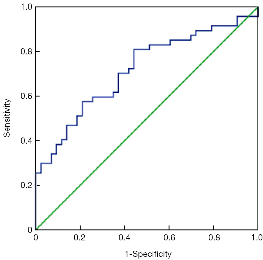
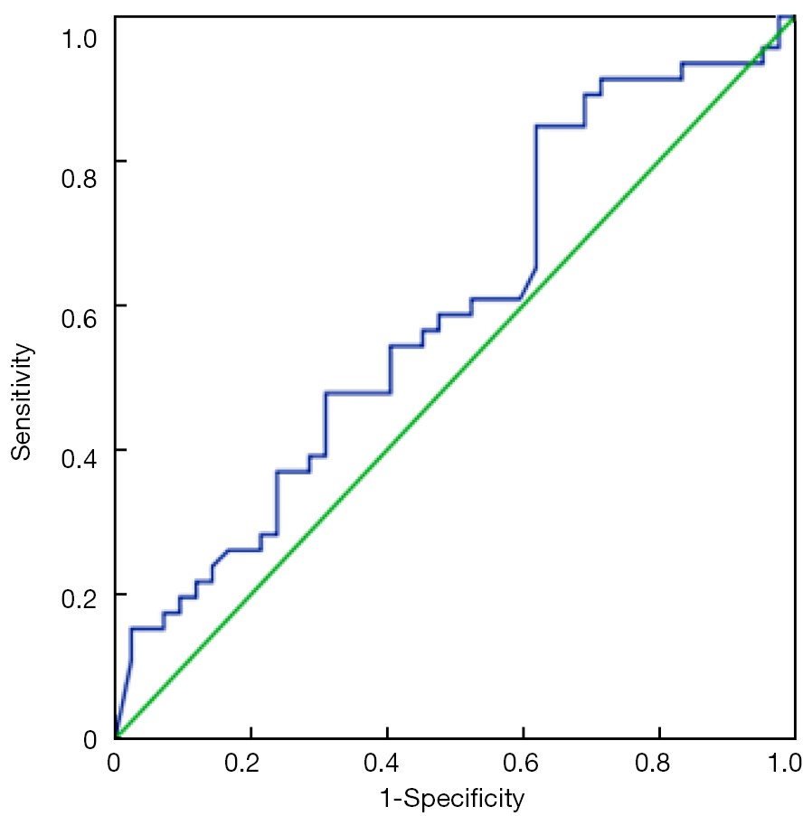
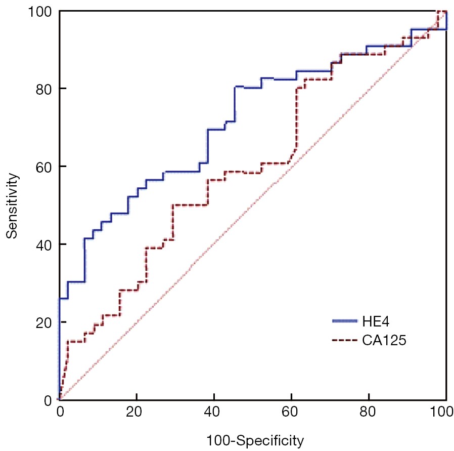
2015, 27(3): 309-317.
doi: 10.3978/j.issn.1000-9604.2015.06.01
Abstract:
ObjectiveHuman epididymis protein 4 (HE4) is a promising biomarker of epithelial ovarian cancer (EOC). But its role in assessing the primary optimal debulking (OD) of EOC remains unknown. The purpose of this study is to elucidate the ability of preoperative HE4 in predicting the primary cytoreductive outcomes in advanced EOC, tubal or peritoneal carcinoma. MethodsWe reviewed the records of 90 patients with advanced ovarian, tubal or peritoneal carcinoma who underwent primary cytoreduction at the Department of Obstetrics and Gynecology of Peking University People’s Hospital between November 2005 and October 2010. Preoperative serum HE4 and CA125 levels were detected with EIA kit. A receiver operating characteristic (ROC) curve was used to determine the most useful HE4 cut-off value. Logistic regression analysis was performed to identify significant preoperative clinical characteristics to predict optimal primary cytoreduction. ResultsOD was achieved in 47.7% (43/48) of patients. The median preoperative HE4 level for patients with OD vs. suboptimal debulking was 423 and 820 pmol/L, respectively (P<0.001). The areas under the ROC curve for HE4 and CA125 were 0.716 and 0.599, respectively (P=0.080). The most useful HE4 cut-off value was 473 pmol/L. Suboptimal cytoreduction was obtained in 66.7% (38/57) of cases with HE4 ≥473 pmol/L compared with only 27.3% (9/33) of cases with HE4 <473 pmol/L. At this threshold, the sensitivity, specificity, positive predictive value (PPV) and negative predictive value (NPV) for diagnosing suboptimal debulking were 81%, 56%, 67%, and 73%, respectively. Logistic regression analysis showed that the patients with HE4 ≥473 pmol/L were less likely to achieve OD (odds ratio =5.044, P=0.002). ConclusionsPreoperative serum HE4 may be helpful to predict whether optimal cytoreductive surgery could be obtained or whether extended cytoreduction would be needed by an interdisciplinary team.