2015 Vol.27(5)
Display Mode: |
2015, 27(5): 437-449.
doi: 10.3978/j.issn.1000-9604.2015.04.08
 Abstract
 FullText HTML
 PDF 842KB
Abstract:
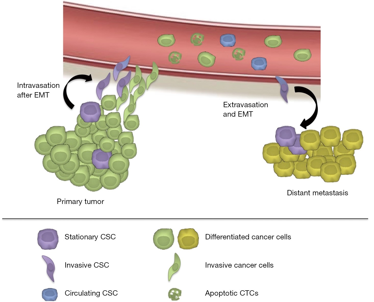
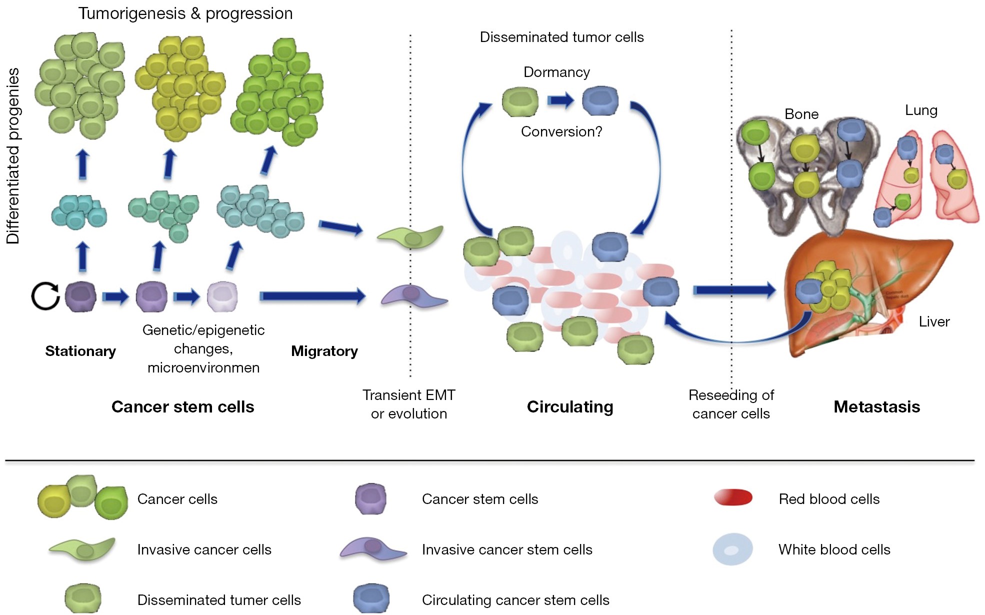
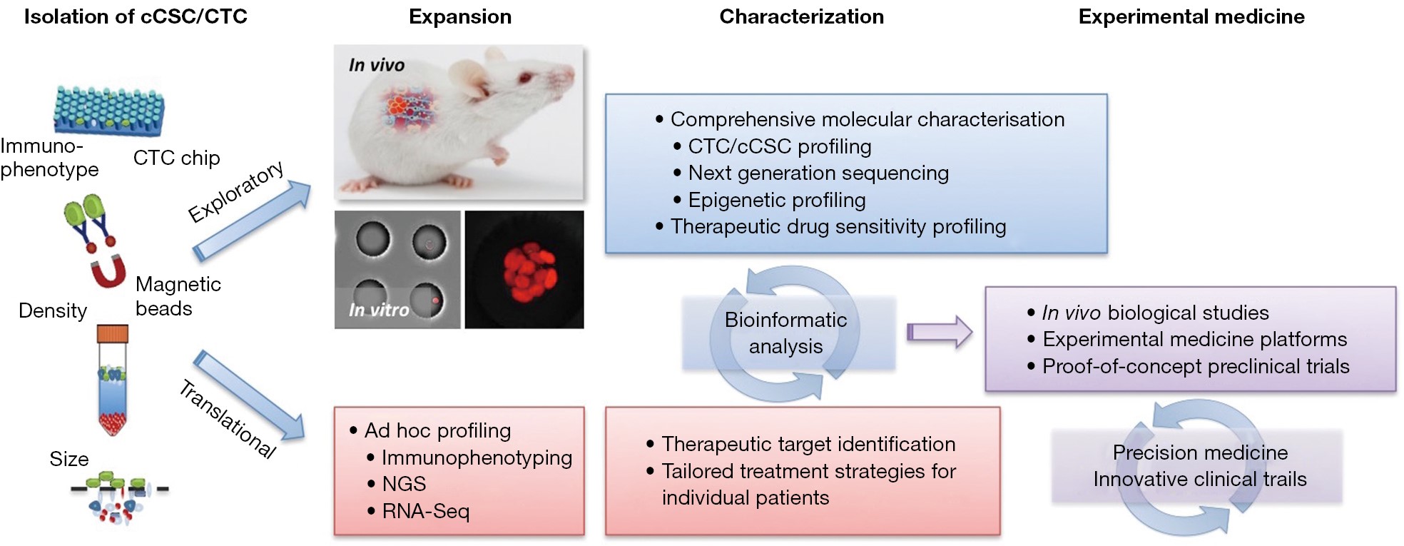
It has been demonstrated that even localized tumors without clinically apparent metastasis give rise to circulating tumor cells (CTCs). A growing number of technically diverse platforms are being developed for detecting/isolating CTCs in the circulating blood. Despite the technical challenges of isolating rare CTCs from blood, recent studies have already shown the predictive value of CTCs enumeration. Thus, it is becoming increasingly accepted that CTC numbers are linked to patients’ outcome and may also be used to monitor treatment response and disease relapse, respectively. Further CTCs provide a non-invasive source for tumor material, ‘liquid biopsy’, which is particularly important for patients, where no biopsy material can be obtained or where serial biopsies of the tumor, e.g., following treatment, are practically impossible. On the other hand the molecular and biological characterization of CTCs has still remained at a rather experimental stage. Future studies are necessary to define CTC heterogeneity to establish the crucial role of circulating cancer stem cells for driving metastasis, which represent a distinct subpopulation of CTCs that bear metastasis-initiating capabilities based on their stemness properties and invasiveness and thus are critical for the patients’ clinical outcome. As compared to non-tumorigenic/metastatic bulk CTCs, circulating cancer stem cells may not only be capable of evading from the primary tumor, but also escape from immune surveillance, survive in the circulating blood and subsequently form metastases in distant organs. Thus, circulating cancer stem cells represent a subset of exclusively tumorigenic cancer stem cells characterized by their invasive characteristics and are potential therapeutic targets for preventing disease progression. To date, only a few original reports and reviews have been published focusing on circulating cancer stem cells. This review discusses the potential importance of isolating and characterizing these circulating cancer stem cells, but also highlights current technological limitations.
It has been demonstrated that even localized tumors without clinically apparent metastasis give rise to circulating tumor cells (CTCs). A growing number of technically diverse platforms are being developed for detecting/isolating CTCs in the circulating blood. Despite the technical challenges of isolating rare CTCs from blood, recent studies have already shown the predictive value of CTCs enumeration. Thus, it is becoming increasingly accepted that CTC numbers are linked to patients’ outcome and may also be used to monitor treatment response and disease relapse, respectively. Further CTCs provide a non-invasive source for tumor material, ‘liquid biopsy’, which is particularly important for patients, where no biopsy material can be obtained or where serial biopsies of the tumor, e.g., following treatment, are practically impossible. On the other hand the molecular and biological characterization of CTCs has still remained at a rather experimental stage. Future studies are necessary to define CTC heterogeneity to establish the crucial role of circulating cancer stem cells for driving metastasis, which represent a distinct subpopulation of CTCs that bear metastasis-initiating capabilities based on their stemness properties and invasiveness and thus are critical for the patients’ clinical outcome. As compared to non-tumorigenic/metastatic bulk CTCs, circulating cancer stem cells may not only be capable of evading from the primary tumor, but also escape from immune surveillance, survive in the circulating blood and subsequently form metastases in distant organs. Thus, circulating cancer stem cells represent a subset of exclusively tumorigenic cancer stem cells characterized by their invasive characteristics and are potential therapeutic targets for preventing disease progression. To date, only a few original reports and reviews have been published focusing on circulating cancer stem cells. This review discusses the potential importance of isolating and characterizing these circulating cancer stem cells, but also highlights current technological limitations.
2015, 27(5): 450-460.
doi: 10.3978/j.issn.1000-9604.2015.04.10
Abstract:
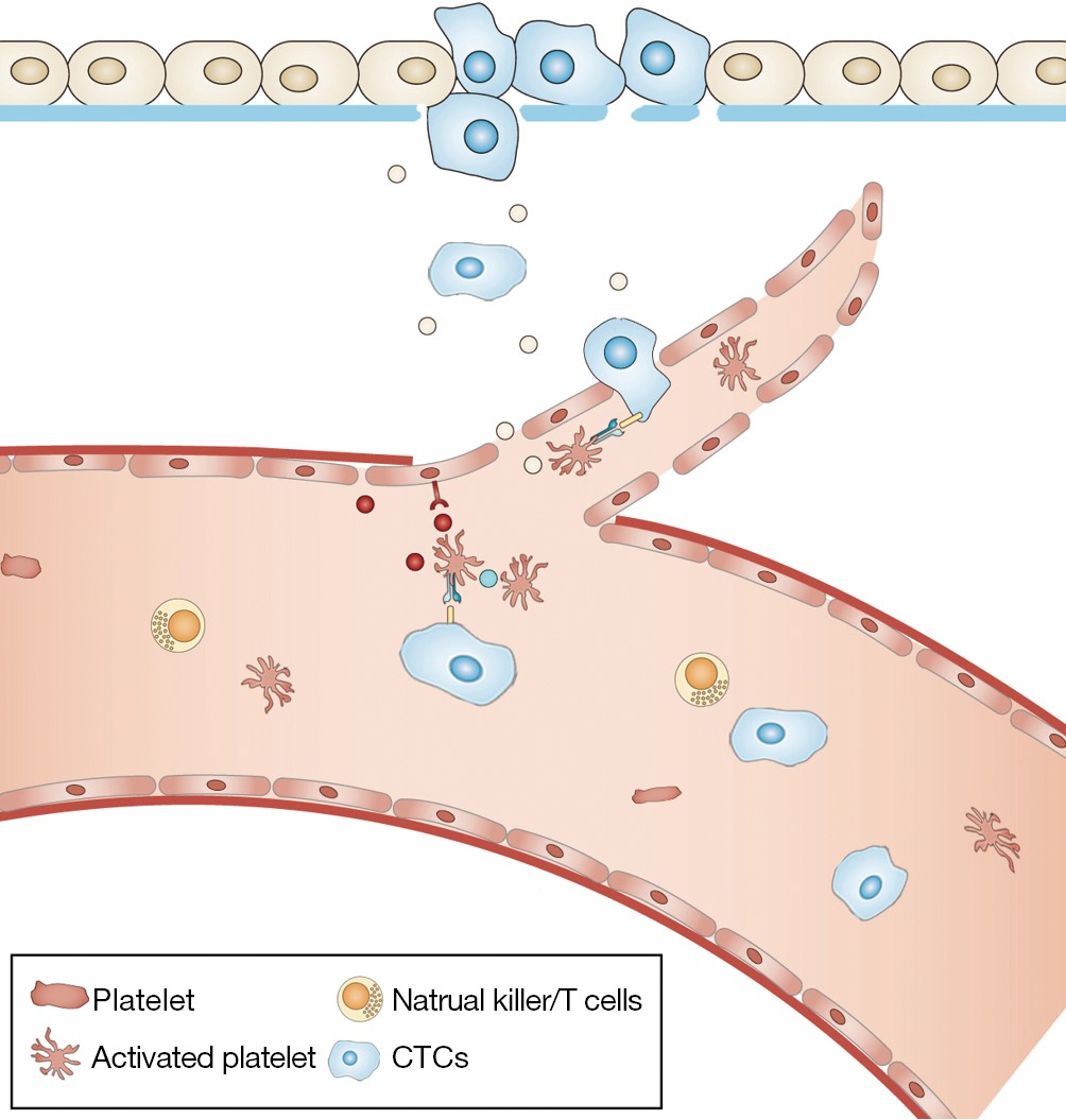
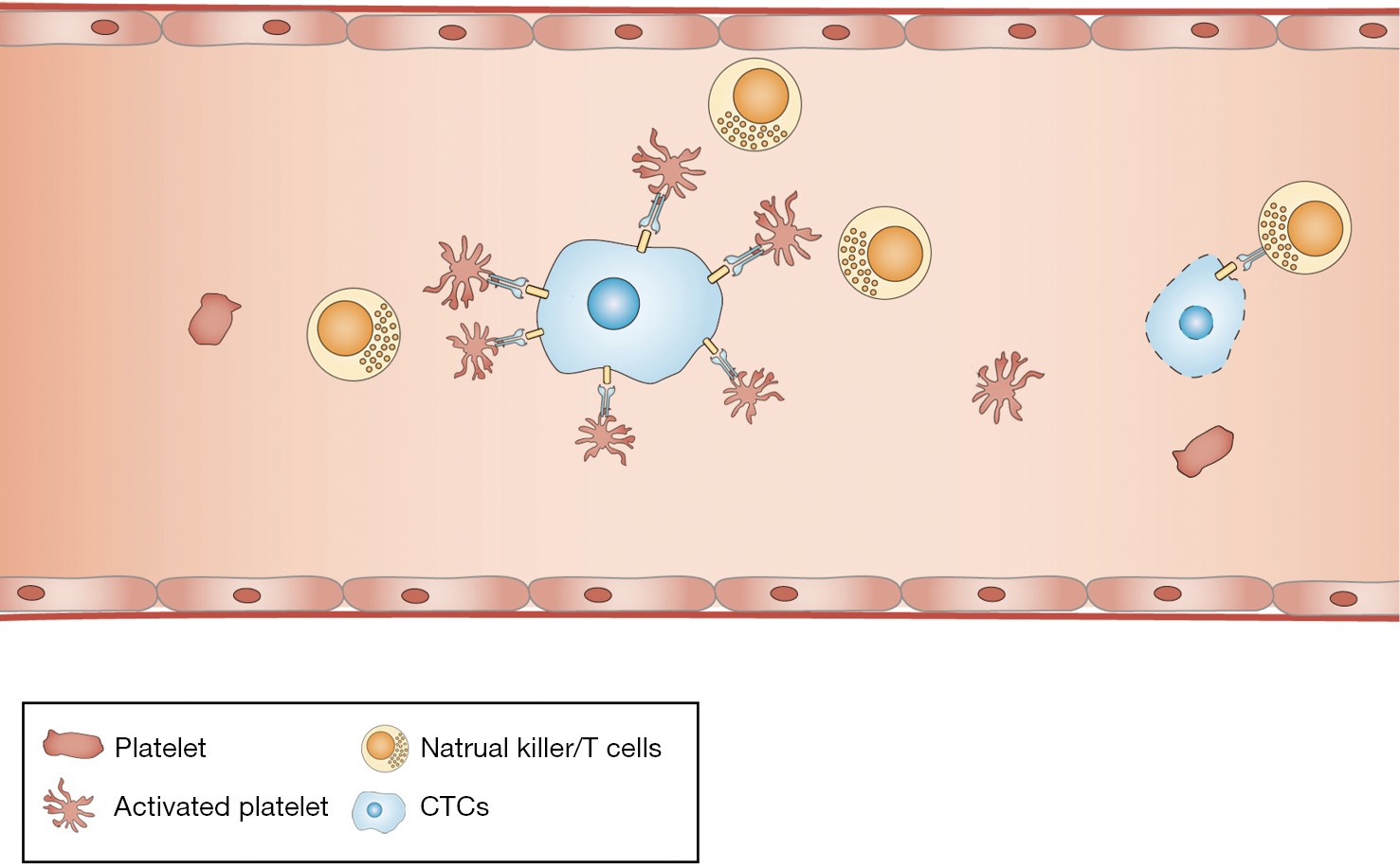
Metastasis is the main cause of cancer-associated mortality. During this complicated process, some cancer cells, also called circulating tumor cells (CTCs), detach from primary sites, enter bloodstream and extravasate at metastatic site. Thrombocytosis is frequently observed in patients with metastatic cancers suggesting the important role of platelets in metastasis. Therefore this review focuses on how platelets facilitate the generation of CTCs, protect them from various host attacks, such as immune assaults, apoptosis and shear stress, and regulate CTCs intravasation/extravasation. Platelet-derived cytokines and receptors are involved in this cascade. Identification the mechanisms underlie platelet-CTCs interactions could lead to the development of new platelet-targeted therapeutic strategy to reduce metastasis.
Metastasis is the main cause of cancer-associated mortality. During this complicated process, some cancer cells, also called circulating tumor cells (CTCs), detach from primary sites, enter bloodstream and extravasate at metastatic site. Thrombocytosis is frequently observed in patients with metastatic cancers suggesting the important role of platelets in metastasis. Therefore this review focuses on how platelets facilitate the generation of CTCs, protect them from various host attacks, such as immune assaults, apoptosis and shear stress, and regulate CTCs intravasation/extravasation. Platelet-derived cytokines and receptors are involved in this cascade. Identification the mechanisms underlie platelet-CTCs interactions could lead to the development of new platelet-targeted therapeutic strategy to reduce metastasis.

2015, 27(5): 461-470.
doi: 10.3978/j.issn.1000-9604.2015.06.02
Abstract:
Circulating tumor cells (CTCs) represent a submicroscopic fraction detached from a primary tumor and in transit to a secondary site. The prognostic significance of CTCs in metastatic cancer patients was demonstrated for the first time more than ten years ago. To date, it seems clear enough that CTCs are highly heterogeneous and dynamically change their shape. Thus, the inadequacy of epithelial cell adhesion molecule (EpCAM) as universal marker for CTCs detection seems unquestionable and alternative methods able to recognize a broader spectrum of phenotypes are definitely needed. In this review the pleiotropic functions of EpCAM are discussed in detail and the role of the molecule in the biology of CTCs is critically dissected.
Circulating tumor cells (CTCs) represent a submicroscopic fraction detached from a primary tumor and in transit to a secondary site. The prognostic significance of CTCs in metastatic cancer patients was demonstrated for the first time more than ten years ago. To date, it seems clear enough that CTCs are highly heterogeneous and dynamically change their shape. Thus, the inadequacy of epithelial cell adhesion molecule (EpCAM) as universal marker for CTCs detection seems unquestionable and alternative methods able to recognize a broader spectrum of phenotypes are definitely needed. In this review the pleiotropic functions of EpCAM are discussed in detail and the role of the molecule in the biology of CTCs is critically dissected.
2015, 27(5): 471-478.
doi: 10.3978/j.issn.1000-9604.2015.09.02
Abstract:
The proved association between the circulating tumor cell (CTC) levels and the patients’ survival parameters has been growing interest to investigate the molecular profile of these neoplastic cells among which hide out precursors capable of initiating a new distant metastatic lesion. The full characterization of the tumor cells in peripheral blood of cancer patients is expected to be of help for understanding and (prospectively) for counteracting the metastatic process. The major hitch that is hampering the successful gaining of this result is the lack of a consensus onto standard operating procedures (SOPs) for performing what we generally define as the “liquid biopsy”. Here we review the more recent acquisitions in the analysis of CTCs and tumor related nucleic acids, looking to the main open questions that are hampering their definitive employ in the routine clinical practice.
The proved association between the circulating tumor cell (CTC) levels and the patients’ survival parameters has been growing interest to investigate the molecular profile of these neoplastic cells among which hide out precursors capable of initiating a new distant metastatic lesion. The full characterization of the tumor cells in peripheral blood of cancer patients is expected to be of help for understanding and (prospectively) for counteracting the metastatic process. The major hitch that is hampering the successful gaining of this result is the lack of a consensus onto standard operating procedures (SOPs) for performing what we generally define as the “liquid biopsy”. Here we review the more recent acquisitions in the analysis of CTCs and tumor related nucleic acids, looking to the main open questions that are hampering their definitive employ in the routine clinical practice.
2015, 27(5): 479-487.
doi: 10.3978/j.issn.1000-9604.2015.09.01
Abstract:
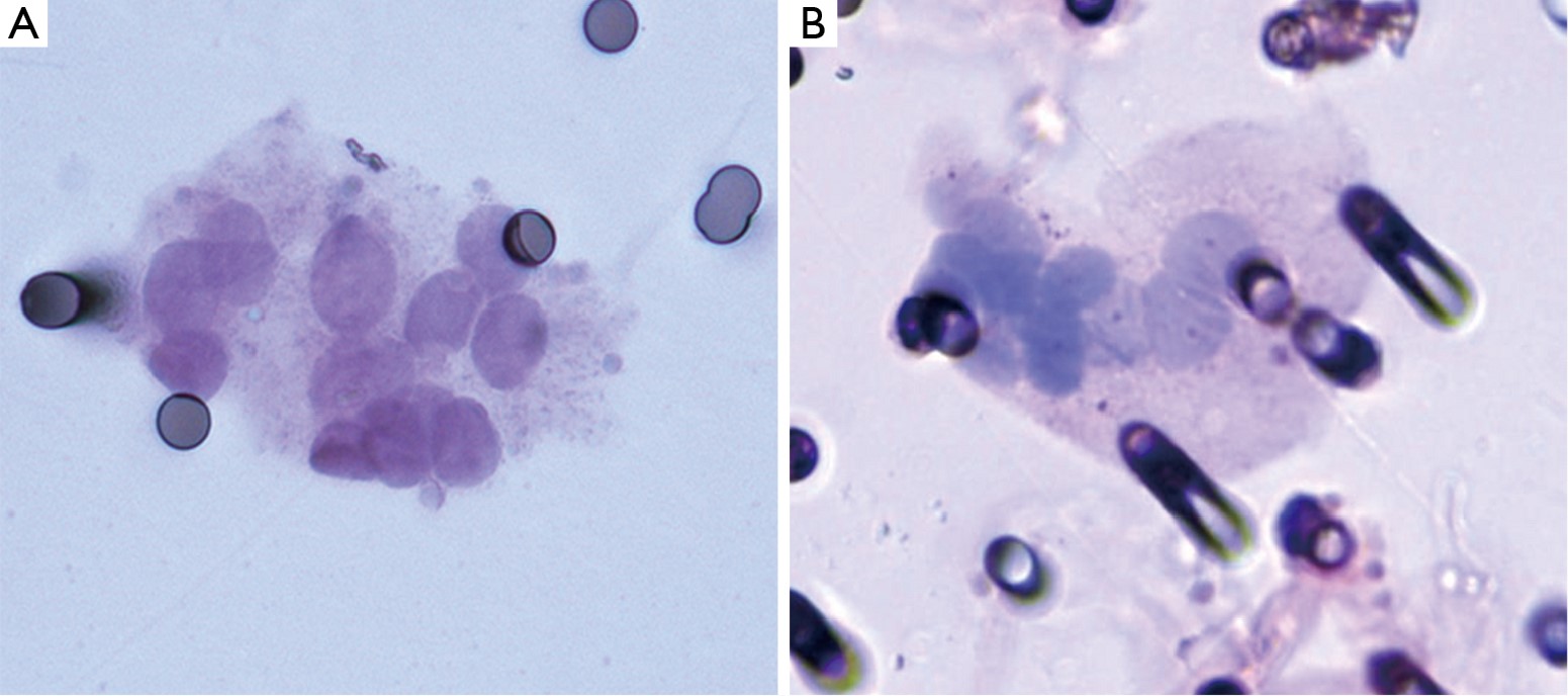
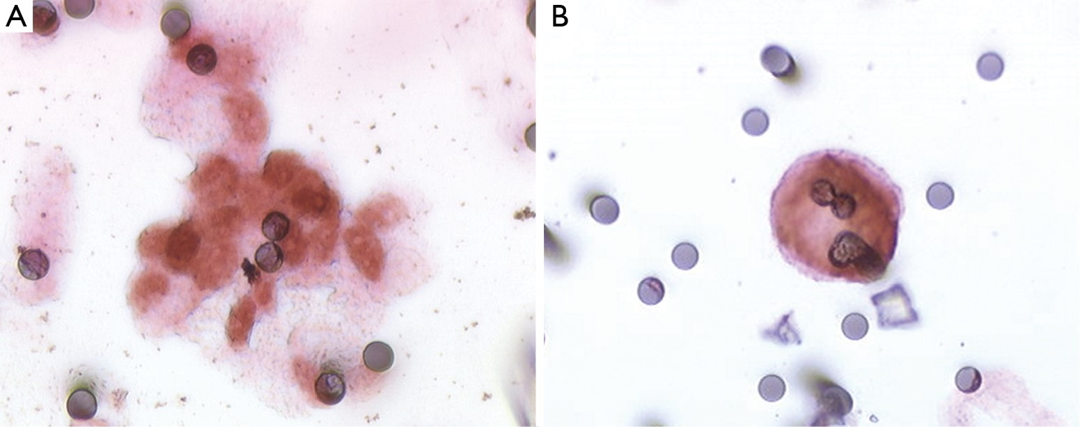
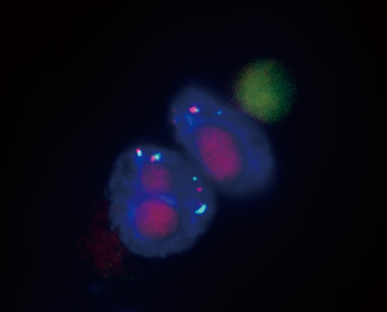
Circulating tumor cells (CTCs) arise from primary or secondary tumors and enter the bloodstream by active or passive intravasation. Given the low number of CTCs, enrichment is necessary for detection. Filtration methods are based on selection of CTCs by size using a filter with 6.5 to 8 µm pores. After coloration, collected CTCs are evaluated according to morphological criteria. Immunophenotyping and fluorescence in situ hybridization techniques may be used. Selected CTCs can also be cultivated in vitro to provide more material. Analysis of genomic mutations is difficult because it requires adapted techniques due to limited DNA materials. Filtration-selected CTCs have shown prognostic value in many studies but multicentric validating trials are mandatory to strengthen this assessment. Other clinical applications are promising such as follow-up, therapy response prediction and diagnosis. Microfluidic emerging systems could optimize filtration-selected CTCs by increasing selection accuracy.
Circulating tumor cells (CTCs) arise from primary or secondary tumors and enter the bloodstream by active or passive intravasation. Given the low number of CTCs, enrichment is necessary for detection. Filtration methods are based on selection of CTCs by size using a filter with 6.5 to 8 µm pores. After coloration, collected CTCs are evaluated according to morphological criteria. Immunophenotyping and fluorescence in situ hybridization techniques may be used. Selected CTCs can also be cultivated in vitro to provide more material. Analysis of genomic mutations is difficult because it requires adapted techniques due to limited DNA materials. Filtration-selected CTCs have shown prognostic value in many studies but multicentric validating trials are mandatory to strengthen this assessment. Other clinical applications are promising such as follow-up, therapy response prediction and diagnosis. Microfluidic emerging systems could optimize filtration-selected CTCs by increasing selection accuracy.
2015, 27(5): 488-490.
doi: 10.3978/j.issn.1000-9604.2015.10.01
Abstract:
The clinical utility of liquid biopsy in cancer treatment will increase as circulating tumor cells (CTCs) analysis move from the enumeration to the real-time measurement of tumor characteristics. Intratumor heterogeneity is becoming increasingly recognized as a major drawback to the shift to personalized medicine. Spatial and temporal heterogeneity might be reflected by the serial assessment of CTCs. Indeed, the developing technologies for CTCs analysis now allow digital genomic and next-generation sequencing approaches, able to differentiate molecular subtypes of the disease and to monitor genetic variation over time. The liquid biopsy of cancer might offer a real-time assessment of tumor biology, providing the opportunity to serially evaluate patients most likely to benefit from targeted drugs based on a dynamic characterization of the disease at the molecular level. Although hurdles remain before liquid biopsy is seen in routine clinical practice, the information derived from CTCs may facilitate the real-time identification of actionable mutations in cancer leading the way toward personalized medicine.
The clinical utility of liquid biopsy in cancer treatment will increase as circulating tumor cells (CTCs) analysis move from the enumeration to the real-time measurement of tumor characteristics. Intratumor heterogeneity is becoming increasingly recognized as a major drawback to the shift to personalized medicine. Spatial and temporal heterogeneity might be reflected by the serial assessment of CTCs. Indeed, the developing technologies for CTCs analysis now allow digital genomic and next-generation sequencing approaches, able to differentiate molecular subtypes of the disease and to monitor genetic variation over time. The liquid biopsy of cancer might offer a real-time assessment of tumor biology, providing the opportunity to serially evaluate patients most likely to benefit from targeted drugs based on a dynamic characterization of the disease at the molecular level. Although hurdles remain before liquid biopsy is seen in routine clinical practice, the information derived from CTCs may facilitate the real-time identification of actionable mutations in cancer leading the way toward personalized medicine.
2015, 27(5): 491-496.
doi: 10.3978/j.issn.1000-9604.2015.04.09
Abstract:
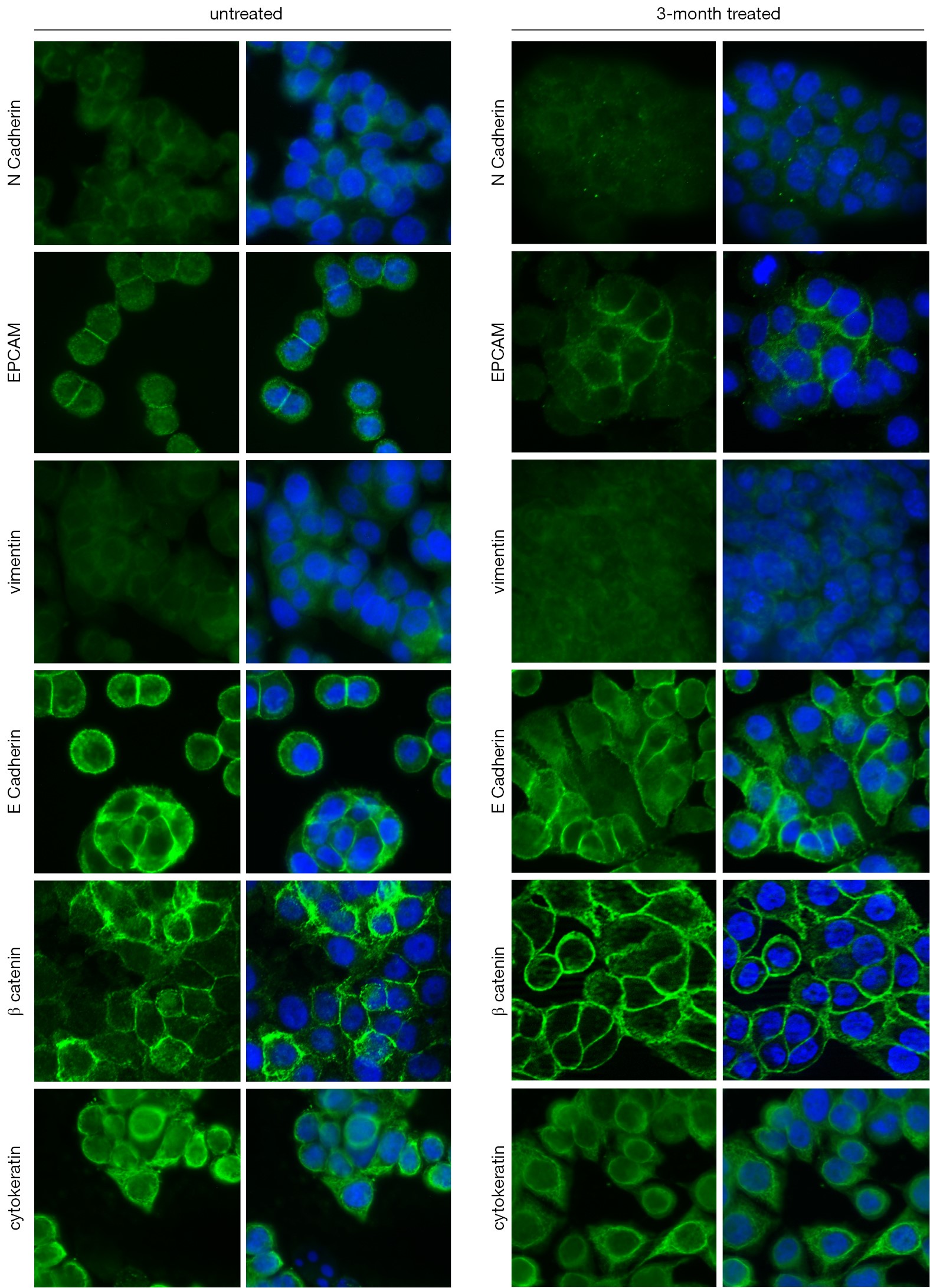
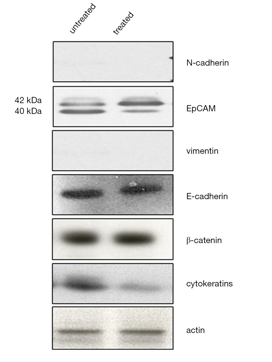
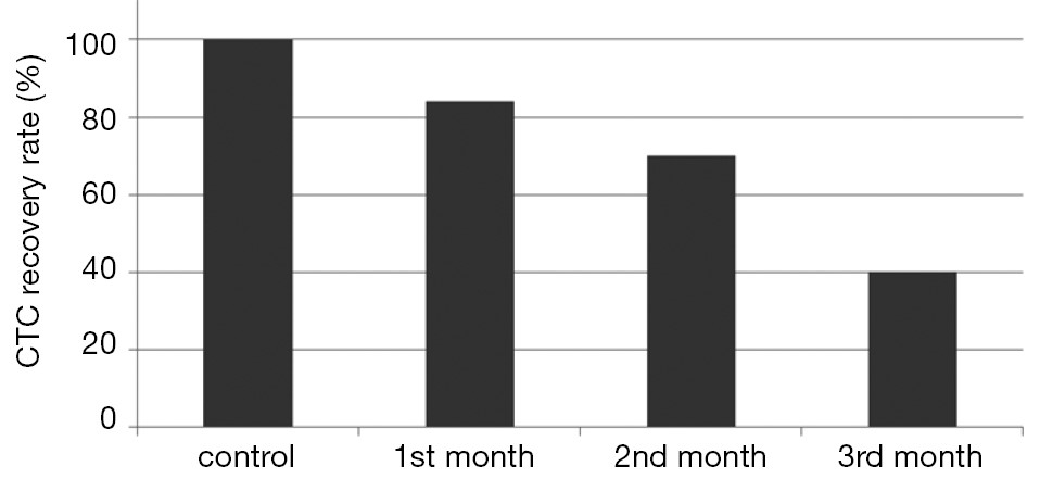
BackgroundCirculating tumor cells (CTCs) are often undetected through the immunomagnetic epithelial cell adhesion molecule (EpCAM)-based CellSearch® System in breast and colorectal cancer (CRC) patients treated with bevacizumab (BEV), where low CTC numbers have been reported even in patients with evidence of progression of disease. To date, the reasons for this discrepancy have not been clarified. This study was carried out to investigate the molecular and phenotypic changes in CRC cells after chronic exposure to BEV in vitro. MethodsThe human CRC cell line WiDr was exposed to a clinically relevant dose of BEV for 3 months in vitro. The expression of epithelial and mesenchymal markers and EpCAM isoforms was determined by western blotting and immunofluorescence. To evaluate the impact of EpCAM variant isoforms expression on CTC enumeration by CellSearch®, untreated and treated colon cancer cells were spiked into 7.5 mL of blood from a healthy donor and enumerated by CellSearch®. ResultsChronic exposure of CRC cell line to BEV induced decreased expression of EpCAM 40 kDa isoform and increased expression EpCAM 42 kDa isoform, together with a decreased expression of cytokeratins (CK), while no evidence of epithelial to mesenchymal transition (EMT) in treated cells was observed. The recovery rate of cells through CellSearch® was gradually reduced in course of treatment with BEV, being 84%, 70% and 40% at 1, 2 and 3 months, respectively. ConclusionsWe hypothesize that BEV may prevent CellSearch® from capturing CTCs through altering EpCAM isoforms.
2015, 27(5): 497-508.
doi: 10.3978/j.issn.1000-9604.2015.10.05
Abstract:
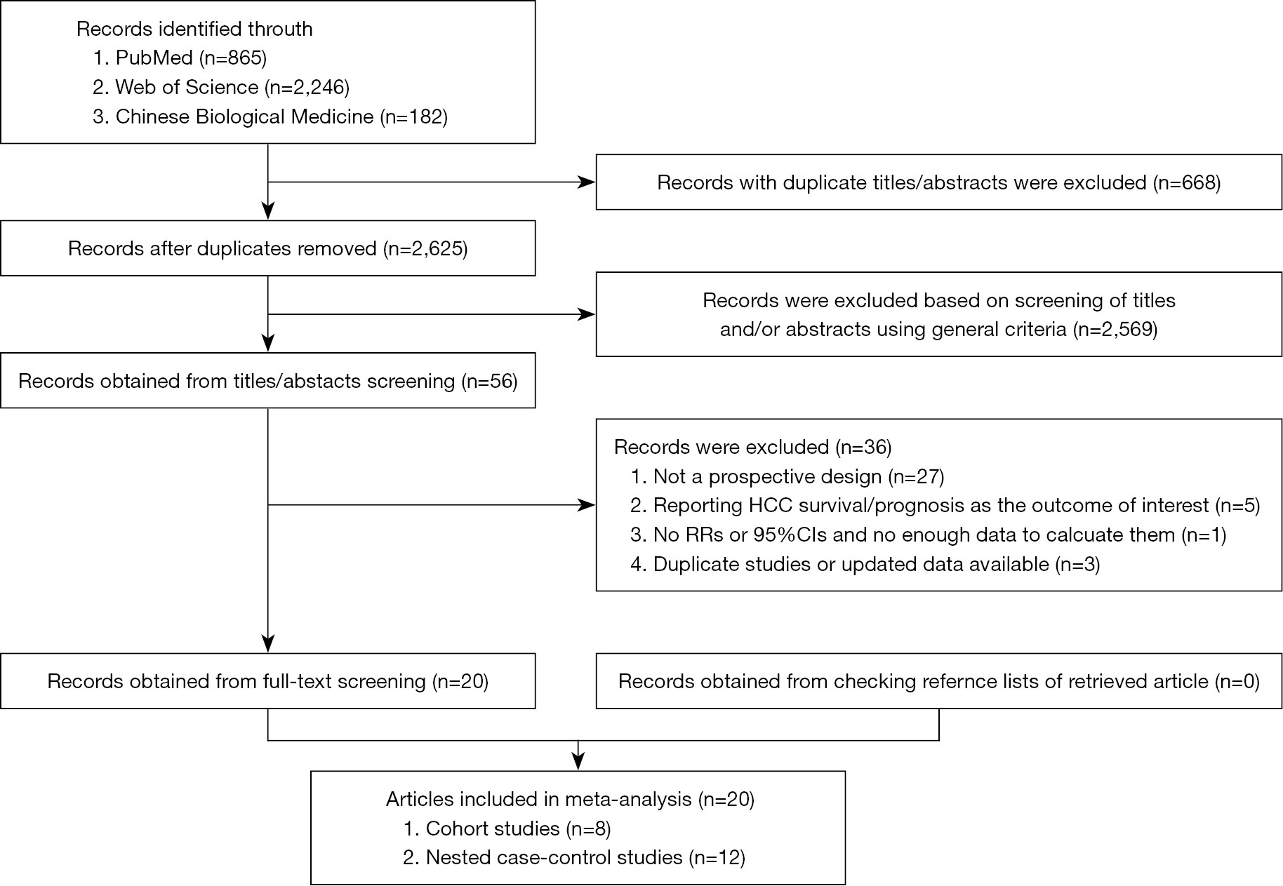
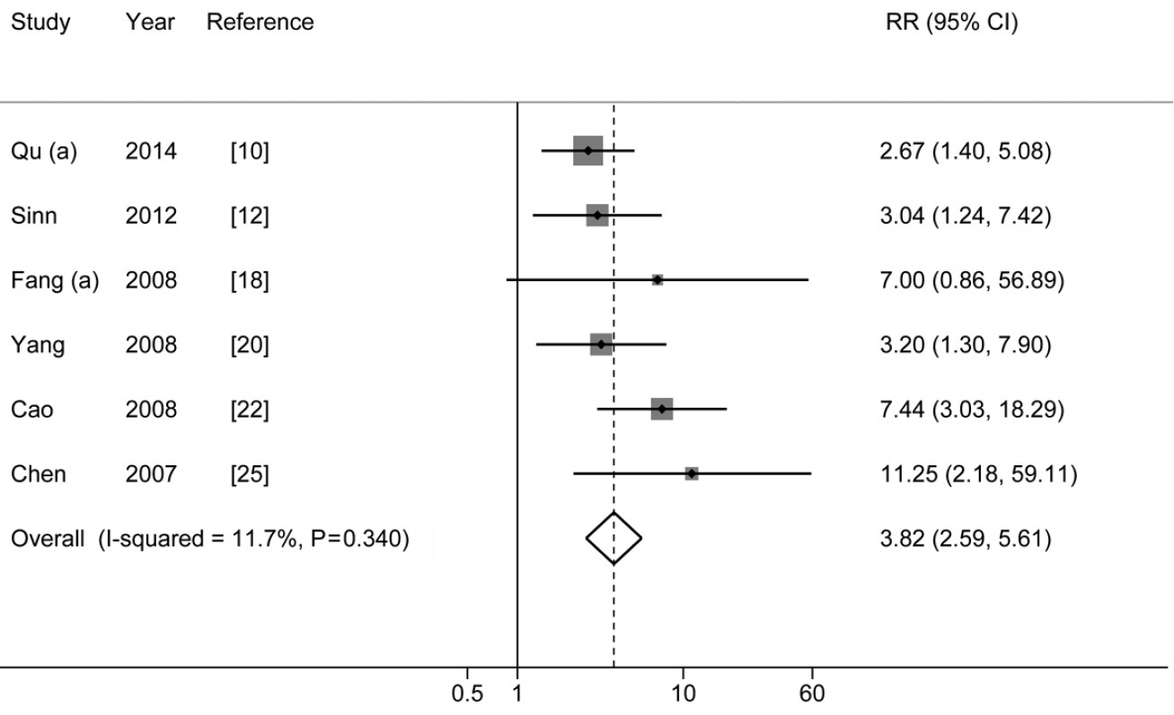
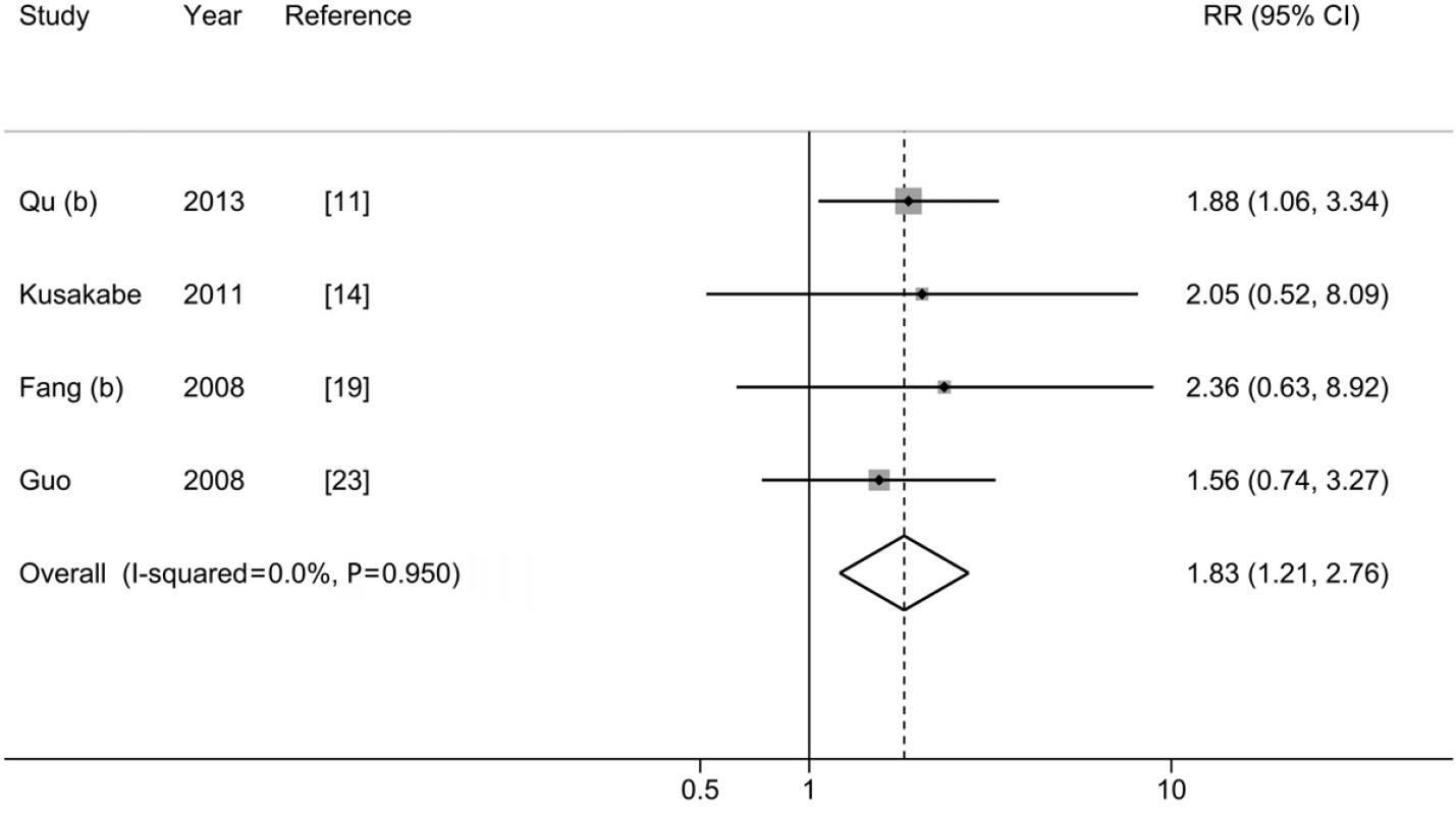
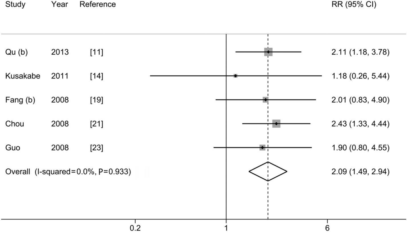
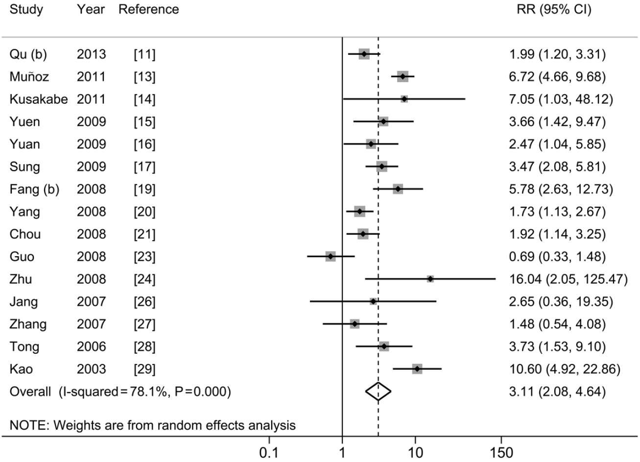
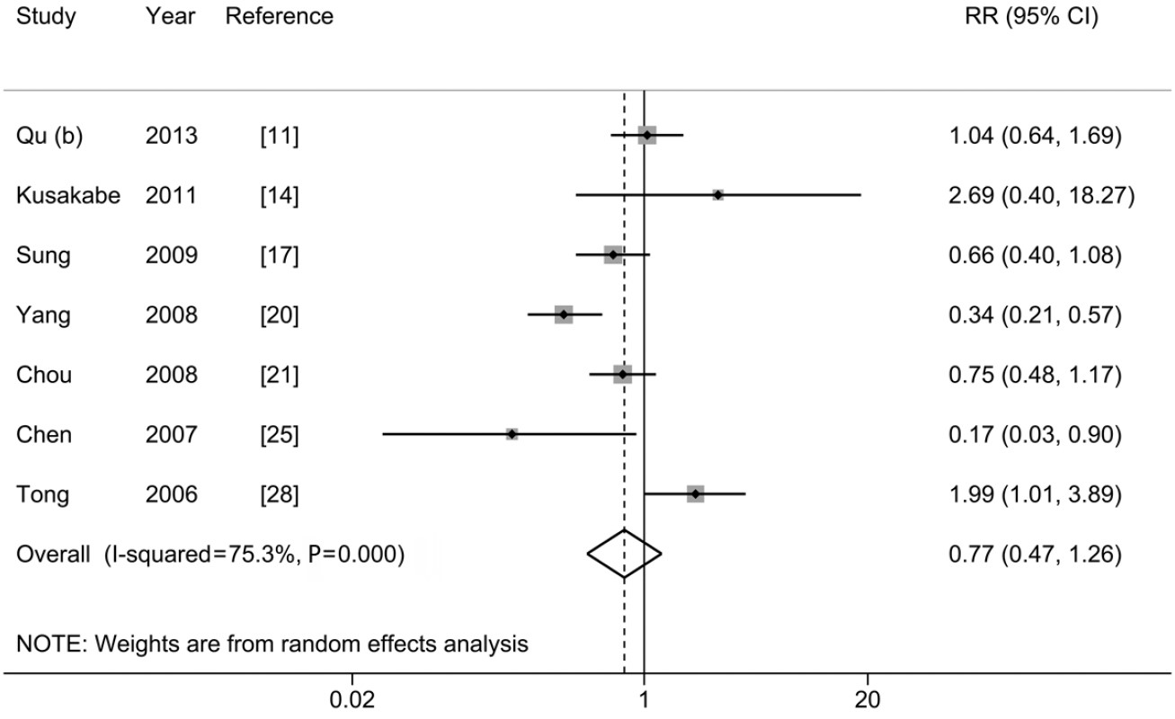
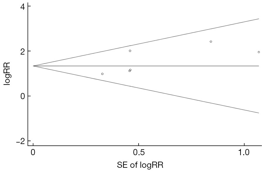
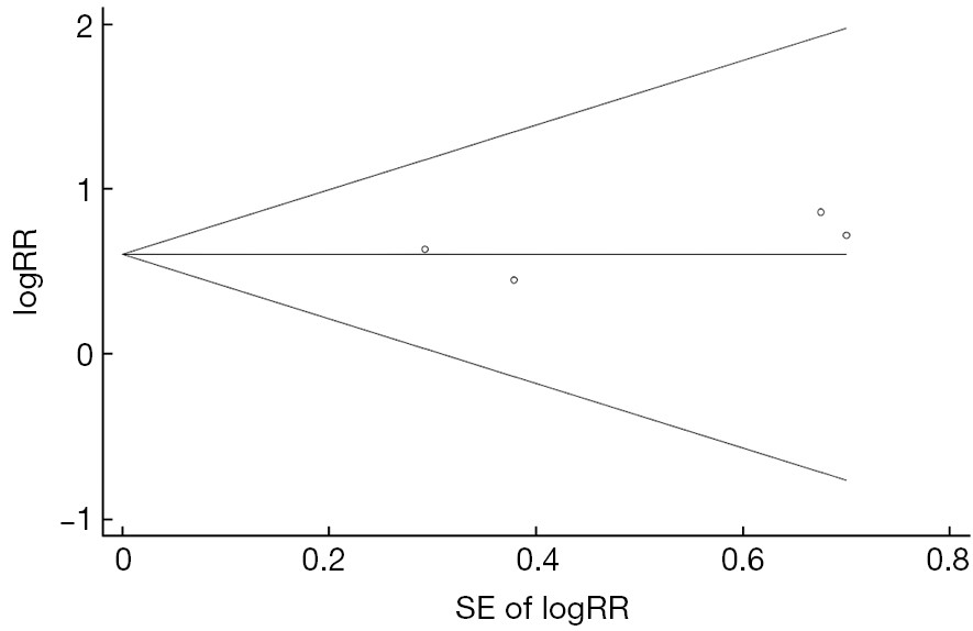
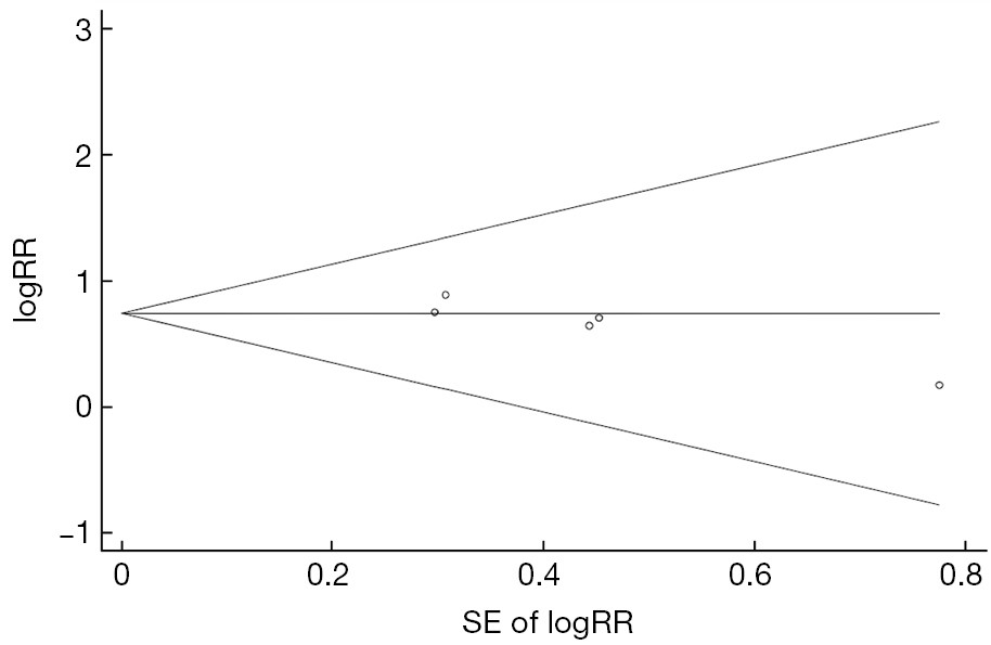
BackgroundThe temporal relationship between hepatitis B virus (HBV) mutations and hepatocellular carcinoma (HCC) remains unclear. MethodsWe conducted a meta-analysis including cohort and nested case-control studies to prospectively examine the HCC risk associated with common variants of HBV in the PreS, Enhancer II, basal core promoter (BCP) and precore regions. Pertinent studies were identified by searching PubMed, Web of Science and the Chinese Biological Medicine databases through to November 2014. Study-specific risk estimates were combined using fixed or random effects models depending on whether significant heterogeneity was detected. ResultsTwenty prospective studies were identified, which included 8 cohort and 12 nested case-control studies. There was an increased risk of HCC associated with any PreS mutations with a pooled relative risk (RR) of 3.82 [95% confidence interval (CI): 2.59-5.61]. The pooled-RR for PreS deletion was 3.98 (95% CI: 2.28-6.95), which was higher than that of PreS2 start codon mutation (pooled-RR=2.63, 95% CI: 1.30-5.34). C1653T in Enhancer II was significantly associated with HCC risk (pooled-RR=1.83; 95% CI: 1.21-2.76). For mutations in BCP, statistically significant pooled-RRs of HCC were obtained for T1753V (pooled-RR=2.09; 95% CI: 1.49-2.94) and A1762T/G1764A double mutations (pooled-RR=3.11; 95% CI: 2.08-4.64). No statistically significant association with HCC risk was observed for G1896A in the precore region (pooled-RR=0.77; 95% CI: 0.47-1.26). ConclusionsThis study demonstrated that PreS mutations, C1653T, T1753V, and A1762T/G1764A, were associated with an increased risk of HCC. Clinical practices concerning the HCC risk prediction and diagnosis may wish to focus on patients with these mutations.
2015, 27(5): 509-515.
doi: 10.3978/j.issn.1000-9604.2015.06.03
Abstract:
BackgroundThe purpose of this study was to analyze the effects of all clinical characteristics on the overall survival time, in order to provide a basis for determining the prognostic factor of patients with pancreatic cancer. MethodsA total of 103 pancreatic cancer patients were admitted to the Department of Radiotherapy and Chemotherapy of the Ruijin Hospital, Shanghai Jiaotong University School of Medicine, between January 2002 and December 2012. There were 68 men and 35 women; the median age was 62 years. Diagnoses of pancreatic cancer in all patients were confirmed by histopathology, cytology, or clinical diagnosis. The Kaplan-Meier method was performed to calculate the overall survival rate. The log-rank method was used to examine the univariate analysis. The Cox regression model was performed for multivariate analysis. ResultsThe median survival time was 293 days, the 1-, 2-, and 3-year survival rates were 27.18%, 5.83%, and 1.94%, respectively. Cox regression analysis revealed that age (P=0.015), Karnofsky performance status (PS) (P=0.002), surgical types (P<0.001), and platelet counts (P<0.001) were independent prognostic factors affecting the overall survival of patients with pancreatic cancer. ConclusionsPancreatic cancer had a poor prognosis, the general physical condition, age, the availability of radical surgery, and platelet counts were factors influencing the overall survival of patients with pancreatic cancer.
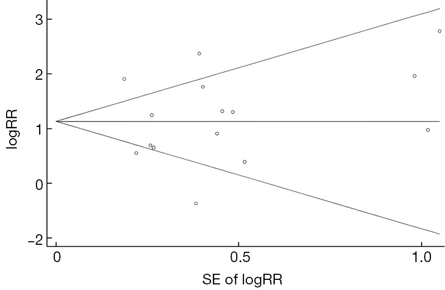
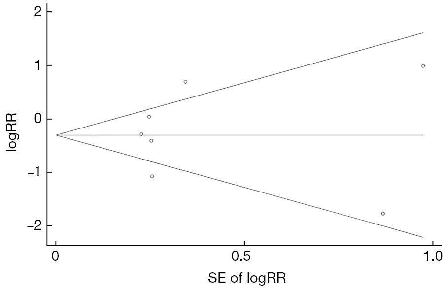
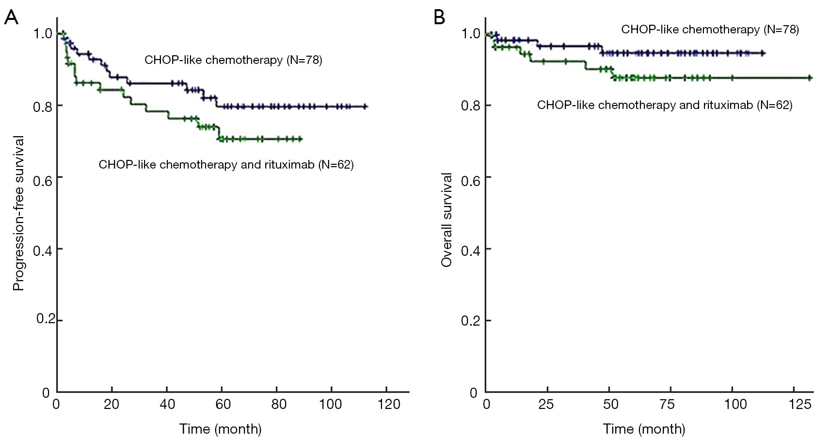
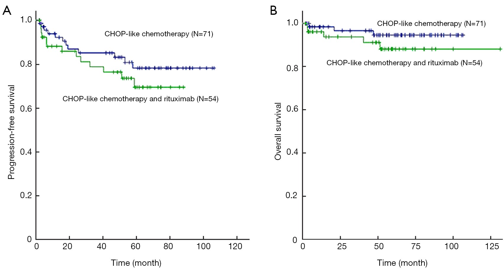
2015, 27(5): 516-523.
doi: 10.3978/j.issn.1000-9604.2015.10.04
Abstract:
BackgroundThe role of rituximab in combination with CHOP regimen in patients with stage I diffuse large B-cell lymphoma (DLBCL) remains to be defined. We aimed to compare CHOP plus rituximab (R-CHOP) with CHOP alone and determine the value of radiotherapy in these patients. MethodsBetween 2003 and 2009, 140 untreated patients with stage I DLBCL were retrospectively analyzed in this study. ResultsSeventy-eight patients were treated in R-CHOP group and 62 in CHOP group. Ninety-one patients received additional radiotherapy at the end of chemotherapy. The different treatment groups were well-balanced with respect to baseline characteristics. Complete response (CR) rate was 77% both in R-CHOP and CHOP groups (P=0.945). After a median follow-up period of 56 months, patients received R-CHOP regimen had similar 5-year progression-free survival (PFS) (76% vs. 85%; log-rank P=0.215) and 5-year overall survival (OS) (90% vs. 96%; log-rank P=0.175) compared with those with CHOP alone. Patients with radiotherapy had significantly increased 5-year PFS compared with those who had chemotherapy alone (86% vs. 71%; log-rank P=0.005). At multivariate analysis, patients who had CR (P=0.008) and received radiotherapy (P=0.003) were significantly associated with superior PFS. ConclusionsCHOP alone could be as effective as R-CHOP regimen and additional radiotherapy would be necessary for stage I or stage I non-bulky DLBCL patients.

2015, 27(5): 524-532.
doi: 10.3978/j.issn.1000-9604.2015.10.03
Abstract:
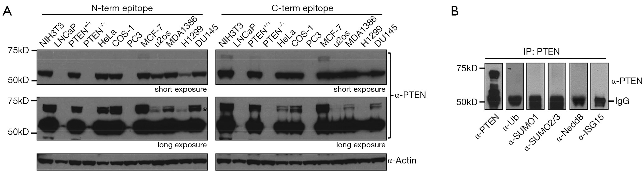
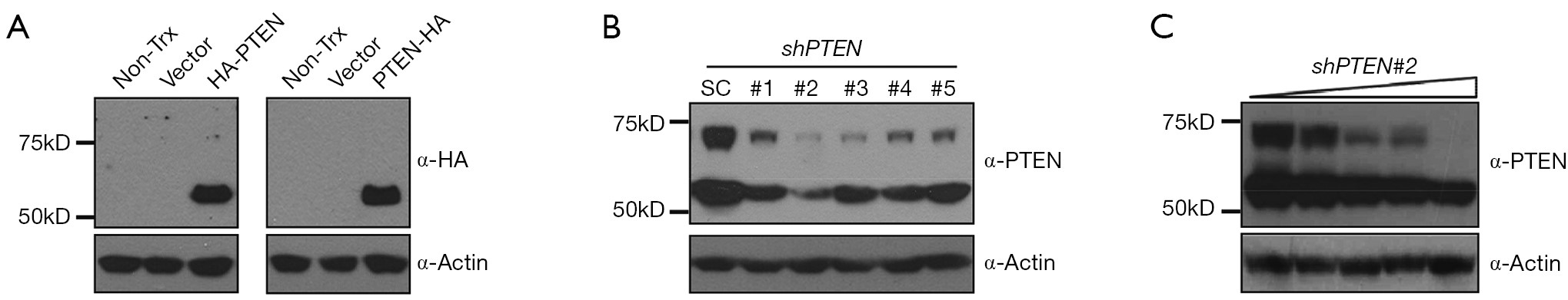
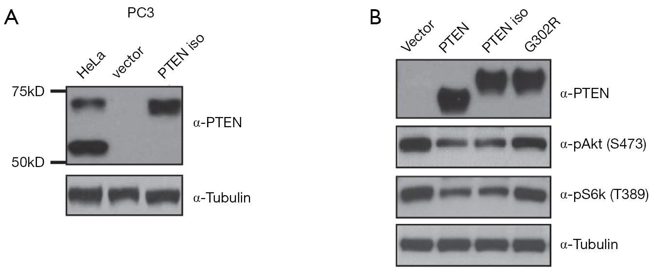
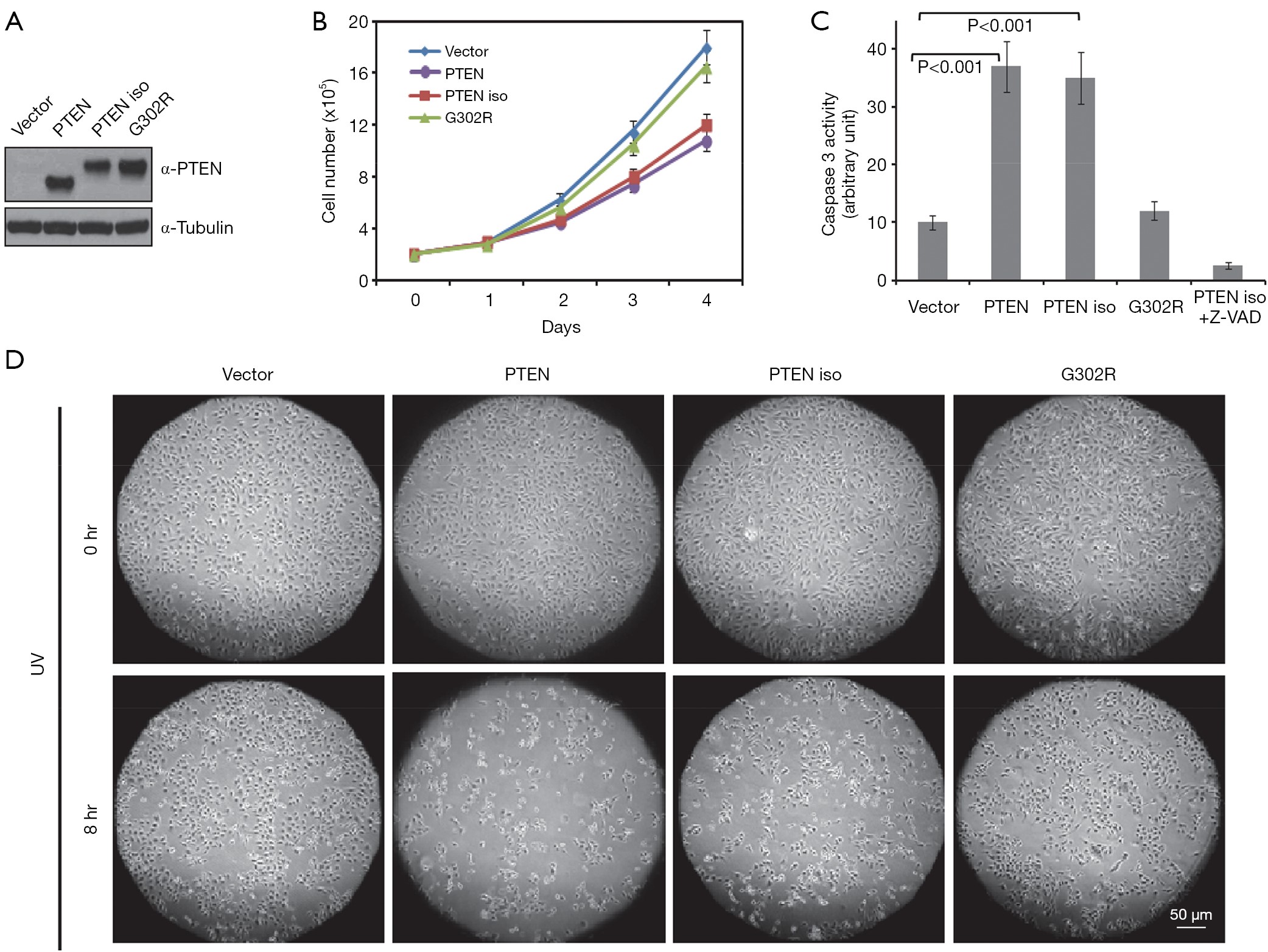
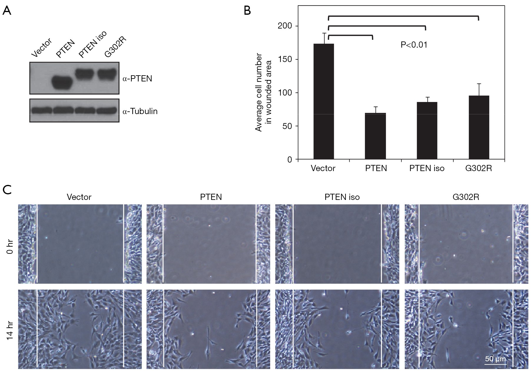
BackgroundTo identify PTEN isoform and explore its potential role in tumor suppression. MethodsWestern blotting, over-expression, shRNA mediated knocking-down, and bioinformatic analysis were used to identify PTEN isoform and test its effect on PI3K-Akt signaling pathway. Cell proliferation, apoptosis, and migration assays were used to test PTEN isoform’s biological activities. ResultsThe PTEN isoform is about 15 kDa bigger than PTEN and its expression is dependent on PTEN status. Immunoprecipitation for PTEN isoform followed by screening with antibodies against ISG15, SUMO1/2/3, Ubiquitin, and Nedd8 showed the identified PTEN isoform is not a general proteinaceous post-translational modification. In addition, overexpression of PTEN cDNA in cells did not generate PTEN isoform whereas knocking-down of PTEN reduced the protein levels of both PTEN and PTEN isoform in a proportional manner. Analysis of PTEN DNA sequence disclosed an alternative translational starting code (CTG) upstream of canonical PTEN coding sequence. Expression of cloned PTEN isoform generated a protein with a size about 15 kDa bigger than PTEN and suppressed PI3K-Akt signaling pathway in cells. Overexpression of PTEN isoform also led to decrease in cell growth and enhanced serum starvation—and UV irradiation—induced apoptosis through activation of Caspase 3. Finally, expression of PTEN isoform inhibited cell migration in scratch assay. ConclusionsPTEN isoform has PTEN-like activity and might be a new tumor suppressor.
2015, 27(5): 533-535.
doi: 10.3978/j.issn.1000-9604.2015.09.03
Abstract:


2015, 27(5): 536-537.
doi: 10.3978/j.issn.1000-9604.2015.10.02
Abstract: