2019 Vol.31(2)
Display Mode: |
2019, 31(2): 223-258.
doi: 10.21147/j.issn.1000-9604.2019.02.01
 Abstract
 FullText HTML
 PDF 3597KB
Abstract:


2019, 31(2): 259-277.
doi: 10.21147/j.issn.1000-9604.2019.02.02
Abstract:
2019, 31(2): 278-294.
doi: 10.21147/j.issn.1000-9604.2019.02.03
Abstract:
2019, 31(2): 306-315.
doi: 10.21147/j.issn.1000-9604.2019.02.05
Abstract:
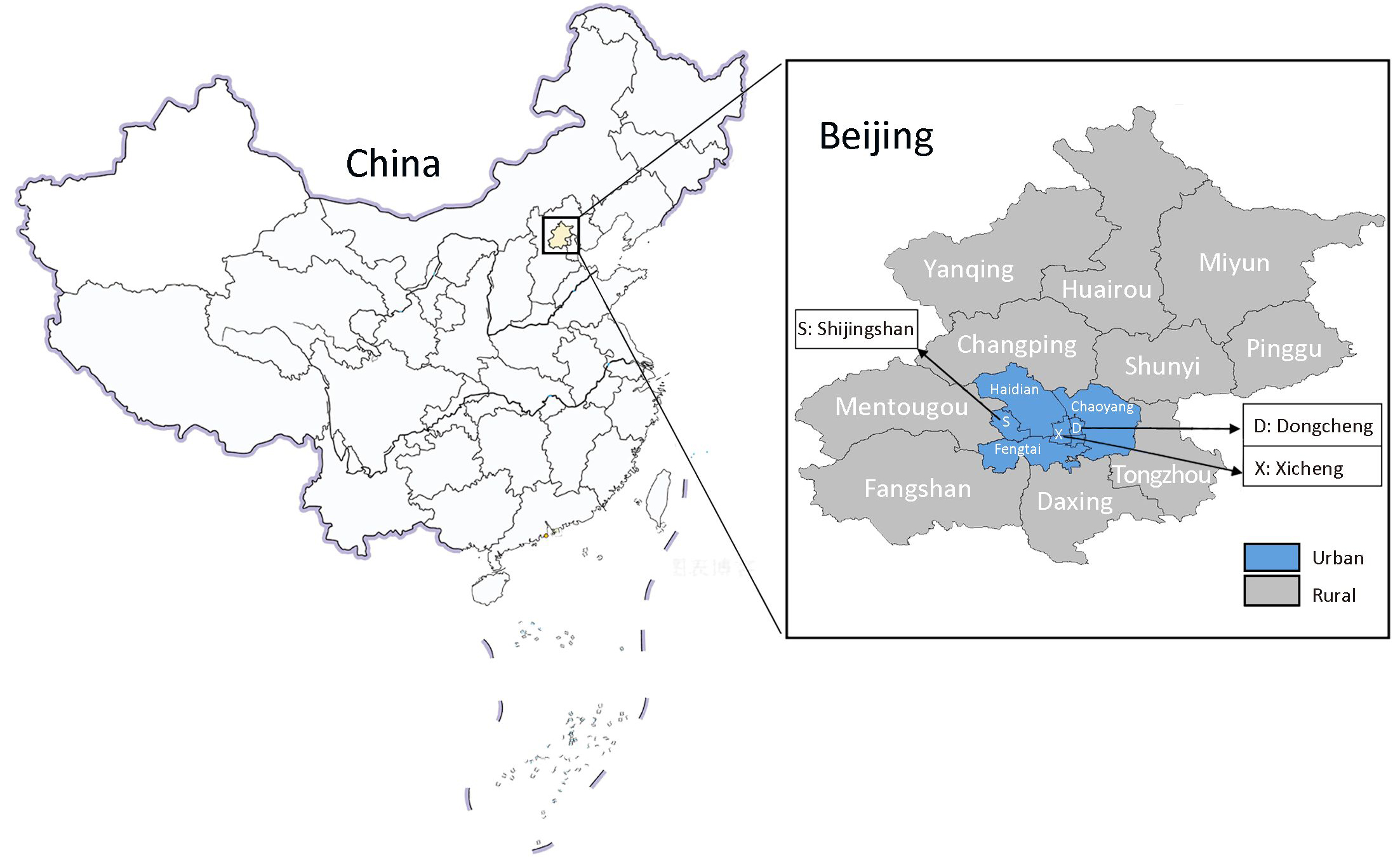
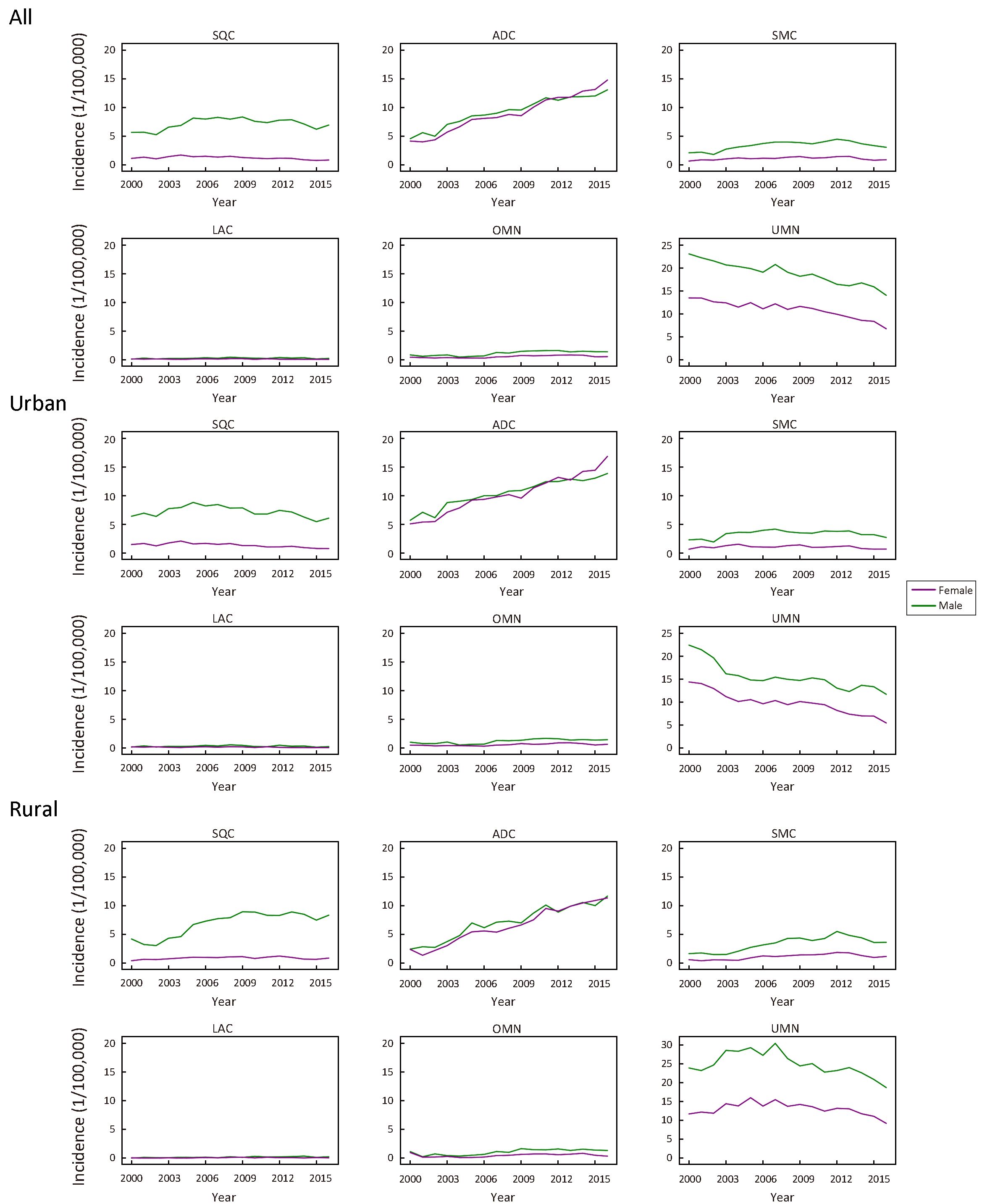
Objective The objective of this study was to characterize secular trends in the sex-specific, age-standardized incidence of lung cancer by histological type in Beijing, China, from 2000 to 2016 based on data from a population-based cancer registry. Methods Data on the incidence of cancer from 2000 to 2016 were obtained from the Beijing Cancer Registry. We examined trends in the sex-specific, age-standardized incidence of lung cancer by histological type using a Joinpoint regression model. Results A total of 117,409 cases of lung cancer were diagnosed from 2000 to 2016. Overall, 73,062 (62.23%) patients were males. The most common histological type among both sexes was adenocarcinoma; however, the proportion of adenocarcinoma differed significantly between males and females (45.36% vs. 77.14%, respectively, P<0.0001). The age-standardized incidence of total lung cancer increased from 2000 to 2010 with an annual percent change (APC) of 2.2% [95% confidence interval (95% CI), 1.5% to 2.9%] and stabilized thereafter. Among males, the incidence of total lung cancer peaked in 2008 and then decreased slightly, with an APC of −1.1% (95% CI, −2.1% to −0.1%). Among females, the incidence increased continuously during the study period, with an APC of 1.4% (95% CI, 0.9% to 1.9%). The incidence of squamous cell carcinoma decreased significantly in recent years among both sexes, with APCs of −2.6% (95% CI, −4.5% to −0.6%) from 2007 to 2016 for males and −5.4% (95% CI, −7.2% to −3.6%) from 2004 to 2016 for females. In contrast, the incidence of adenocarcinoma increased continuously throughout the study period, by APCs of 4.0% (95% CI, 2.6% to 5.4%) for males and 6.2% (95% CI, 4.8% to 7.6%) for females. The incidence of small cell carcinoma peaked in 2007 and stabilized thereafter among males, whereas it peaked in 2012 and then decreased with an APC of −14.7% (95% CI, −25.3% to −2.6%) among females. The incidence of large cell carcinoma and other specified malignant neoplasm did not change much, whereas the incidence of unspecified type decreased among both sexes during the study period. Conclusions Although the incidence of squamous cell carcinoma decreased significantly among both sexes in recent years in Beijing, China, adenocarcinoma increased continuously throughout the study period among both sexes. Knowledge of differences in trends is useful for surveillance and control of lung cancer. However, the reason for the increase in adenocarcinoma remains unclear and warrants investigation.
2019, 31(2): 316-328.
doi: 10.21147/j.issn.1000-9604.2019.02.06
Abstract:
Objective The objective was to systematically assess lung cancer risk prediction models by critical evaluation of methodology, transparency and validation in order to provide a direction for future model development. Methods Electronic searches (including PubMed, EMbase, the Cochrane Library, Web of Science, the China National Knowledge Infrastructure, Wanfang, the Chinese BioMedical Literature Database, and other official cancer websites) were completed with English and Chinese databases until April 30th, 2018. Main reported sources were input data, assumptions and sensitivity analysis. Model validation was based on statements in the publications regarding internal validation, external validation and/or cross-validation. Results Twenty-two studies (containing 11 multiple-use and 11 single-use models) were included. Original models were developed between 2003 and 2016. Most of these were from the United States. Multivariate logistic regression was widely used to identify a model. The minimum area under the curve for each model was 0.57 and the largest was 0.87. The smallest C statistic was 0.59 and the largest 0.85. Six studies were validated by external validation and three were cross-validated. In total, 2 models had a high risk of bias, 6 models reported the most used variables were age and smoking duration, and 5 models included family history of lung cancer. Conclusions The prediction accuracy of the models was high overall, indicating that it is feasible to use models for high-risk population prediction. However, the process of model development and reporting is not optimal with a high risk of bias. This risk affects prediction accuracy, influencing the promotion and further development of the model. In view of this, model developers need to be more attentive to bias risk control and validity verification in the development of models.

2019, 31(2): 329-338.
doi: 10.21147/j.issn.1000-9604.2019.02.07
Abstract:
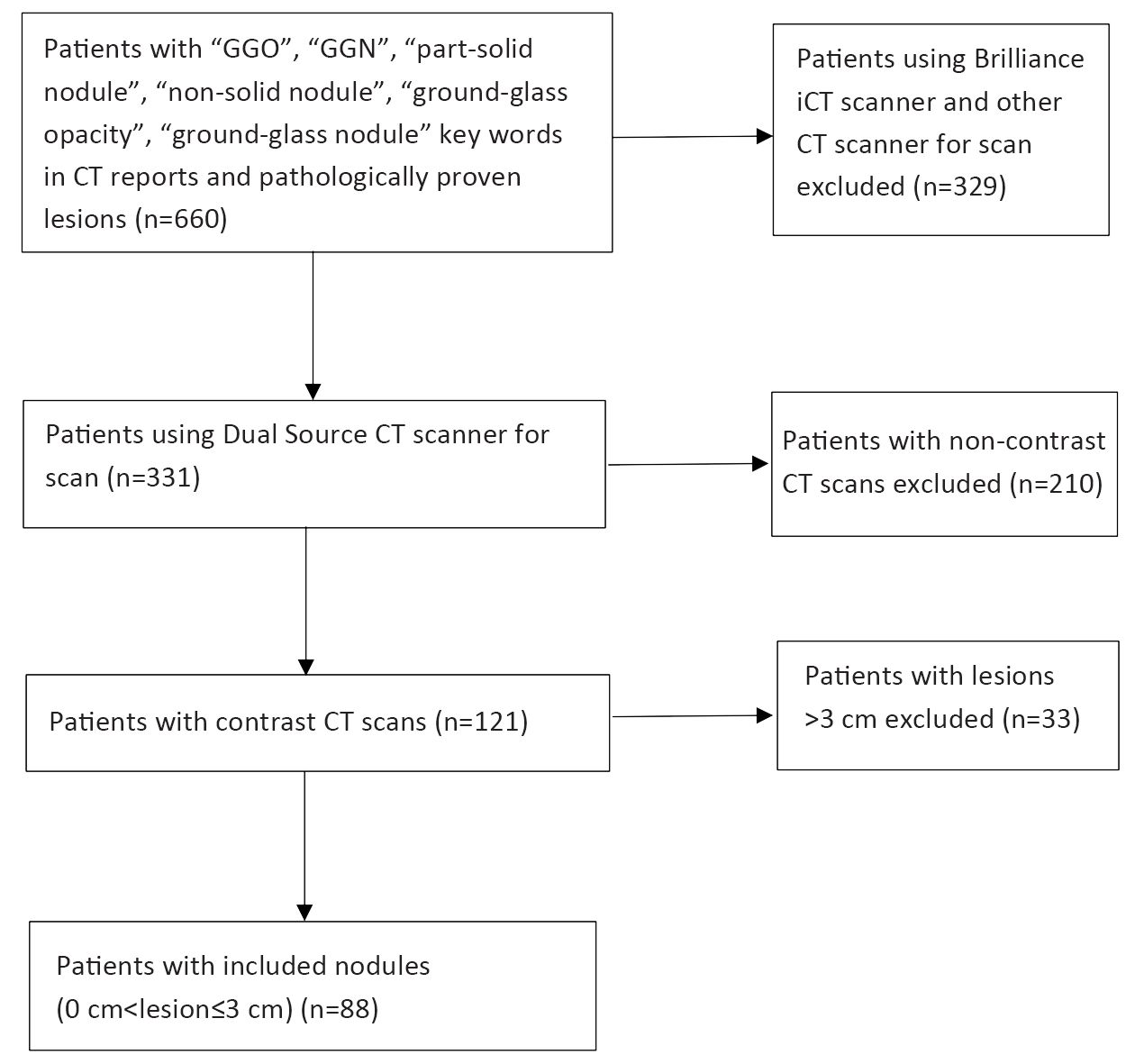
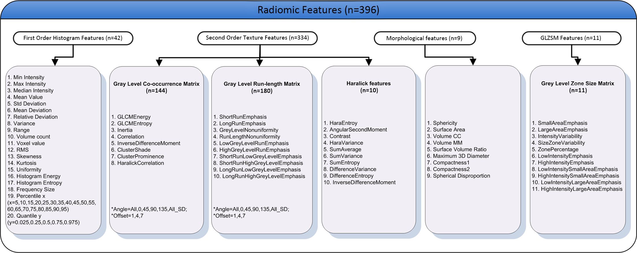
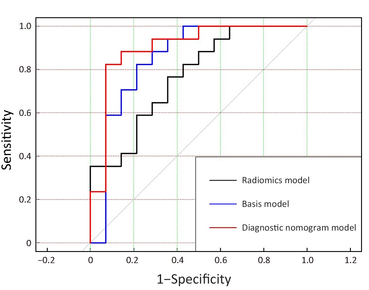
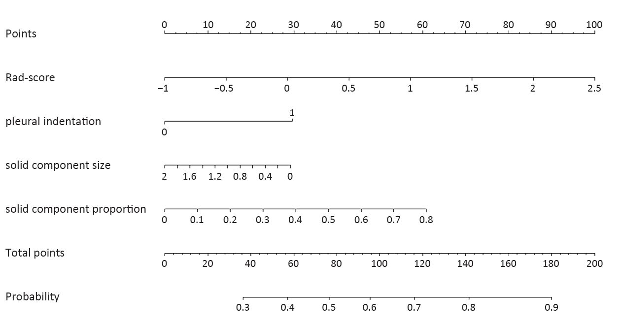
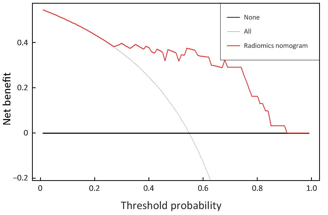
Objective We aim to investigate radiomic imaging features extracted in computed tomography (CT) images to differentiate invasive pulmonary adenocarcinomas (IPAs) from non-IPAs appearing as part-solid ground-glass nodules (GGNs), and to incorporate significant radiomic features with other clinically-assessed features to develop a diagnostic nomogram model for IPAs. Methods This retrospective study was performed, with Institutional Review Board approval, on 88 patients with a total of 100 part-solid nodules (56 IPAs and 44 non-IPAs) that were surgically confirmed between February 2014 and November 2016 in the First Affiliated Hospital of China Medical University. Quantitative radiomic features were computed automatically on 3D nodule volume segmented from arterial-phase contrast-enhanced CT images. A set of regular risk factors and visually-assessed qualitative CT imaging features were compared with the radiomic features using logistic regression analysis. Three diagnostic models, i.e., a basis model using the clinical factors and qualitative CT features, a radiomics model using significant radiomic features, and a nomogram model combining all significant features, were built and compared in terms of receiver operating characteristic (ROC) curves. Decision curve analysis was performed for the nomogram model to explore its potential clinical benefit. Results In addition to three visually-assessed qualitative imaging features, another three quantitative features selected from hundreds of radiomic features were found to be significantly (all P<0.05) associated with IPAs. The diagnostic nomogram model showed a significantly higher performance [area under the ROC curve (AUC) =0.903] in differentiating IPAs from non-IPAs than either the basis model (AUC=0.853, P=0.0009) or the radiomics model (AUC=0.769, P<0.0001). Decision curve analysis indicates a potential benefit of using such a nomogram model in clinical diagnosis. Conclusions Quantitative radiomic features provide additional information over clinically-assessed qualitative features for differentiating IPAs from non-IPAs appearing as GGNs, and a diagnostic nomogram model including all these significant features may be clinically useful in preoperative strategy planning.
2019, 31(2): 339-348.
doi: 10.21147/j.issn.1000-9604.2019.02.08
Abstract:
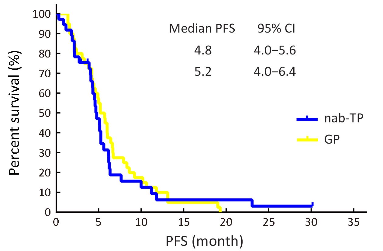
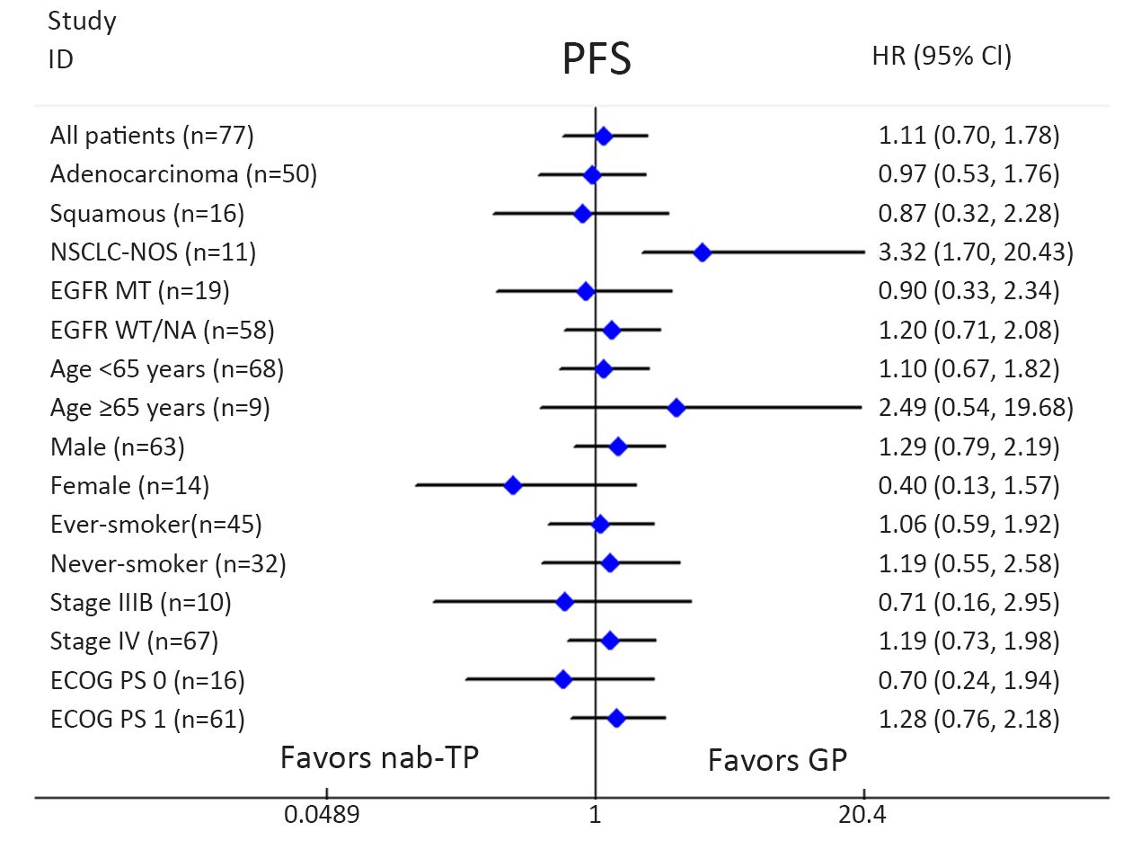
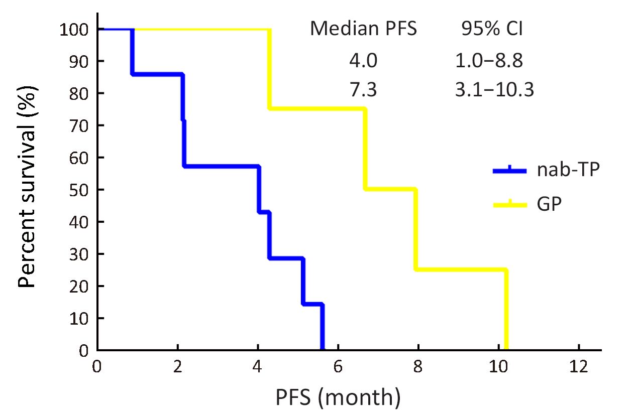
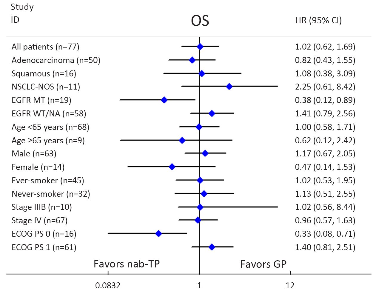
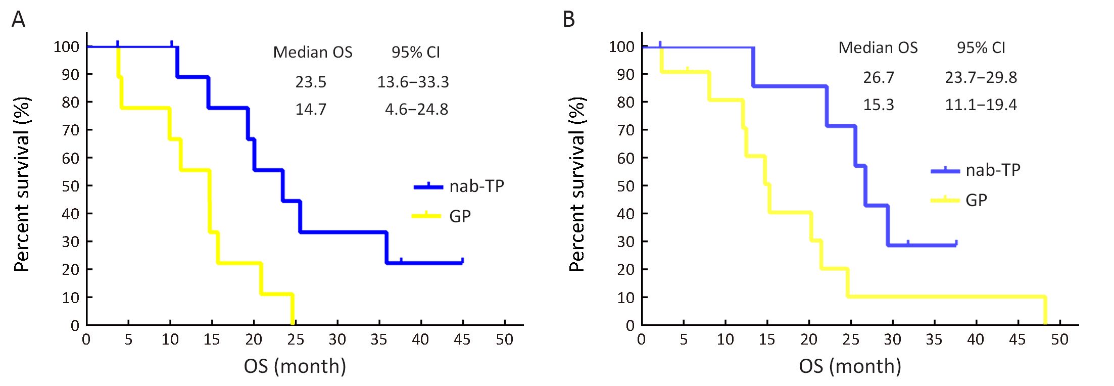
Objective The aim of this trial was to compare both the efficacy and the safety of a weekly nanoparticle albumin-bound paclitaxel (nab-paclitaxel) plus cisplatin vs. gemcitabine plus cisplatin in patients with advanced non-small-cell lung cancer (NSCLC). Methods A total of 84 participants received either 100 mg/m2 nab-paclitaxel each week on d 1, 8 and 15 of a 28 day cycle, as well as cisplatin 75 mg/m2 on d 1 every three weeks (nab-TP arm); or gemcitabine 1,000 mg/m2 on d 1 and 8, plus cisplatin 75 mg/m2 on d 1 every three weeks (GP arm). The primary end point was progression-free survival (PFS). The secondary end points were overall response rate (ORR) and overall survival (OS). Results According to our analysis, the median PFS was 4.8 months for the nab-TP arm vs. 5.2 months for the GP arm (P=0.55). Analysis showed the median OS was 14.6 months for participants who were in the nab-TP arm vs. 15.1 months for those in the GP arm (P=0.94). Besides, nab-TP showed OS advantages over GP in patients harboring epidermal growth factor receptor (EGFR) mutation (26.7 vs. 15.3 months, P=0.046) and patients with a performance status of 0 (23.5 vs. 14.7 months, P=0.020). It was found that incidences of drug-related grade 3 or 4 toxicities were comparable between the two treatment arms. Conclusions Therefore, it can be seen that weekly nab-TP treatment has a similar efficacy and tolerability to GP treatment for patients who are undergoing their first-line treatment for NSCLC. It could be that survival differences among platinum doublets in the context of both EGFR mutation and performance status have the potential to be the basis for our further clinical trials.

2019, 31(2): 349-356.
doi: 10.21147/j.issn.1000-9604.2019.02.09
Abstract:
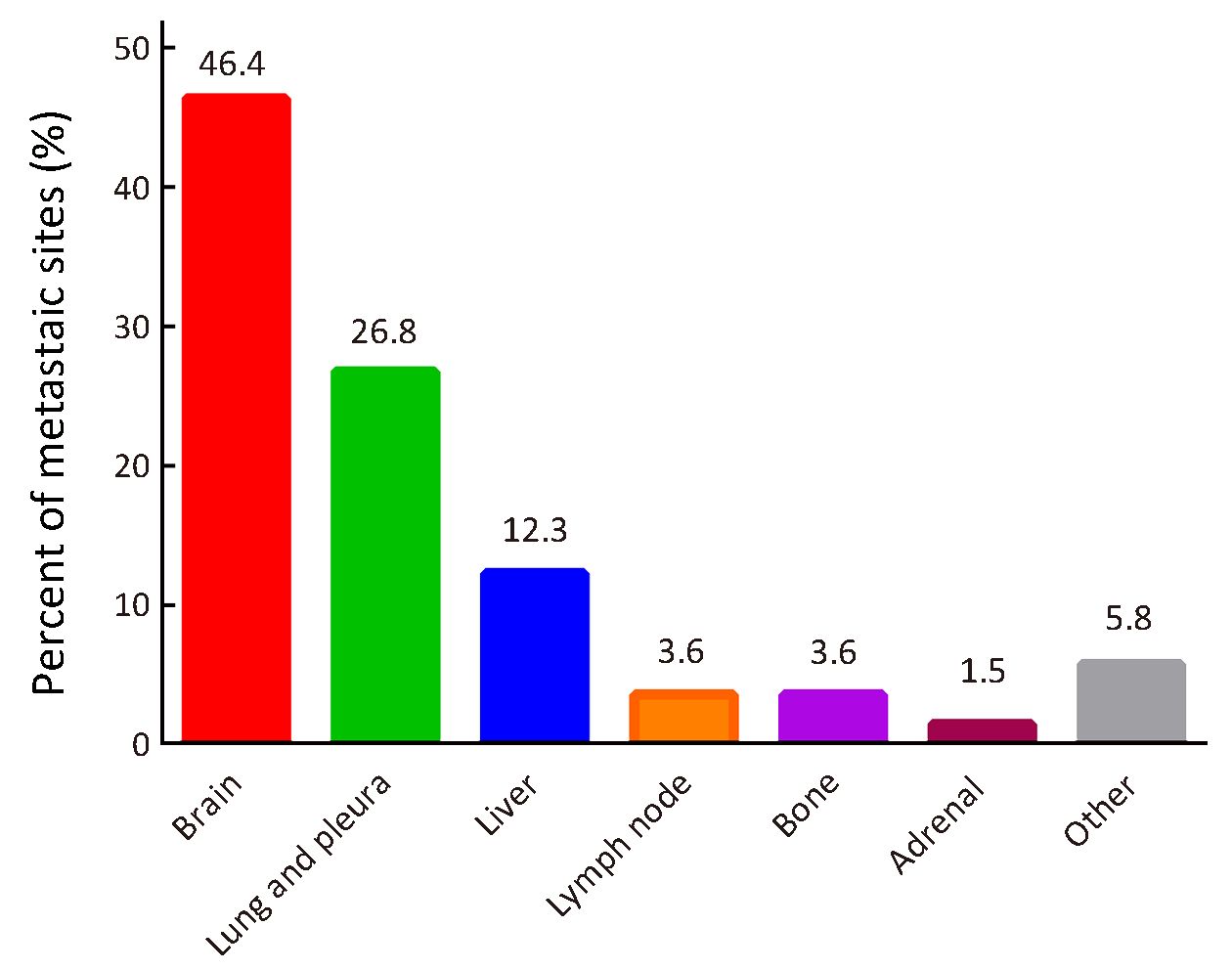
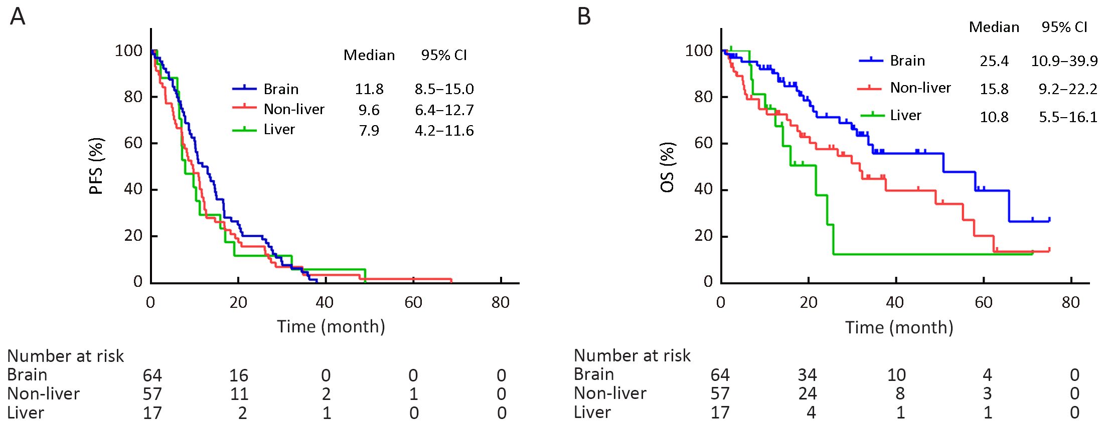
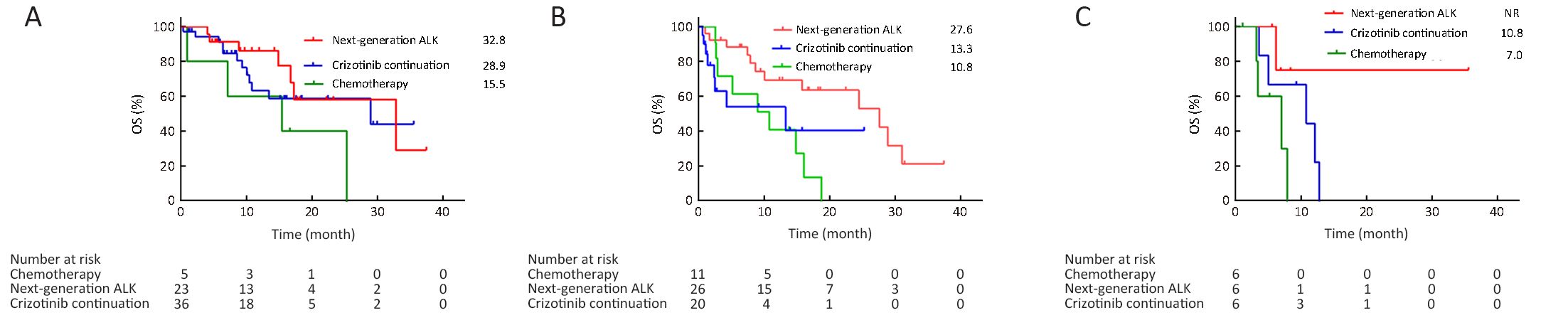
Objective Crizotinib is recommended as the first-line therapy for advanced anaplastic lymphoma kinase (ALK)-positive non-small-cell lung cancer (NSCLC). Despite its initial efficacy, patients ultimately acquire resistance to crizotinib within 1 year. In such patients, the optimal sequential therapy after crizotinib treatment remains unknown. This study explored which sequential therapy option confers the greatest benefit. Methods A total of 138 patients with advanced ALK-positive NSCLC resistant to crizotinib were studied. Based on patterns of disease progression of metastases, patients were divided into 3 groups: brain progression, non-liver progression, and liver progression. Sequential therapies included crizotinib continuation plus local therapy, next-generation ALK inhibitors (ALKi’s), and chemotherapy. The primary endpoint was overall survival (OS) from the time of crizotinib resistance to death or last follow-up. Results The 138 patients included 64 cases with progression in brain, 57 cases in non-liver sites and 17 cases in liver. A significant difference in OS was observed among the distinct progression pattern (median OS, 25.4 months in brain, 15.8 months in non-liver, and 10.8 months in liver, respectively, P=0.020). The difference in OS among sequential therapies was statistically significant in the non-liver progression group (median OS, 27.6 months with next-generation ALKi’s, 13.3 months with crizotinib continuation, and 10.8 months with chemotherapy, respectively, P=0.019). However, crizotinib continuation plus local therapy seems to provide non-inferior median OS compared with next-generation ALKi’s for patients with brain progression (median OS, 28.9 months vs. 32.8 months, P=0.204). And no significant differences in OS were found in patients with progression in liver (P=0.061). Conclusions Crizotinib continuation together with local therapy might be a feasible strategy for patients with progression in brain beyond crizotinib resistance, as well as next-generation ALKi’s. Next-generation ALKi’s tended to provide a survival benefit in patients with non-liver progression.
2019, 31(2): 357-365.
doi: 10.21147/j.issn.1000-9604.2019.02.10
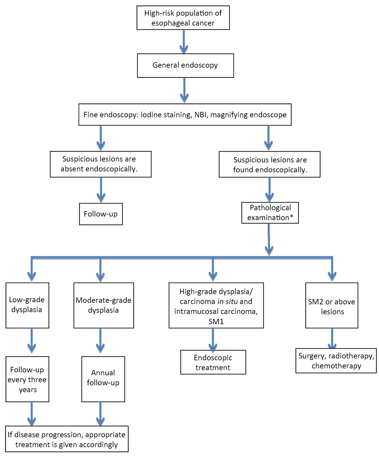
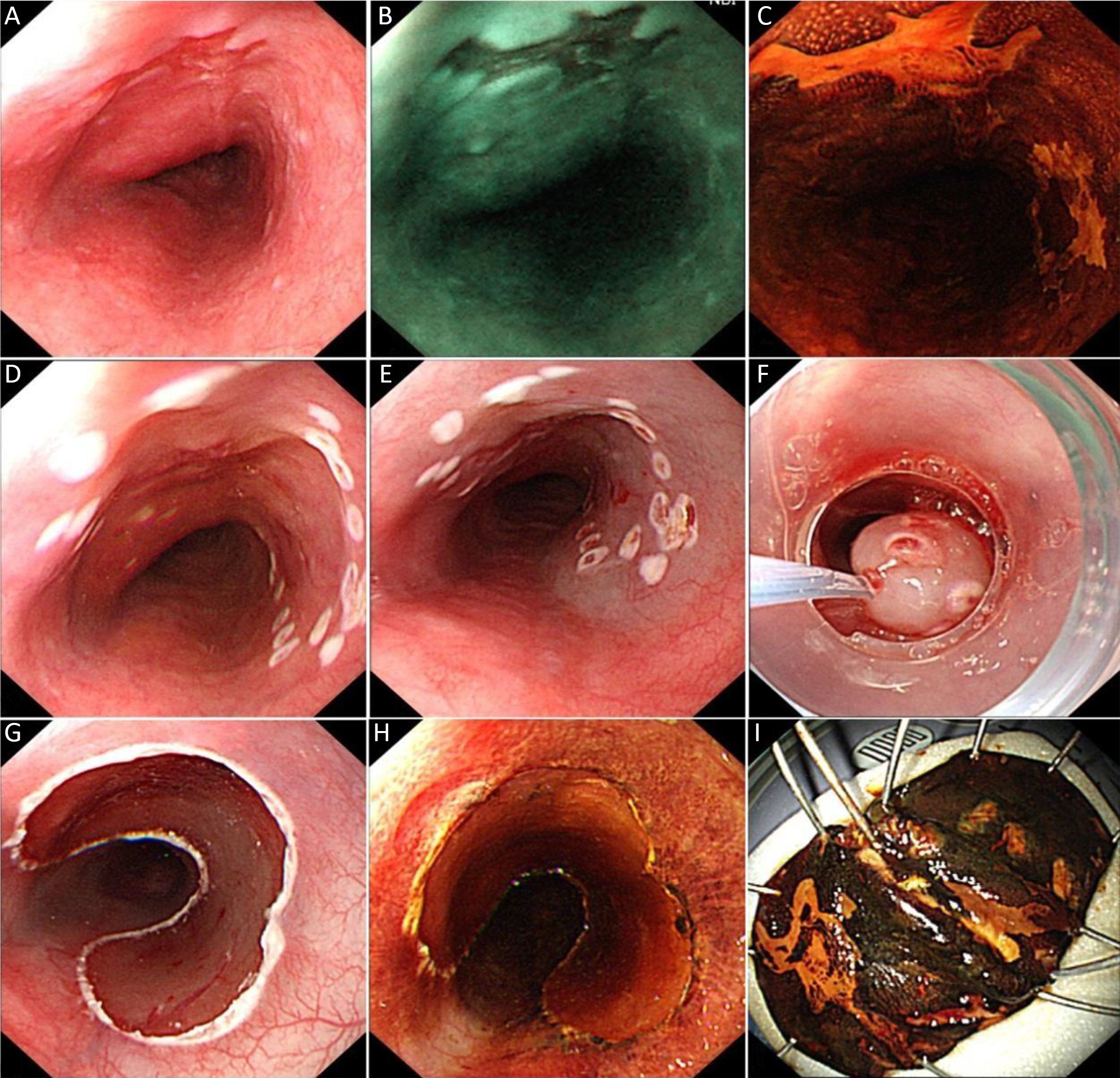
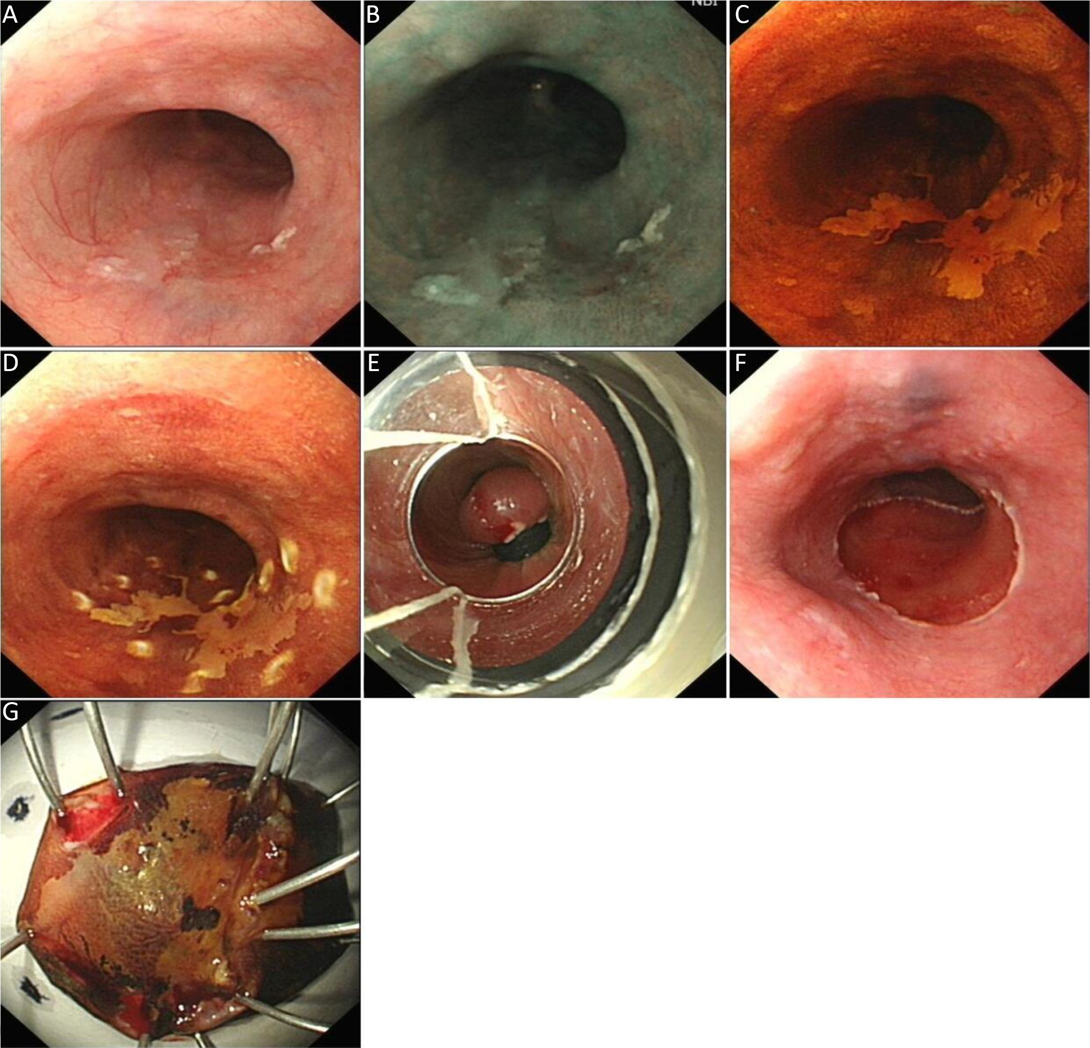
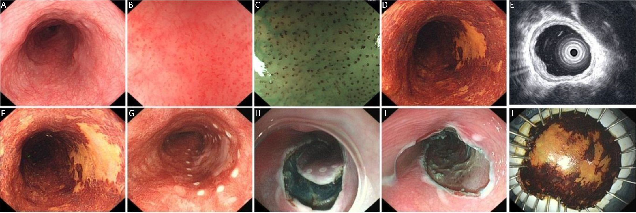
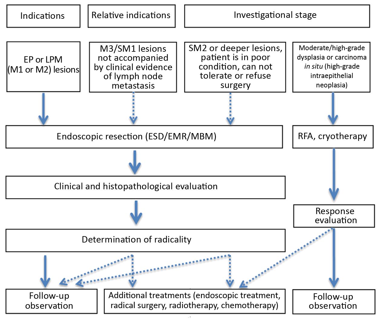
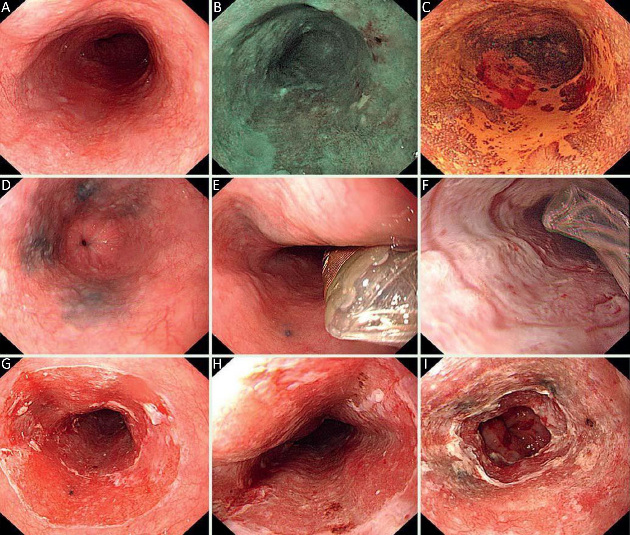
Abstract:
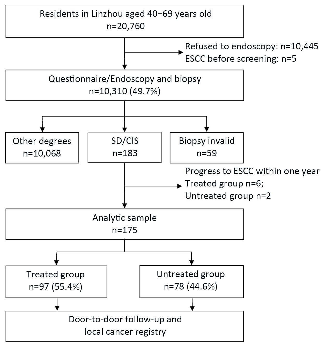
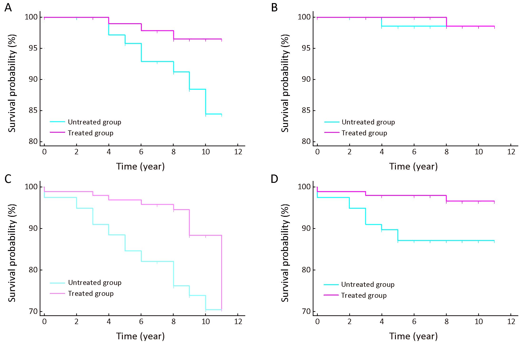
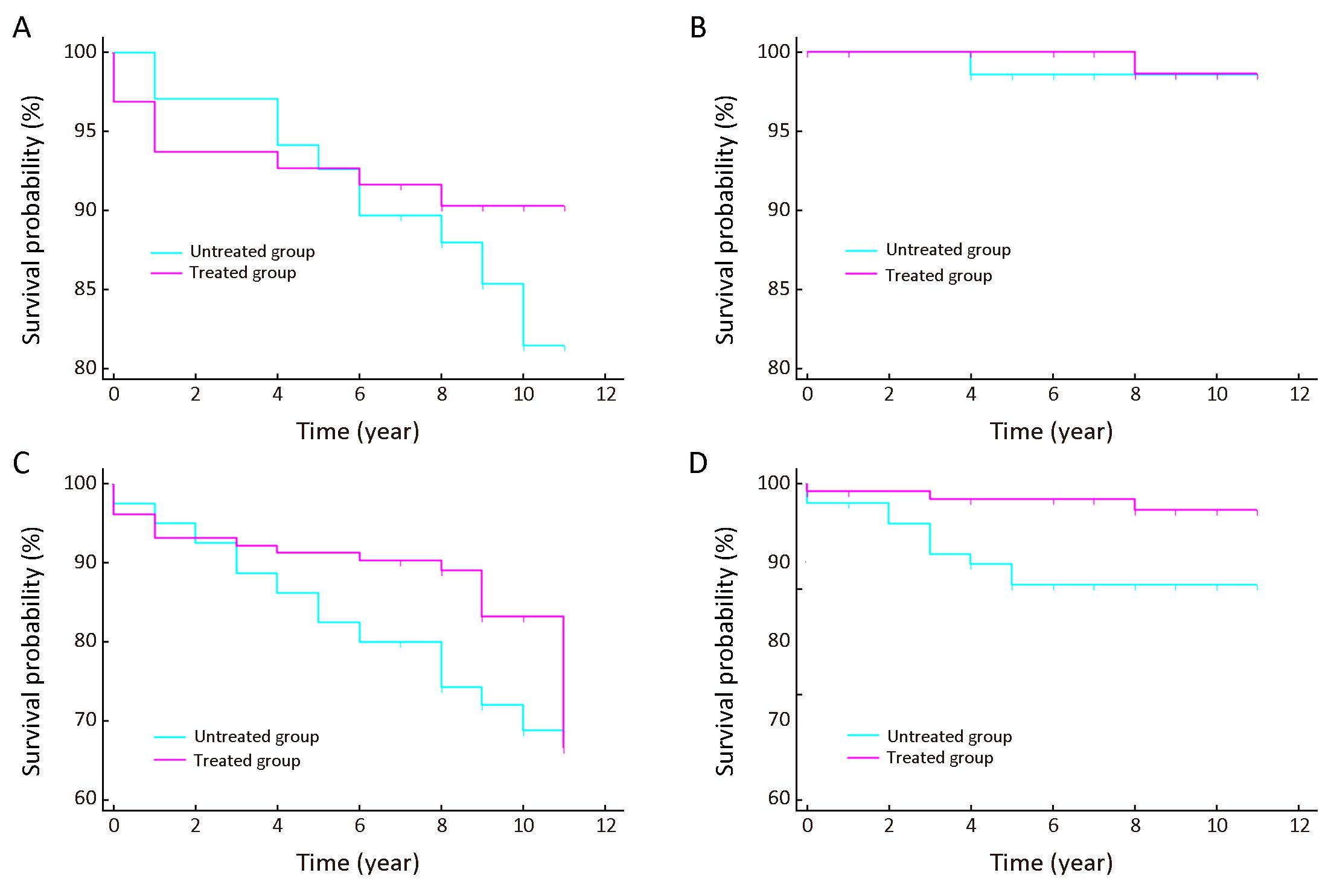
Objective To explore the natural history of severe dysplasia/carcinoma in situ (SD/CIS) patients and to evaluate the efficacy of endoscopic treatment to SD/CIS patients. Methods Between January 2005 and December 2009, a population-based prospective screening program on esophageal squamous cell carcinoma (ESCC) was performed in Linzhou, China, with endoscopic screening plus iodine staining. All the eligible histologically confirmed SD/CIS patients were followed up through the door-to-door follow-up and local cancer registry. The endpoint was diagnosed as ESCC or the December 31st, 2016. Kaplan-Meier survival analysis and Log-rank test were used to compare the survival rates among treated and untreated patients. Results A total of 175 SD/CIS patients were enrolled and grouped by whether they received endoscopic treatment. Eleven-year cumulative incidence rates for untreated and treated SD/CIS patients were 10.7% [95% confidence interval (95% CI): 6.9−16.1] and 3.2% (95% CI: 1.4−7.0), respectively. The ESCC incidence free survival rate, and all-cause incidence and mortality free survival rates were all significantly higher in the treated patients vs. untreated patients (P=0.043, P=0.008 and P=0.015, respectively). The ESCC mortality free survival rate showed no significant differences between the two groups (P=0.847). Conclusions The cumulative incidence rate of SD/CIS patients to ESCC was much lower than previously reported. The Kaplan-Meier survival analysis showed that endoscopic treatment could increase the ESCC and all-cause disease-free survival rates of SD/CIS patients significantly.
2019, 31(2): 366-374.
doi: 10.21147/j.issn.1000-9604.2019.02.11
Abstract:
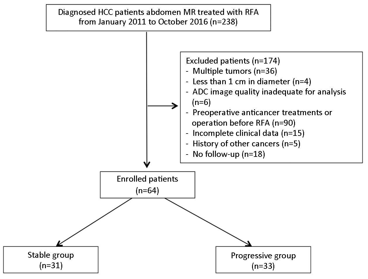
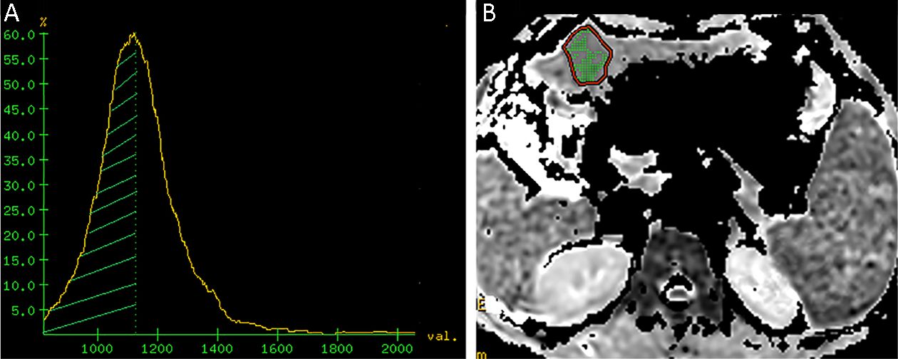
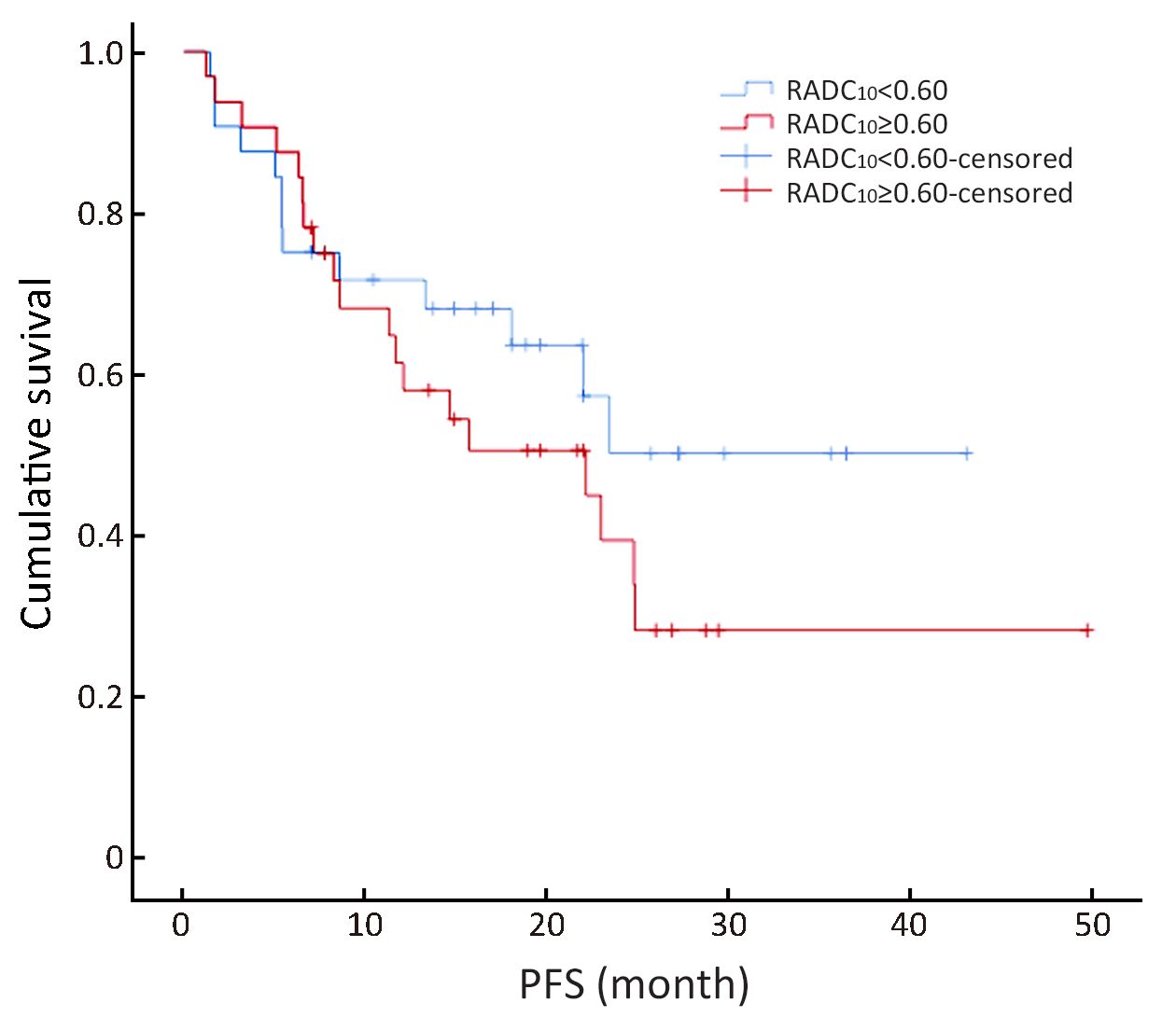
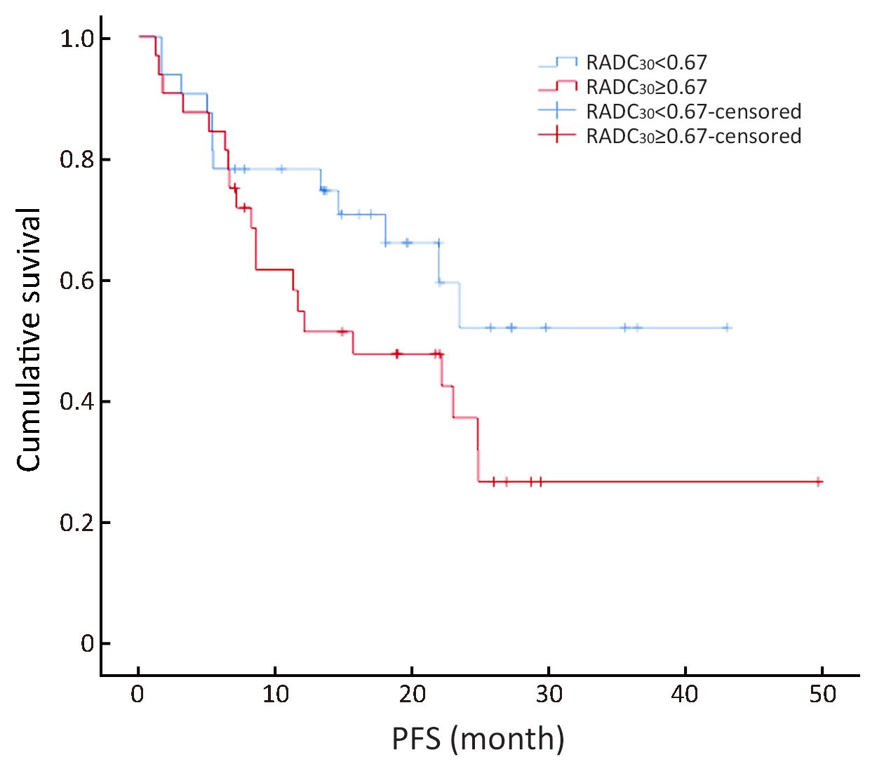
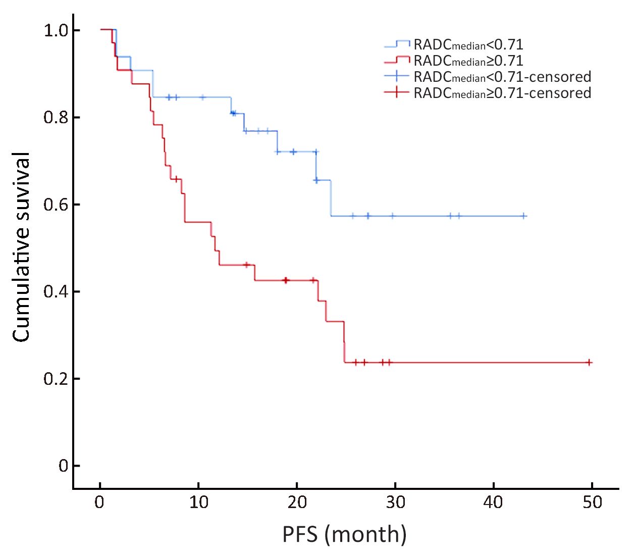
Objective The aim of this study was to predict tumor progression in patients with hepatocellular carcinoma (HCC) treated with radiofrequency ablation (RFA) using histogram analysis of apparent diffusion coefficients (ADC). Methods Breath-hold diffusion weighted imaging (DWI) was performed in 64 patients (33 progressive and 31 stable) with biopsy-proven HCC prior to RFA. All patients had pre-treatment magnetic resonance imaging (MRI) and follow-up computed tomography (CT) or MRI. The ADC values (ADC10, ADC30, ADCmedian and ADCmax) were obtained from the histogram’s 10th, 30th, 50th and 100th percentiles. The ratios of ADC10, ADC30, ADCmedian and ADCmax to the mean non-lesion area-ADC (RADC10, RADC30, RADCmedian, and RADCmax) were calculated. The two patient groups were compared. Key predictive factors for survival were determined using the univariate and multivariate analysis of the Cox model. The Kaplan-Meier survival analysis was performed, and pairs of survival curves based on the key factors were compared using the log-rank test. Results The ADC30, ADCmedian, ADCmax, RADC30, RADCmedian, and RADCmax were significantly larger in the progressive group than in the stable group (P<0.05). The median progression-free survival (PFS) was 22.9 months for all patients. The mean PFS for the stable and progressive groups were 47.7±1.3 and 9.8±1.3 months, respectively. Univariate analysis indicated that RADC10, RADC30, and RADCmedian were significantly correlated with the PFS [hazard ratio (HR)=31.02, 43.84, and 44.29, respectively, P<0.05 for all]. Multivariate analysis showed that RADCmedian was the only independent predictor of tumor progression (P=0.04). And the cutoff value of RADCmedian was 0.71. Conclusions Pre-RFA ADC histogram analysis might serve as a useful biomarker for predicting tumor progression and survival in patients with HCC treated with RFA.

2019, 31(2): 375-388.
doi: 10.21147/j.issn.1000-9604.2019.02.12
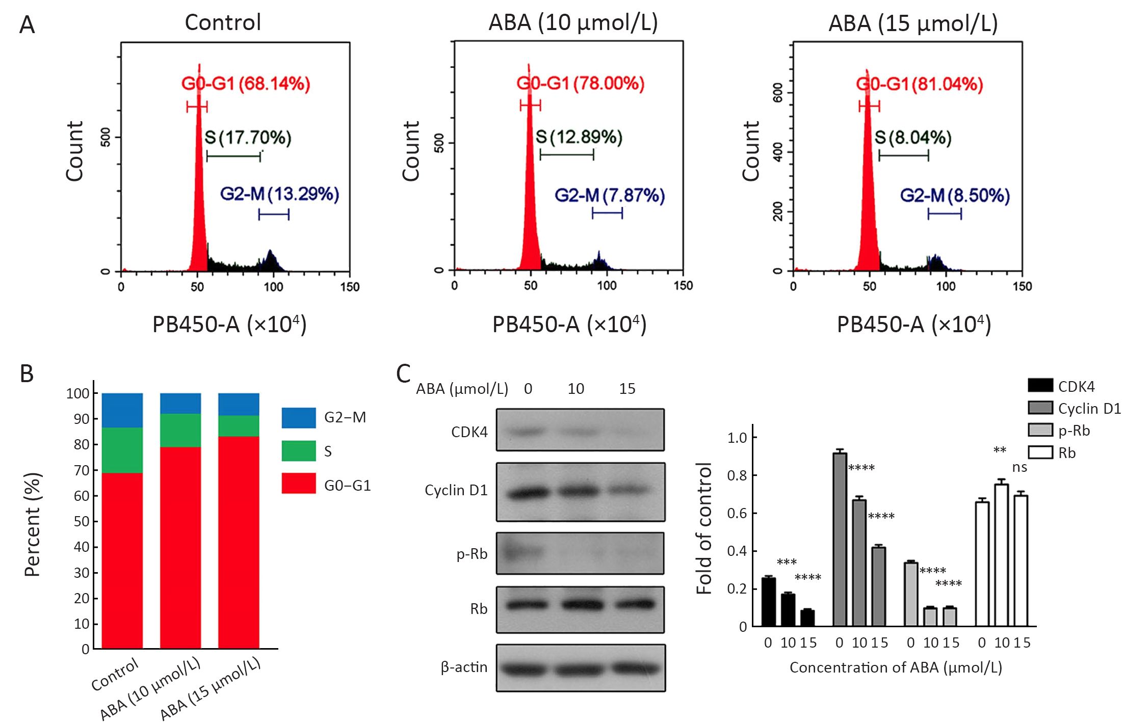
Abstract:
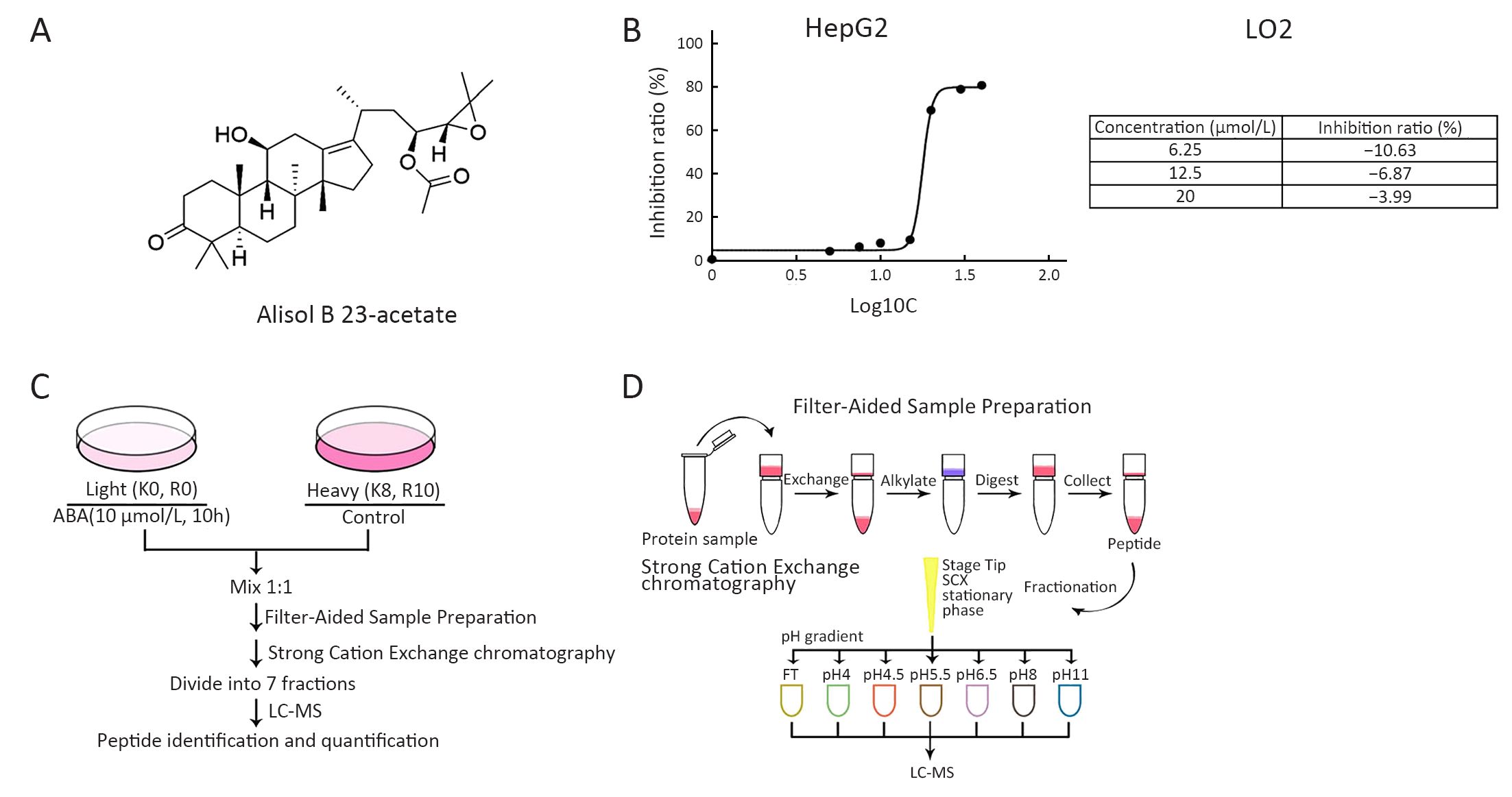
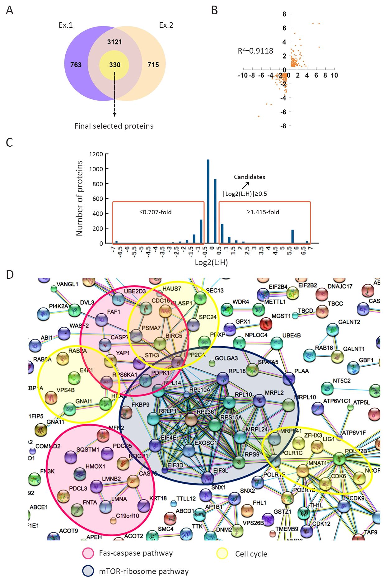
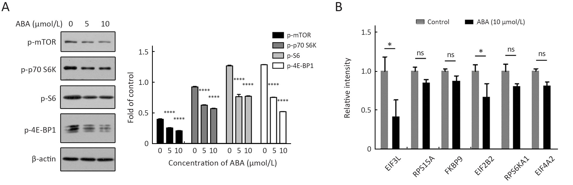
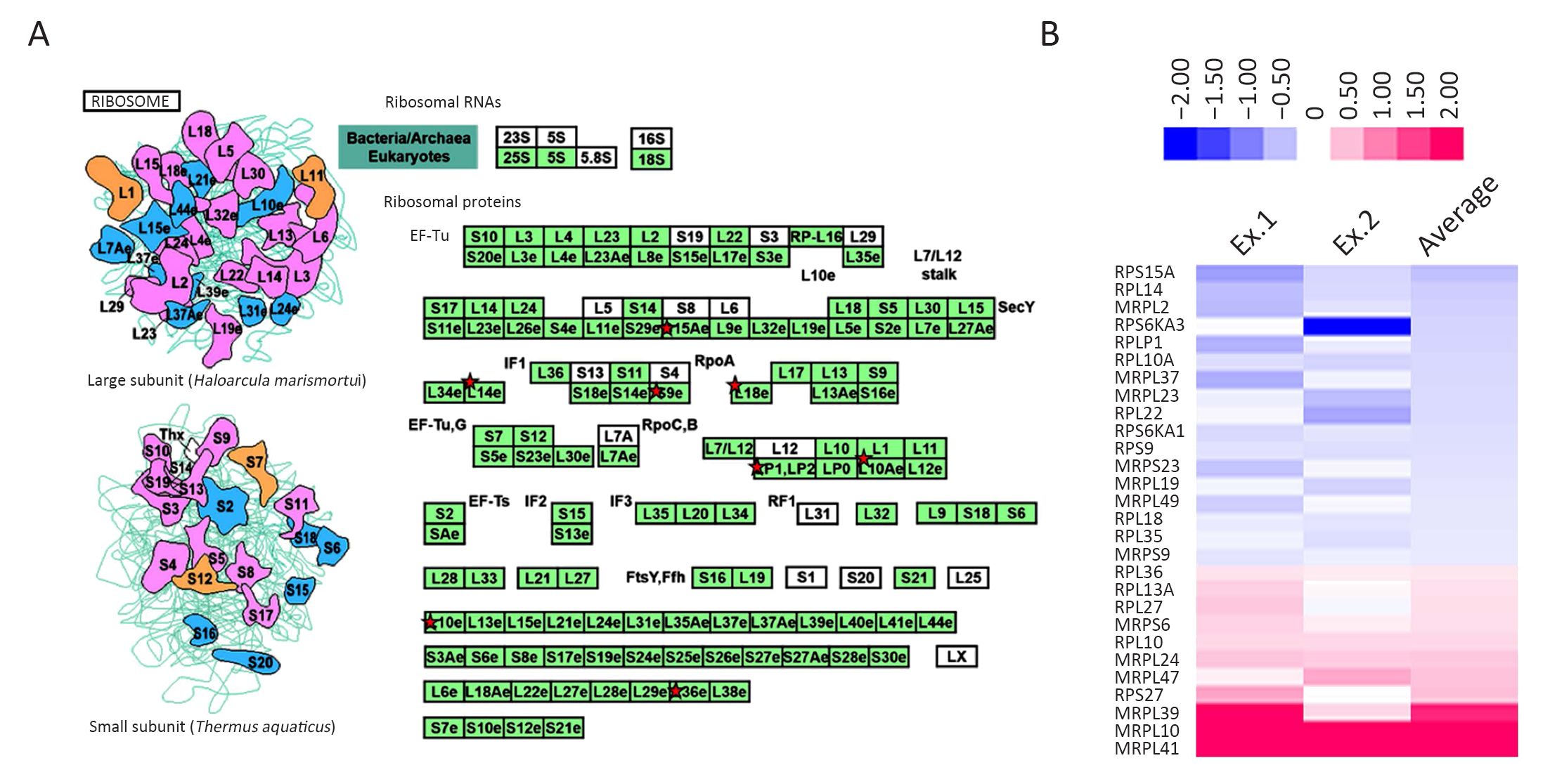
Objective The present study aimed to investigate the molecular events in alisol B 23-acetate (ABA) cytotoxic activity against a liver cancer cell line. Methods First, we employed a quantitative proteomics approach based on stable isotope labeling by amino acids in cell culture (SILAC) to identify the different proteins expressed in HepG2 liver cancer cells upon exposure to ABA. Next, bioinformatics analyses through DAVID and STRING on-line tools were used to predict the pathways involved. Finally, we applied functional validation including cell cycle analysis and Western blotting for apoptosis and mTOR pathway-related proteins to confirm the bioinformatics predictions. Results We identified 330 different proteins with the SILAC-based quantitative proteomics approach. The bioinformatics analysis and the functional validation revealed that the mTOR pathway, ribosome biogenesis, cell cycle, and apoptosis pathways were differentially regulated by ABA. G1 cell cycle arrest, apoptosis and mTOR inhibition were confirmed. Conclusions ABA, a potential mTOR inhibitor, induces the disruption of ribosomal biogenesis. It also affects the mTOR-MRP axis to cause G1 cell cycle arrest and finally leads to cancer cell apoptosis.

2019, 31(2): 389-399.
doi: 10.21147/j.issn.1000-9604.2019.02.13
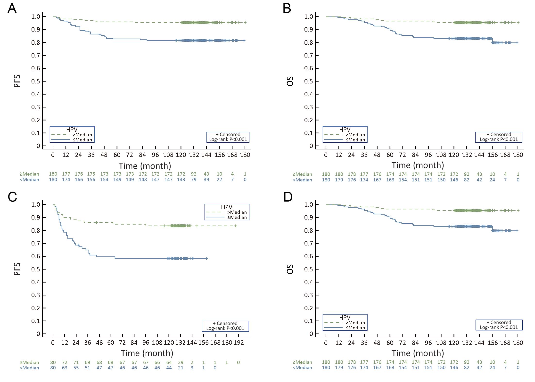
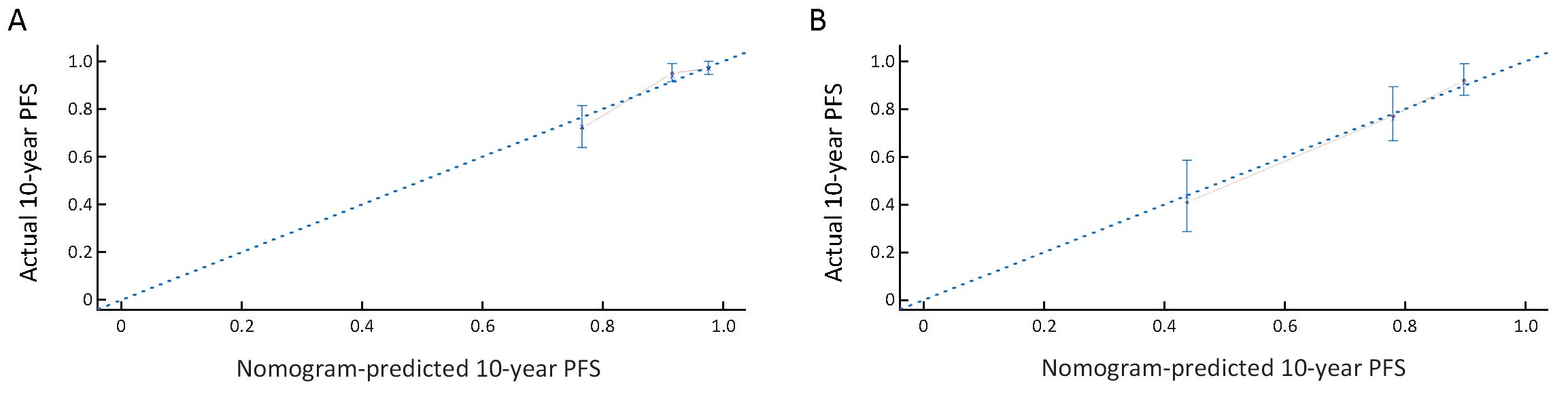
Abstract:
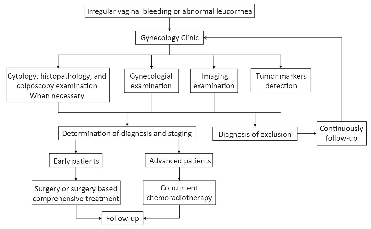
Objective To investigate the prognostic value of pretreatment human papillomavirus (HPV) viral load for cervical cancer, and to develop nomograms based on HPV load and other clinicopathological factors for long-term survival. Methods We conducted a prospective study on cervical squamous cell carcinoma (SCC) patients diagnosed between January 2003 and December 2008. Cervical samples were tested for HPV viral load by the Hybrid Capture II (HCII) assay before treatment and 6 months after treatment. Clinical characteristics and follow-up information were also collected. A multivariable Cox proportional hazards model was used to adjust covariates in both the radical hysterectomy (RH) treatment group and concurrent chemoradiotherapy (CCRT) treatment group to identify relevant covariates, and then nomograms were constructed and used for internal validation. Results A total of 520 SCC patients enrolled in this study with a median follow-up of 127 months, 360 patients received RH, whereas 160 patients received CCRT. The median HPV viral load in RH and CCRT groups was 356.10 and 294.29, respectively. Tumor size was positively correlated with high pretreatment HPV load in both groups. In CCRT group, the advanced International Federation of Gynecology and Obstetrics (FIGO) stage and enlarged retroperitoneal lymph node status determined by computed tomography (LNSCT) were correlated with low HPV load group. Initial HPV viral load, FIGO stage and lymph node metastasis were prognostic factors for RH group, whereas HPV viral load, squamous cell carcinoma antigen (SCC-Ag) level and LNSCT were identified as prognostic factors for CCRT group. Nomograms incorporating these predictors for 10-year progression-free survival (PFS) were constructed [concordance index (C-index): 0.756, 0.749]. Conclusions A low pretreatment HPV viral load is an independent prognostic factor for poor prognosis of cervical SCC and is related to other clinicopathological factors. The survival nomogram based on HPV viral load could predict the long-term prognosis.

2019, 31(2): 400-409.
doi: 10.21147/j.issn.1000-9604.2019.02.14
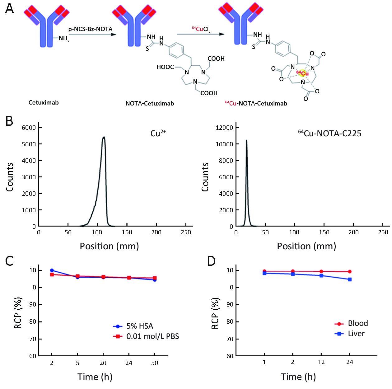
Abstract:
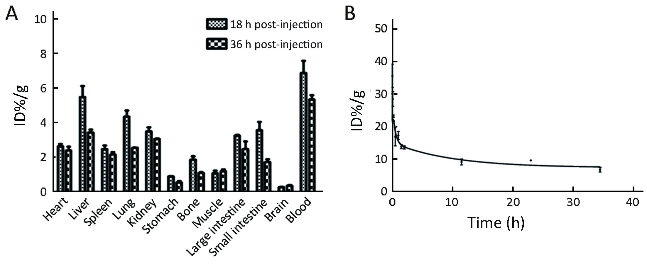
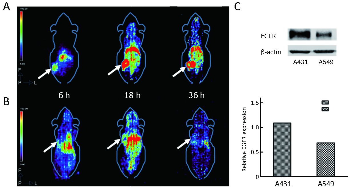
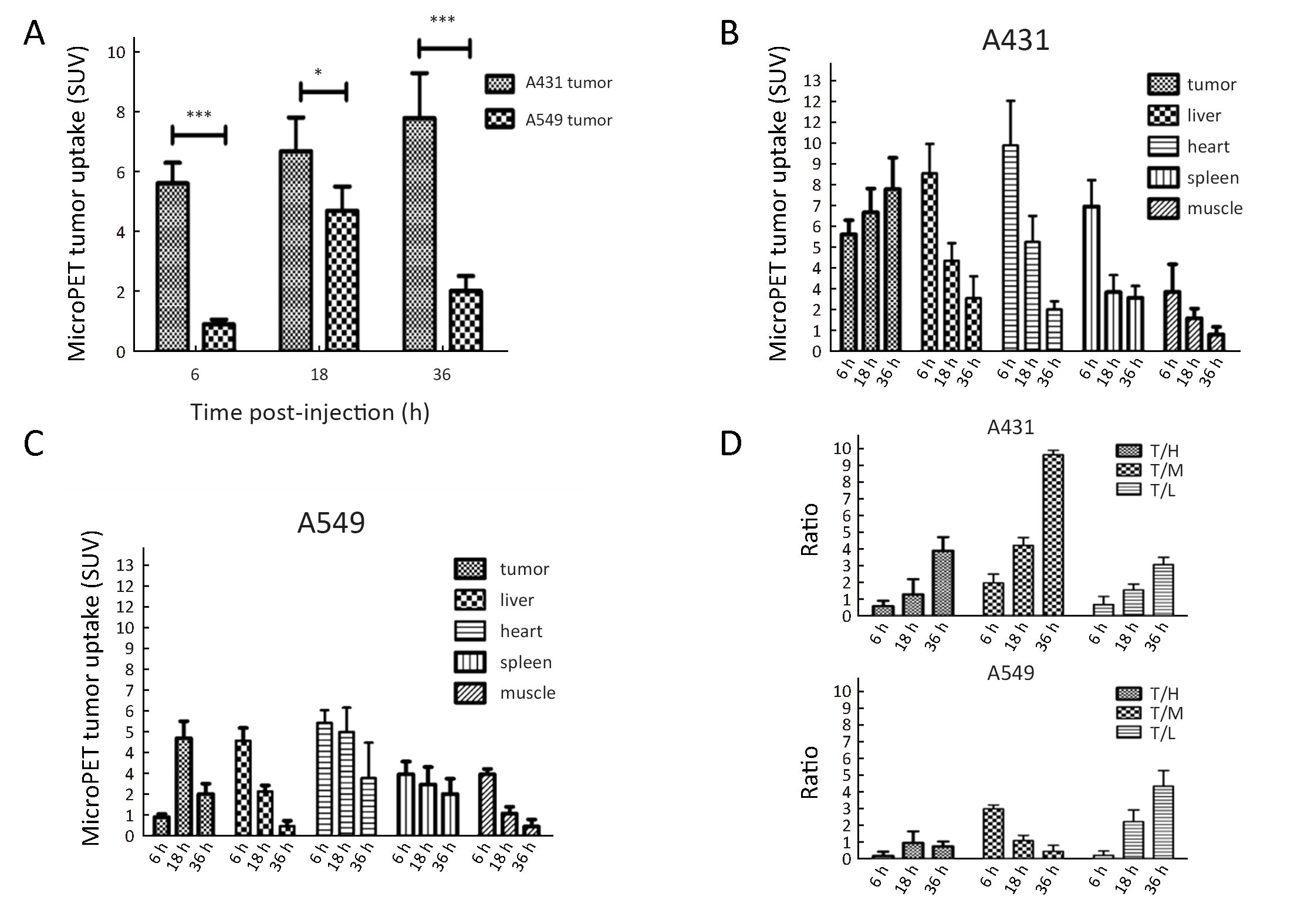
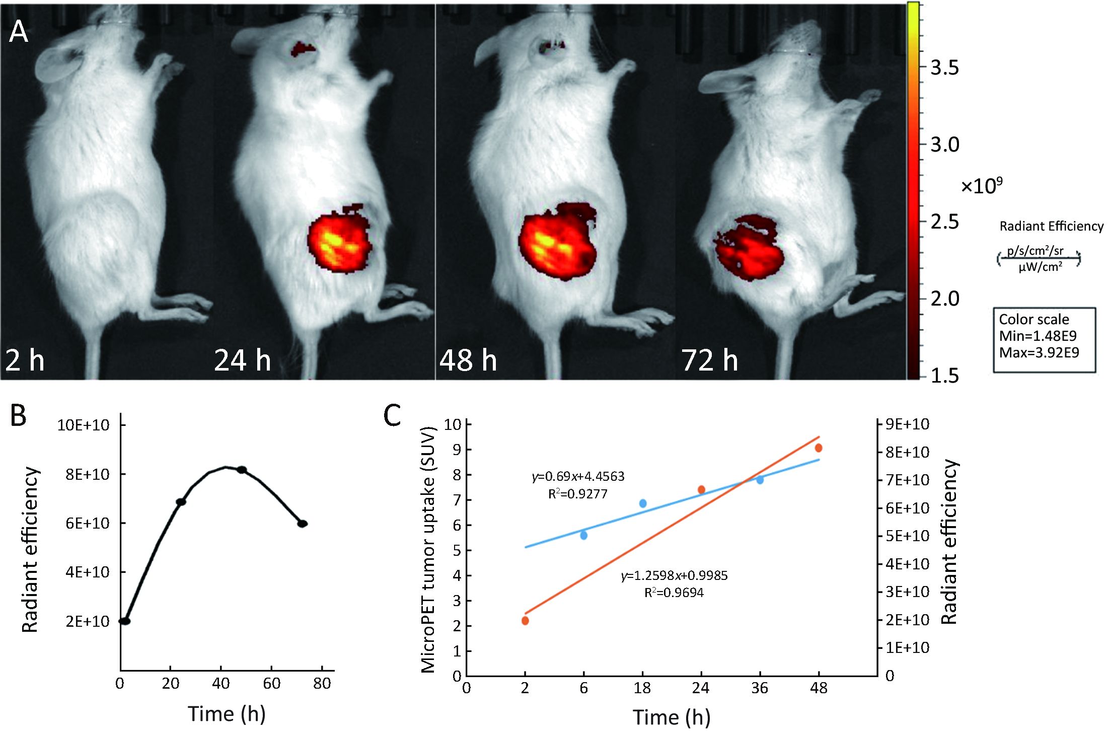
Objective Epidermal growth factor receptor (EGFR) is overexpressed in a wide variety of solid tumors, serving as a well-characterized target for cancer imaging or therapy. In this study, we aimed to design and synthesize a radiotracer, 64Cu-NOTA-C225, targeting EGFR for tumor positron emission tomography (PET) imaging. Methods Cetuximab (C225) was conjugated to a bifunctional chelator, p-isothiocyanatobenzyl-1,4,7-triazacyclononane-1,4,7-triacetic acid (NOTA), and further radiolabeled with copper-64 for PET imaging. 64Cu-NOTA-IgG and Cy5.5-C225 were also synthesized as control probes. A431 and A549 mouse models were established for micro-PET and/or near-infrared fluorescence (NIRF) imaging. Results 64Cu-NOTA-C225 exhibited stability in vivo and in vitro up to 24 h and 50 h post-injection, respectively. A431 tumors with average standard uptake values (SUVs) of 5.61±0.69, 6.68±1.14, 7.80±1.51 at 6, 18 and 36 h post-injection, respectively, which were significantly higher than that of moderate EGFR expressing tumors (A549), with SUVs of 0.89±0.16, 4.70±0.81, 2.01±0.50 at 6, 18 and 36 h post-injection, respectively. The expression levels of A431 and A549 were confirmed by western blotting. Additionally, the tracer uptake in A431 tumors can be blocked by unlabeled cetuximab, suggesting that tracer uptake by tumors was receptor-mediated. Furthermore, NIRF imaging using Cy5.5-C225 showed that the fluorescence intensity in tumors increased with time, with a maximal intensity of 8.17E+10 (p/s/cm2/sr)/(μW/cm2) at 48 h post-injection, which is consistent with the paradigm from micro-PET imaging in A431 tumor-bearing mice. Conclusions The 64Cu-NOTA-C225 PET imaging may be able to specifically and sensitively differentiate tumor models with different EGFR expression levels. It offers potentials as a PET radiotracer for imaging of tracer EGFR-positive tumors.

2019, 31(2): 410-418.
doi: 10.21147/j.issn.1000-9604.2019.02.15
Abstract:
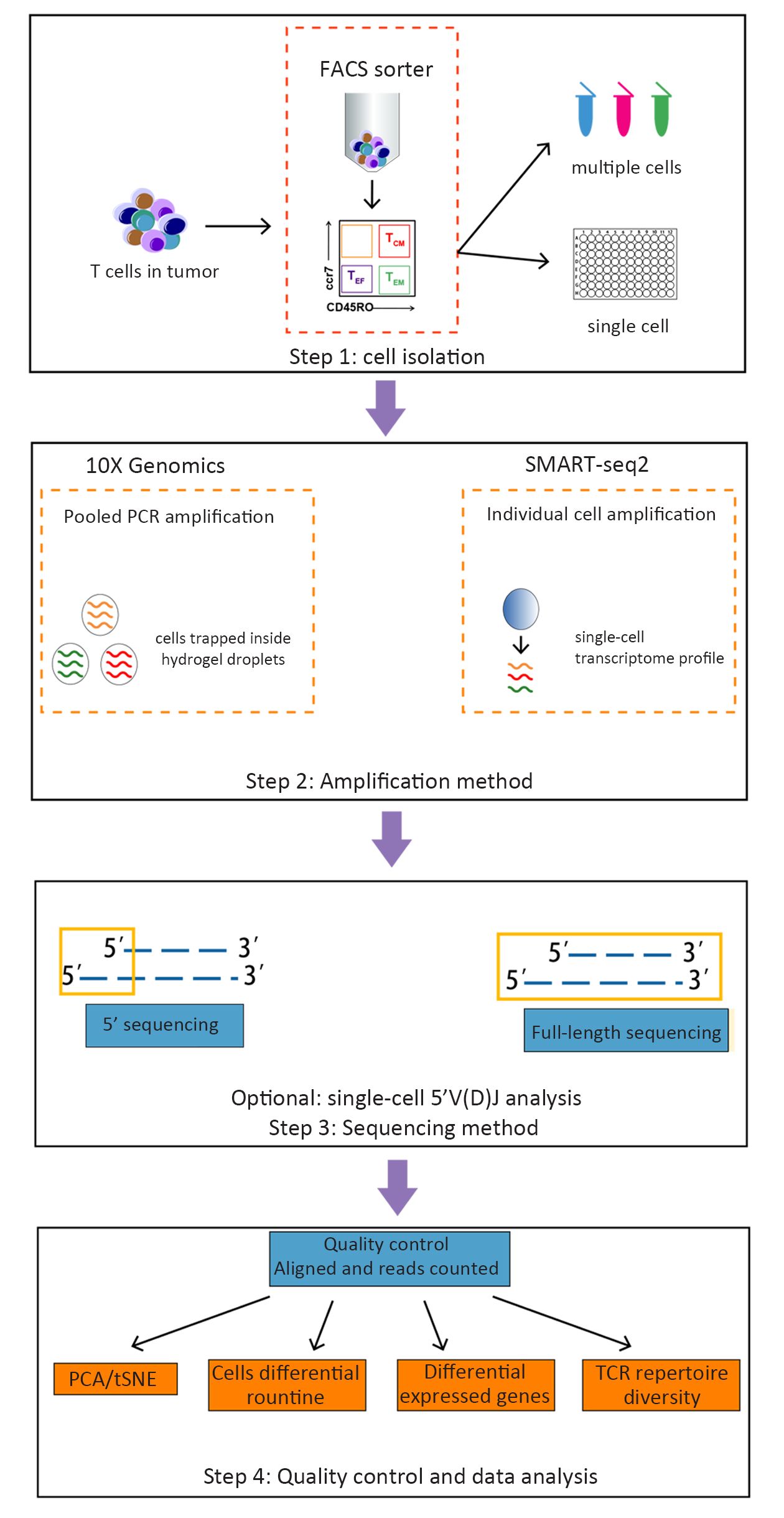
Human T cells are a highly heterogeneous population and can recognize a wide variety of antigens by their T cell receptors (TCRs). Tumor cells display a large repertoire of antigens that serve as potential targets for recognition, thus making T cells in the tumor micro-environment more complicated. Making a connection between TCRs and the transcriptional information of individual T cells will be interesting for investigating clonal expansion within T cell populations under pathologic conditions. Advances in single cell RNA-sequencing (scRNA-seq) have allowed for comprehensive analysis of T cells. In this review, we briefly describe the research progress on tumor micro-environment T cells using single cell RNA sequencing, and then discuss how scRNA-seq can be used to resolve immune system heterogeneity in health and disease. Finally, we point out future directions in this field and potential for immunotherapy.
Human T cells are a highly heterogeneous population and can recognize a wide variety of antigens by their T cell receptors (TCRs). Tumor cells display a large repertoire of antigens that serve as potential targets for recognition, thus making T cells in the tumor micro-environment more complicated. Making a connection between TCRs and the transcriptional information of individual T cells will be interesting for investigating clonal expansion within T cell populations under pathologic conditions. Advances in single cell RNA-sequencing (scRNA-seq) have allowed for comprehensive analysis of T cells. In this review, we briefly describe the research progress on tumor micro-environment T cells using single cell RNA sequencing, and then discuss how scRNA-seq can be used to resolve immune system heterogeneity in health and disease. Finally, we point out future directions in this field and potential for immunotherapy.