2020 Vol.32(4)
Display Mode: |
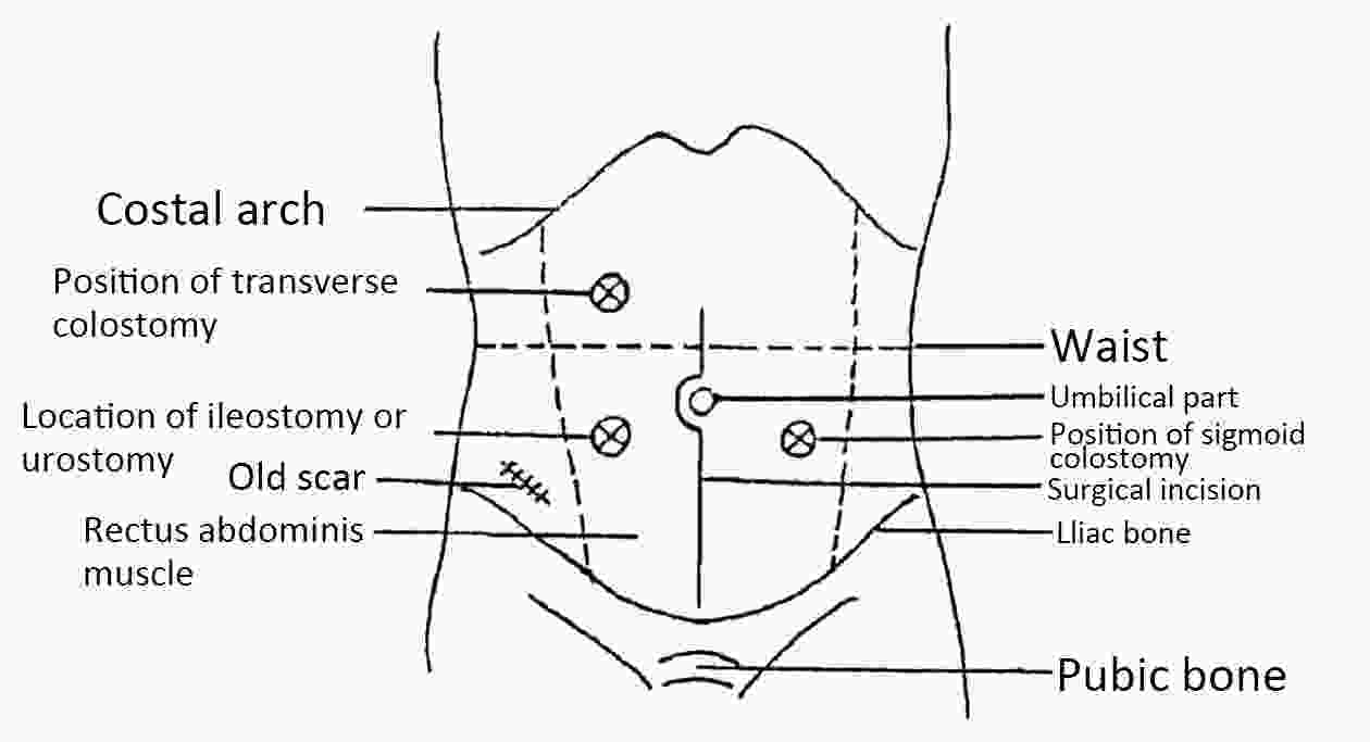
National guidelines for diagnosis and treatment of colorectal cancer 2020 in China (English version)
2020, 32(4): 415-445.
doi: 10.21147/j.issn.1000-9604.2020.04.01
Abstract:
2020, 32(4): 446-447.
doi: 10.21147/j.issn.1000-9604.2020.04.02
Abstract:
2020, 32(4): 448-466.
doi: 10.21147/j.issn.1000-9604.2020.04.03
Abstract:
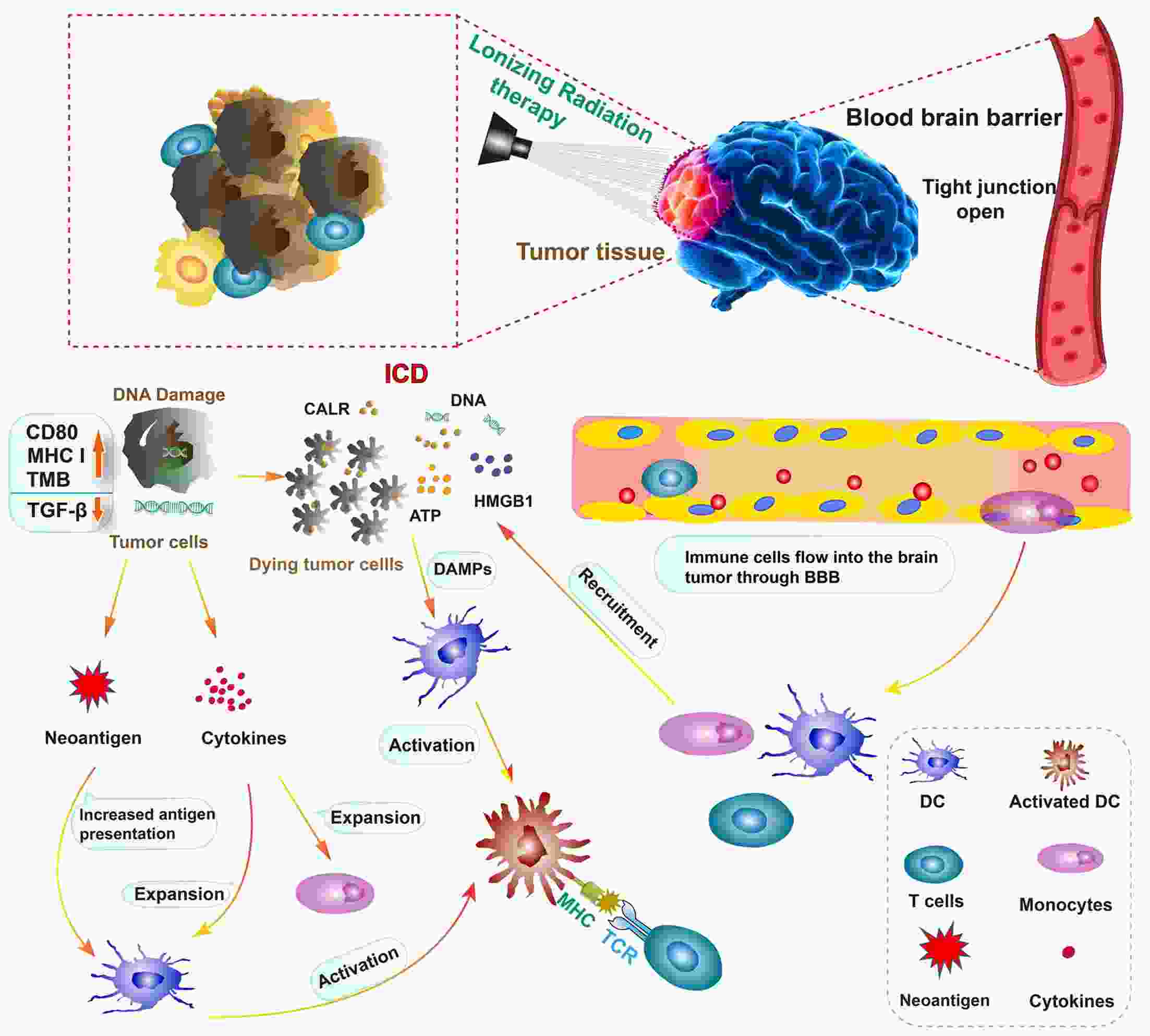
The prognosis of brain metastases (BM) is traditionally poor. BM are mainly treated by local radiotherapy, including stereotactic radiosurgery (SRS) or whole brain radiation therapy (WBRT). Recently, immunotherapy (i.e., immune checkpoint inhibitors, ICI) has demonstrated a survival advantage in multiple malignancies commonly associated with BM. Individually, radiotherapy and ICI both treat BM efficiently; hence, their combination seems logical. In this review, we summarize the existing preclinical and clinical evidence that supports the applicability of radiotherapy as a sensitizer of ICI for BM. Further, we discuss the optimal timing at which radiotherapy and ICI should be administered and review the safety of the combination therapy. Data from a few clinical studies suggest that combining SRS or WBRT with ICI simultaneously rather than consecutively potentially enhances brain abscopal-like responses and survival. However, there is a lack of conclusion about the definition of “simultaneous”; the cumulative toxic effect of the combined therapies also requires further study. Thus, ongoing and planned prospective trials are needed to further explore and validate the effect, safety, and optimal timing of the combination of immunotherapy with radiotherapy for patients with BM.
The prognosis of brain metastases (BM) is traditionally poor. BM are mainly treated by local radiotherapy, including stereotactic radiosurgery (SRS) or whole brain radiation therapy (WBRT). Recently, immunotherapy (i.e., immune checkpoint inhibitors, ICI) has demonstrated a survival advantage in multiple malignancies commonly associated with BM. Individually, radiotherapy and ICI both treat BM efficiently; hence, their combination seems logical. In this review, we summarize the existing preclinical and clinical evidence that supports the applicability of radiotherapy as a sensitizer of ICI for BM. Further, we discuss the optimal timing at which radiotherapy and ICI should be administered and review the safety of the combination therapy. Data from a few clinical studies suggest that combining SRS or WBRT with ICI simultaneously rather than consecutively potentially enhances brain abscopal-like responses and survival. However, there is a lack of conclusion about the definition of “simultaneous”; the cumulative toxic effect of the combined therapies also requires further study. Thus, ongoing and planned prospective trials are needed to further explore and validate the effect, safety, and optimal timing of the combination of immunotherapy with radiotherapy for patients with BM.
2020, 32(4): 467-475.
doi: 10.21147/j.issn.1000-9604.2020.04.04
Abstract:
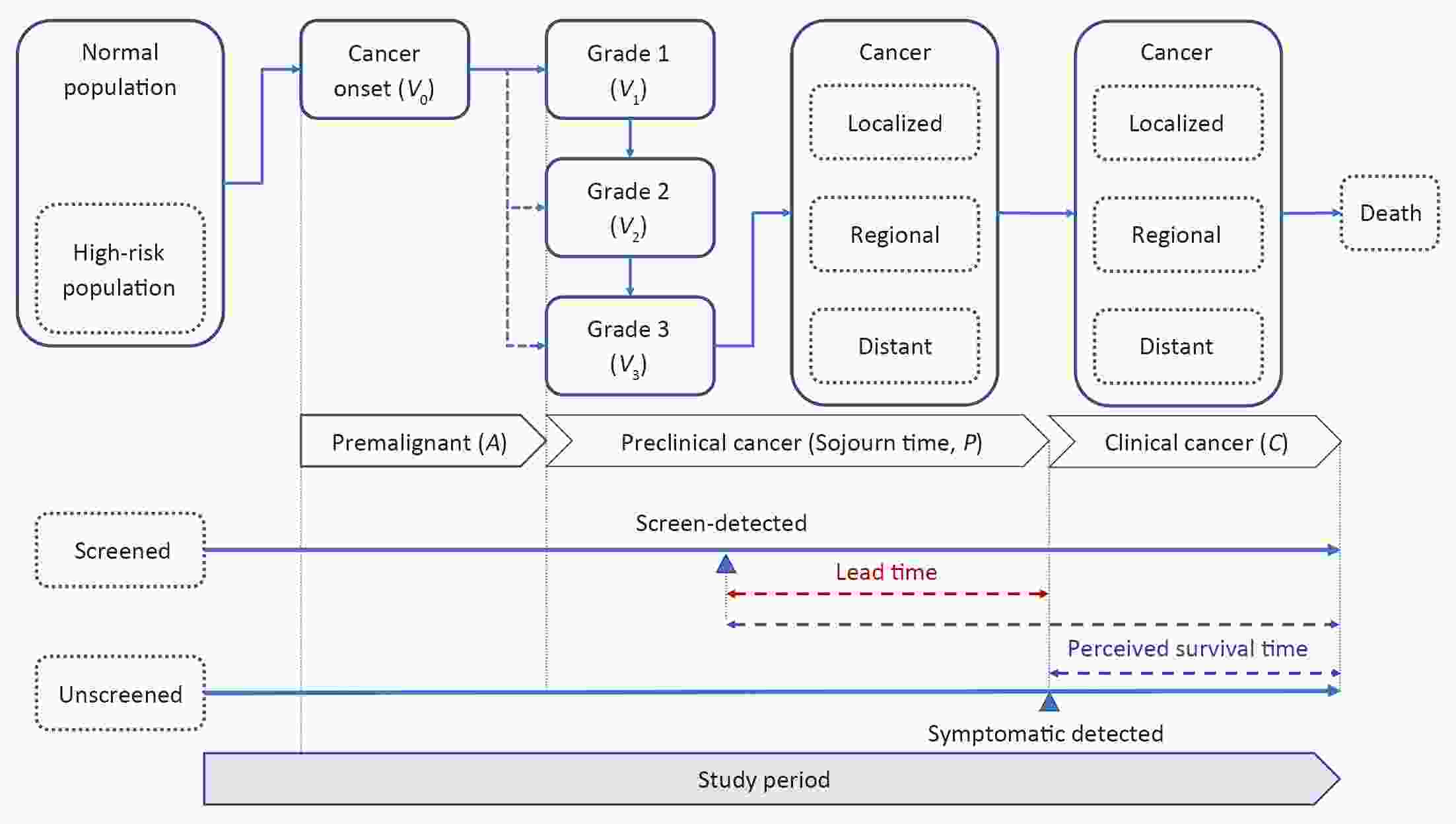
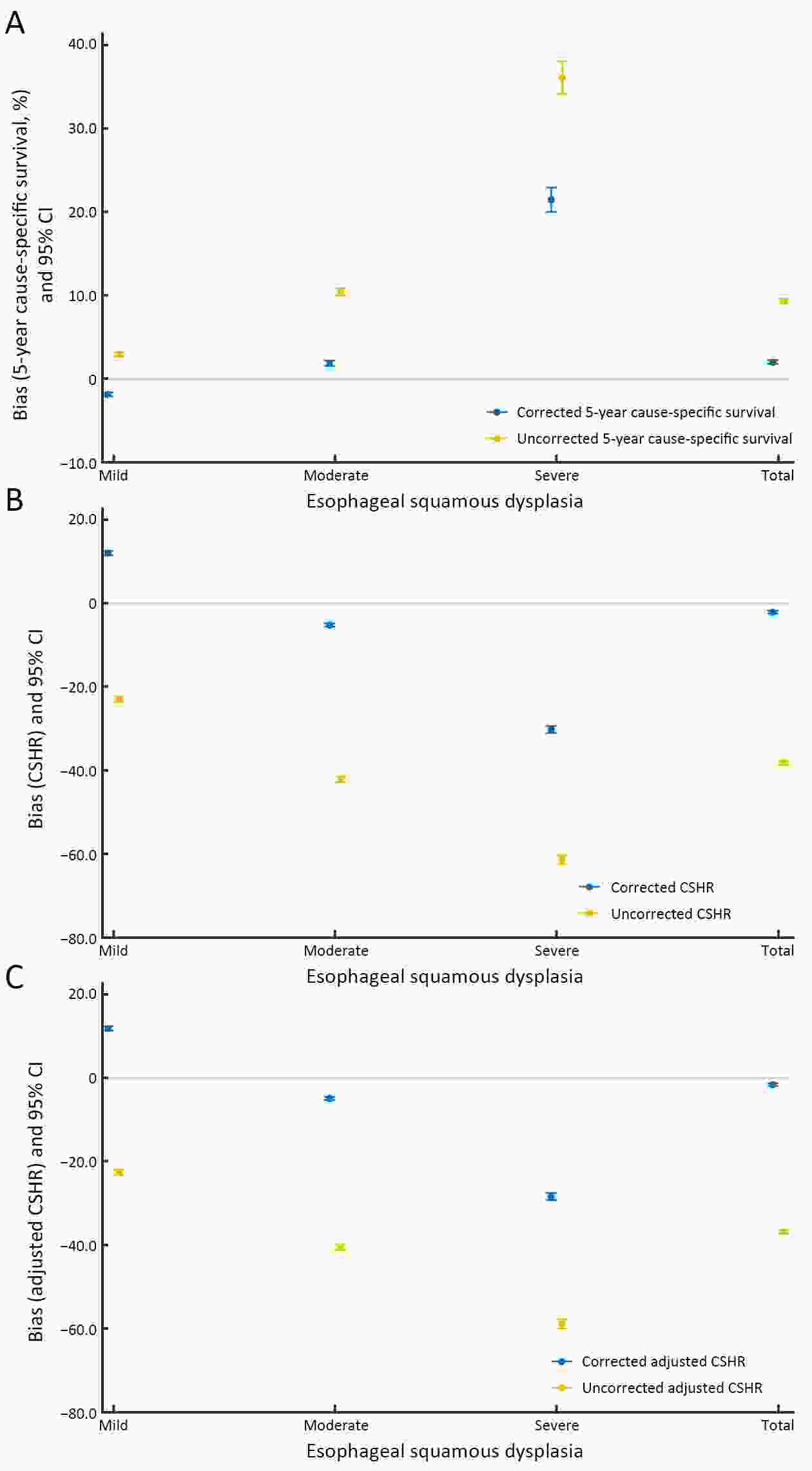
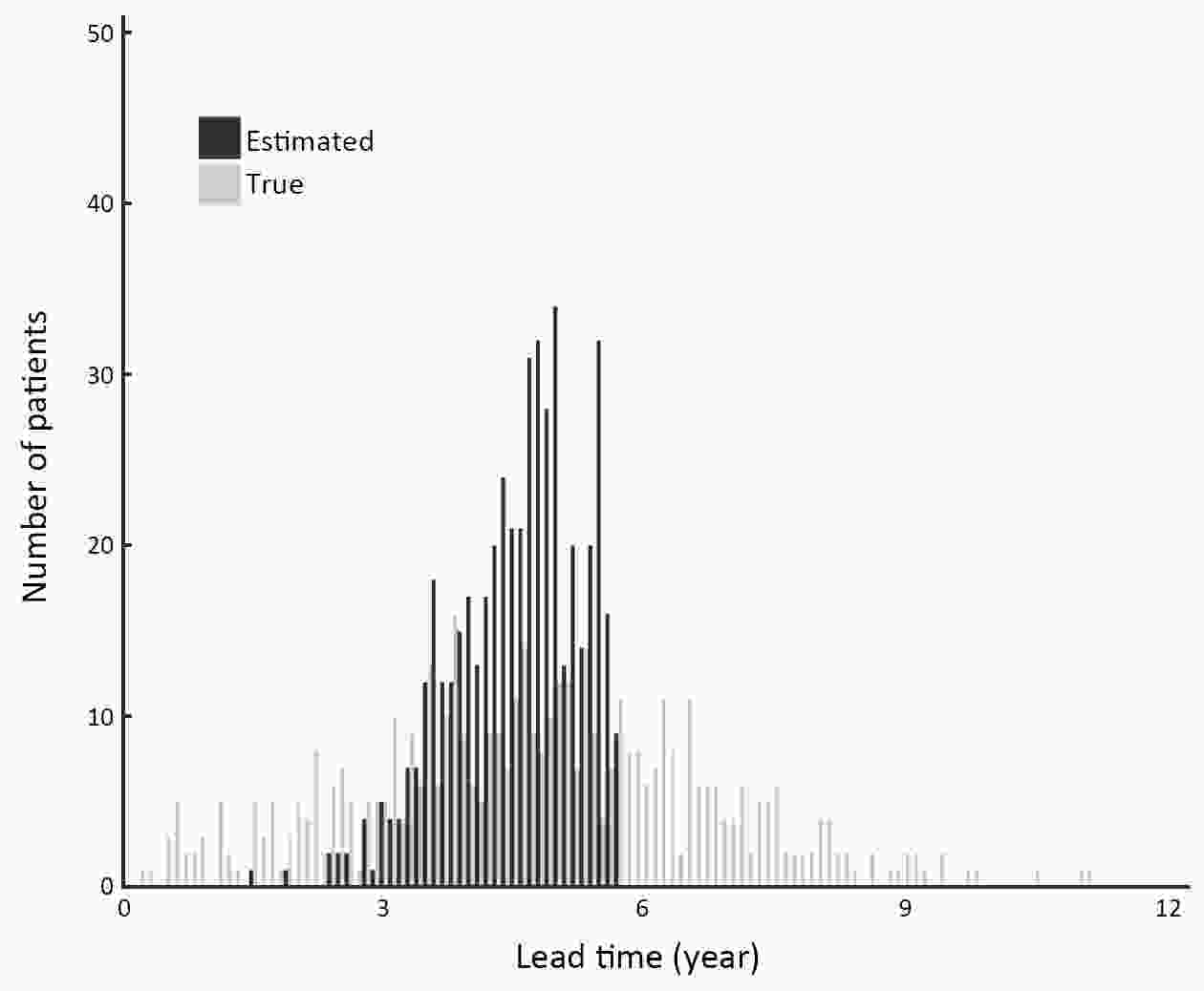
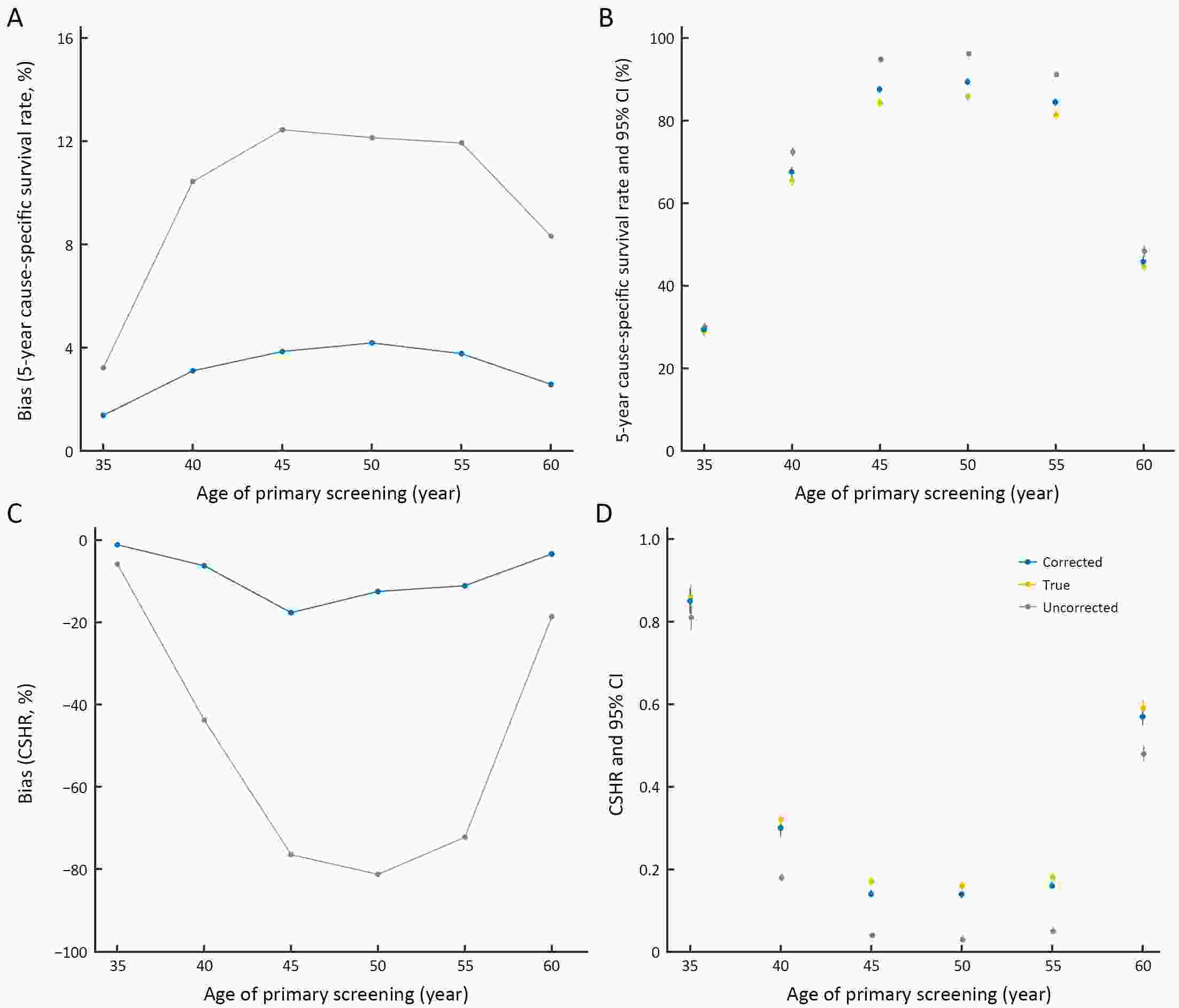
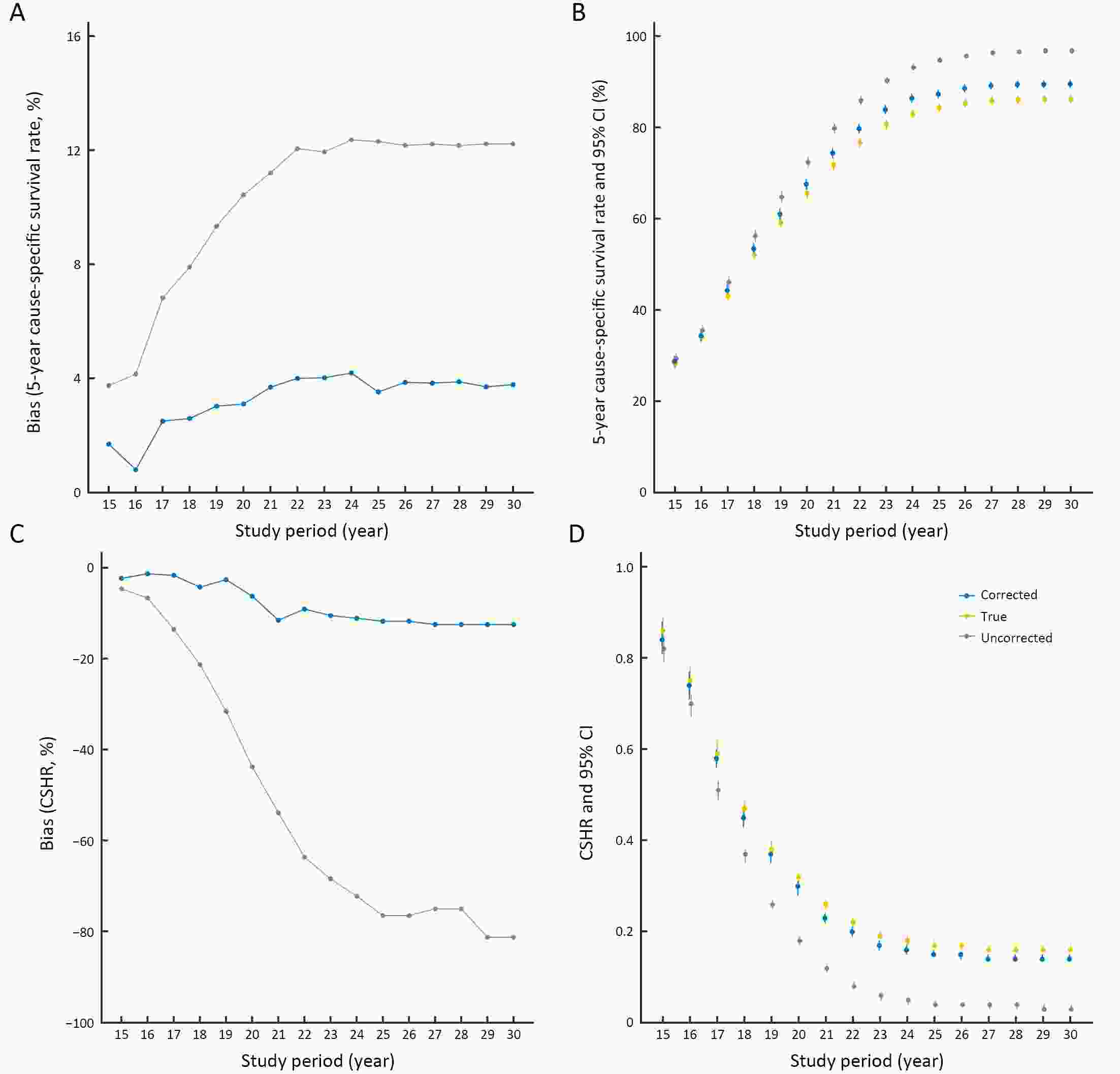
Objective To investigate what extent lead-time bias is likely to affect endoscopic screening effectiveness for esophageal cancer in the high-risk area in China. Methods A screening model based on the epidemiological cancer registry data, yielding a population-level incidence and mortality rates, was carried out to simulate study participants in the high-risk area in China, and investigate the effect of lead-time bias on endoscopic screening with control for length bias. Results Of 100,000 participants, 6,150 (6.15%) were diagnosed with esophageal squamous dysplasia during the 20-year follow-up period. The estimated lead time ranged from 1.67 to 5.78 years, with a median time of 4.62 years [interquartile range (IQR): 4.07−5.11 years] in the high-risk area in China. Lead-time bias exaggerated screening effectiveness severely, causing more than a 10% overestimation in 5-year cause-specific survival rate and around a 43% reduction in cause-specific hazard ratio. The magnitude of lead-time bias on endoscopic screening for esophageal cancer varied depending on the screening strategies, in which an inverted U-shaped and U-shaped effects were observed in the 5-year cause-specific survival rate and cause-specific hazard ratio respectively concerning a range of ages for primary screening. Conclusions Lead-time bias, usually causing an overestimation of screening effectiveness, is an elementary and fundamental issue in cancer screening. Quantification and correction of lead-time bias are essential when evaluating the effectiveness of endoscopic screening in the high-risk area in China.
2020, 32(4): 476-484.
doi: 10.21147/j.issn.1000-9604.2020.04.05
Abstract:
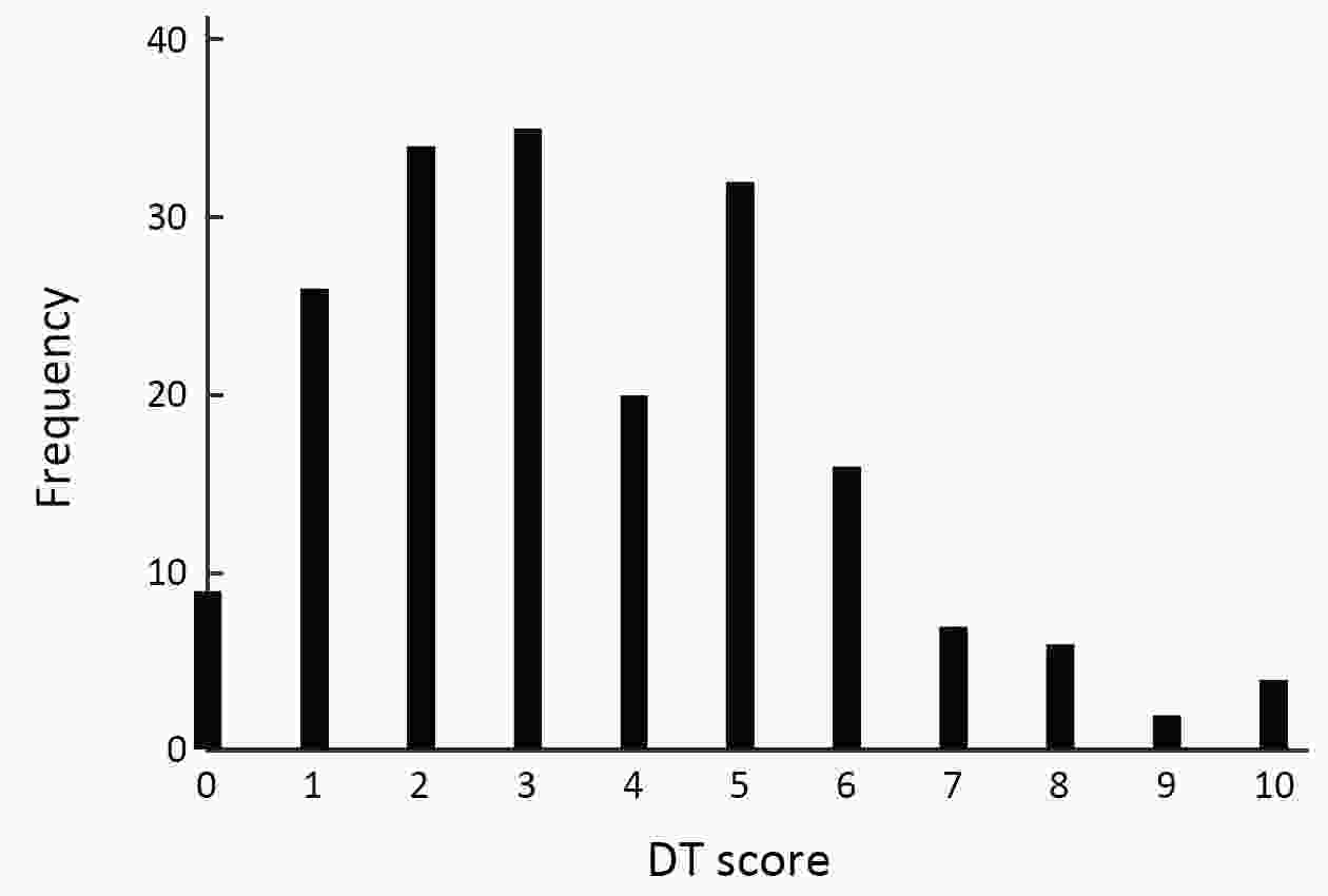
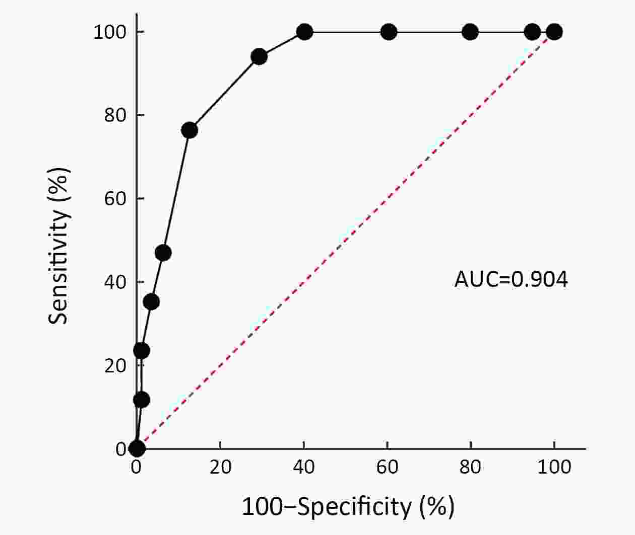
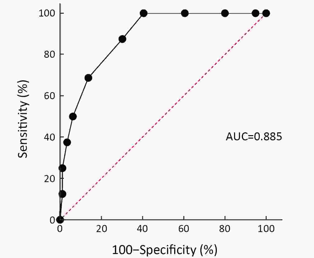
ObjectiveFamily caregivers (FCs) of breast cancer patients play a vital role throughout the treatment process. Psychological distress of FCs is common and often ignored. A simple and effective instrument for screening psychological distress would help in selecting those FCs requiring special attention and intervention. Here, the validity of distress thermometer (DT) in FCs of Chinese breast cancer patients receiving postoperative chemotherapy was assessed, and the prevalence of anxiety and depression was evaluated. MethodsWe recruited 200 FCs of hospitalized breast cancer patients in this cross-sectional descriptive study. Before the first cycle of adjuvant chemotherapy, the levels of anxiety and depression among FCs were assessed using DT and Hospital Anxiety and Depression Scale (HADS). In total, 191 valid cases were analyzed. HADS was used as the diagnostic standard to assess the effectiveness of DT as a screening tool for anxiety and depression as well as to analyze the diagnostic efficiency of DT at various cutoff points. ResultsThe definitive prevalence of both anxiety and depression was 8.90%. The mean level of anxiety and depression among FCs was 5.64±3.69 and 5.09±3.85, respectively, both of which were significantly higher than corresponding Chinese norms (P<0.01). The areas under receiver operating characteristic curves of DT for the diagnoses of FCs’ anxiety and depression were 0.904 and 0.885, respectively. A cutoff value of 5 produced the best diagnostic effects of DT for anxiety and depression. ConclusionsThe levels of both anxiety and depression were higher in the FCs of Chinese breast cancer patients receiving postoperative chemotherapy than the national norm. DT might be an effective tool to initially screen psychological distress among FCs. This process could be integrated into the palliative care of breast cancer patients and warrant further research.
2020, 32(4): 485-496.
doi: 10.21147/j.issn.1000-9604.2020.04.06
Abstract:
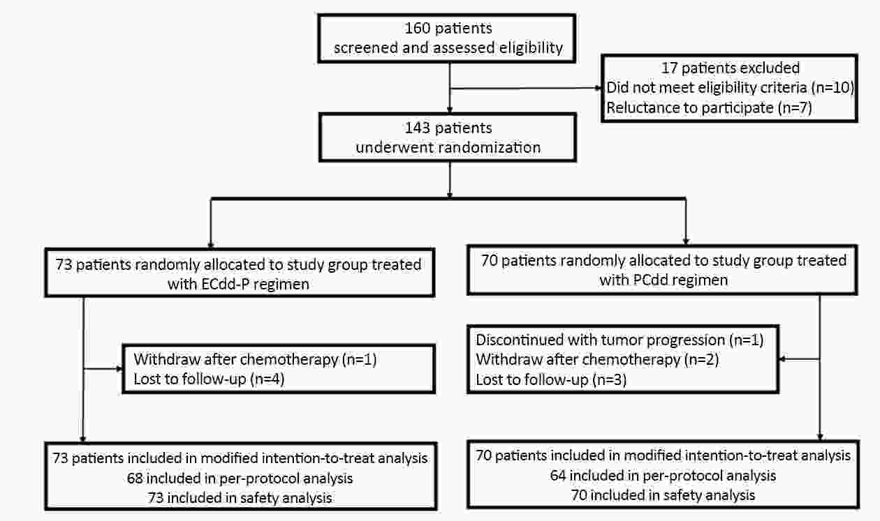
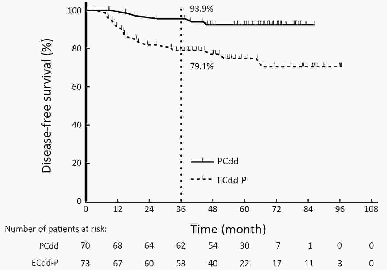
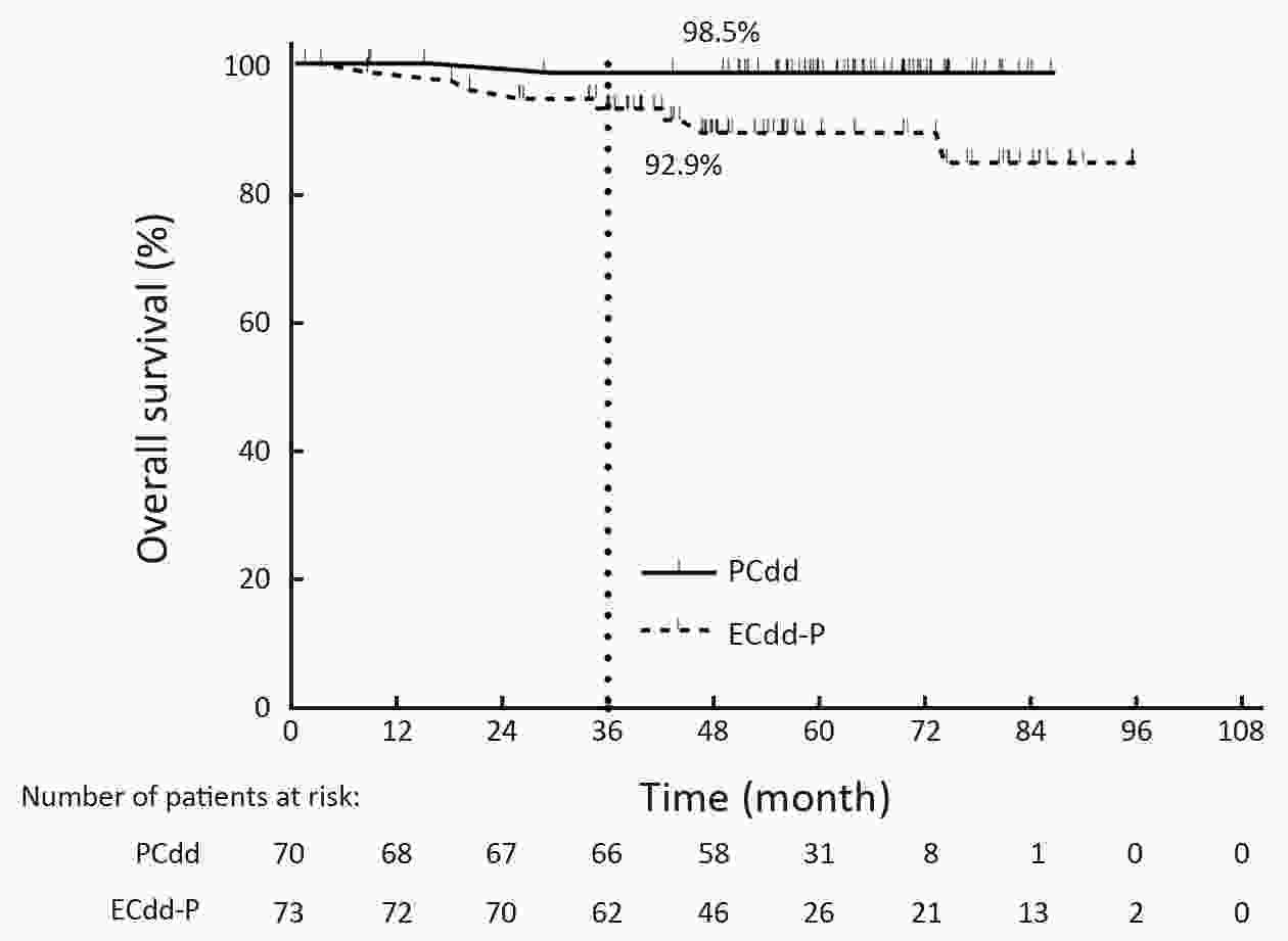
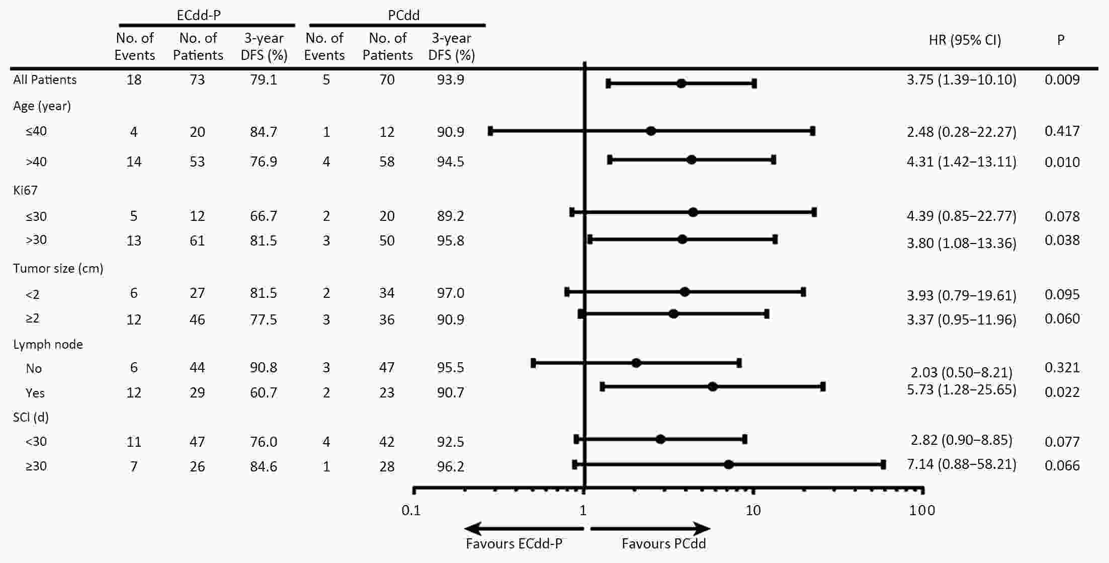
ObjectiveThe objective of this open-label, randomized study was to compare dose-dense paclitaxel plus carboplatin (PCdd) with dose-dense epirubicin and cyclophosphamide followed by paclitaxel (ECdd-P) as an adjuvant chemotherapy for early triple-negative breast cancer (TNBC). MethodsWe included Chinese patients with high recurrence risk TNBC who underwent primary breast cancer surgery. They were randomly assigned to receive PCdd [paclitaxel 150 mg/m2 on d 1 and carboplatin, the area under the curve, (AUC)=3 on d 2] or ECdd-P (epirubicin 80 mg/m2 divided in 2 d and cyclophosphamide 600 mg/m2 on d 1 for 4 cycles followed by paclitaxel 175 mg/m2 on d 1 for 4 cycles) every 2 weeks with granulocyte colony-stimulating factor (G-CSF) support. The primary endpoint was 3-year disease-free survival (DFS); the secondary endpoints were overall survival (OS) and safety. ResultsThe intent-to-treat population included 143 patients (70 in the PCdd arm and 73 in the ECdd-P arm). Compared with the ECdd-P arm, the PCdd arm had significantly higher 3-year DFS [93.9% vs. 79.1%; hazard ratio (HR)=0.310; 95% confidence interval (95% CI), 0.137−0.704; log-rank, P=0.005] and OS (98.5% vs. 92.9%; HR=0.142; 95% CI, 0.060−0.825; log-rank, P=0.028). Worse neutropenia (grade 3/4) was found in the ECdd-P than the PCdd arm (47.9% vs. 21.4%, P=0.001). ConclusionsPCdd was superior to ECdd-P as an adjuvant chemotherapy for early TNBC with respect to improving the 3-year DFS and OS. PCdd also yielded lower hematological toxicity. Thus, PCdd might be a preferred regimen for early TNBC patients with a high recurrence risk.
2020, 32(4): 497-507.
doi: 10.21147/j.issn.1000-9604.2020.04.07
Abstract:
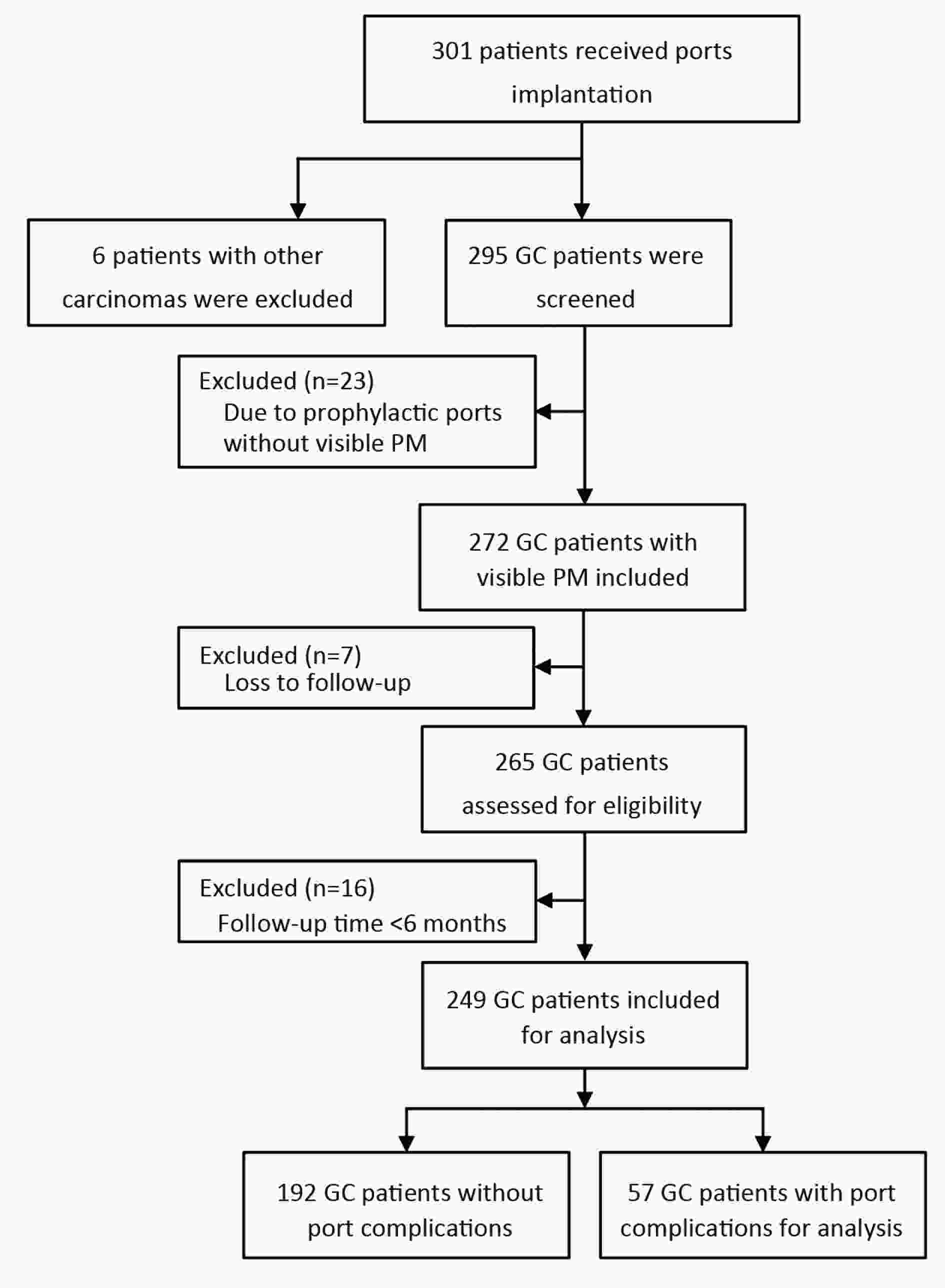
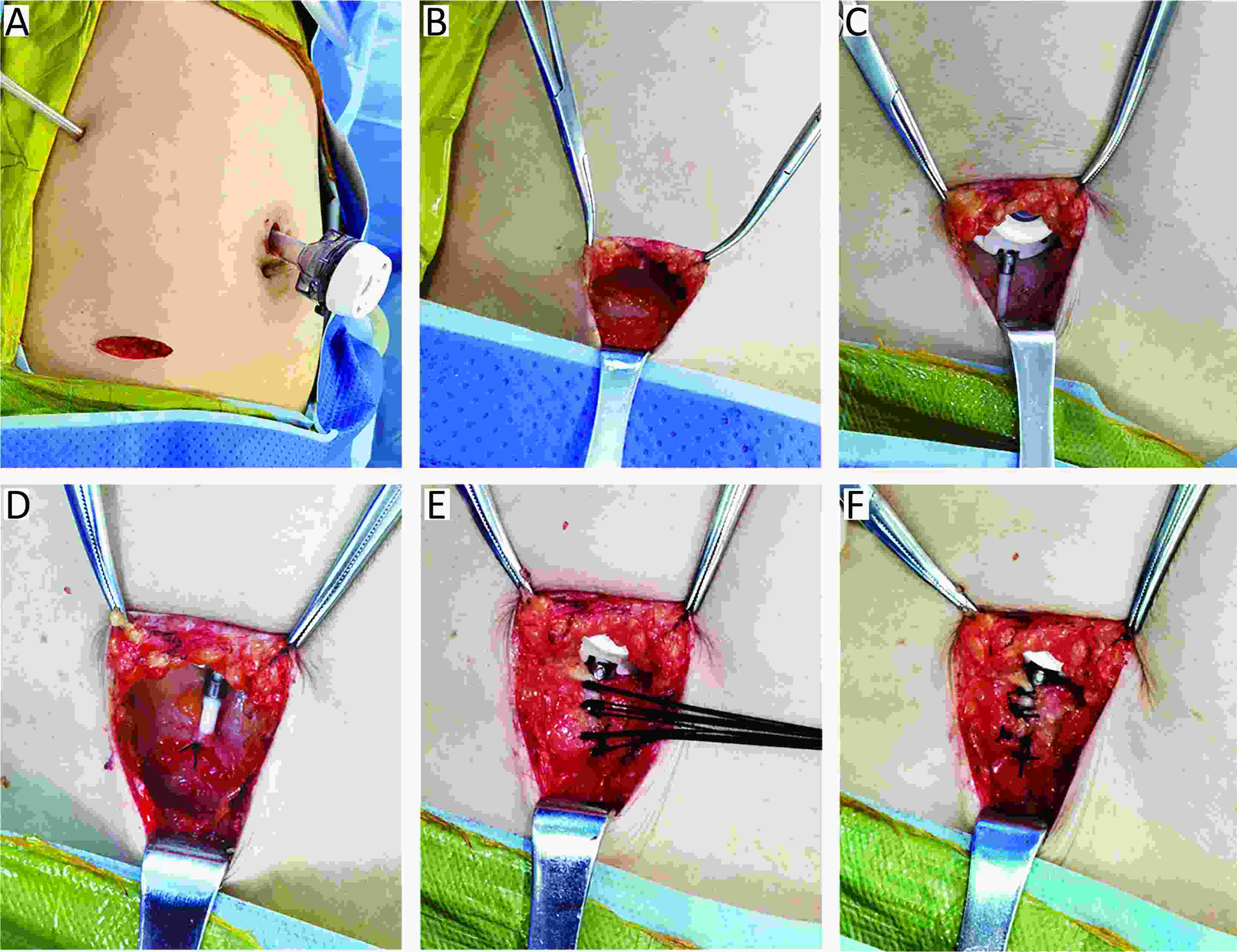
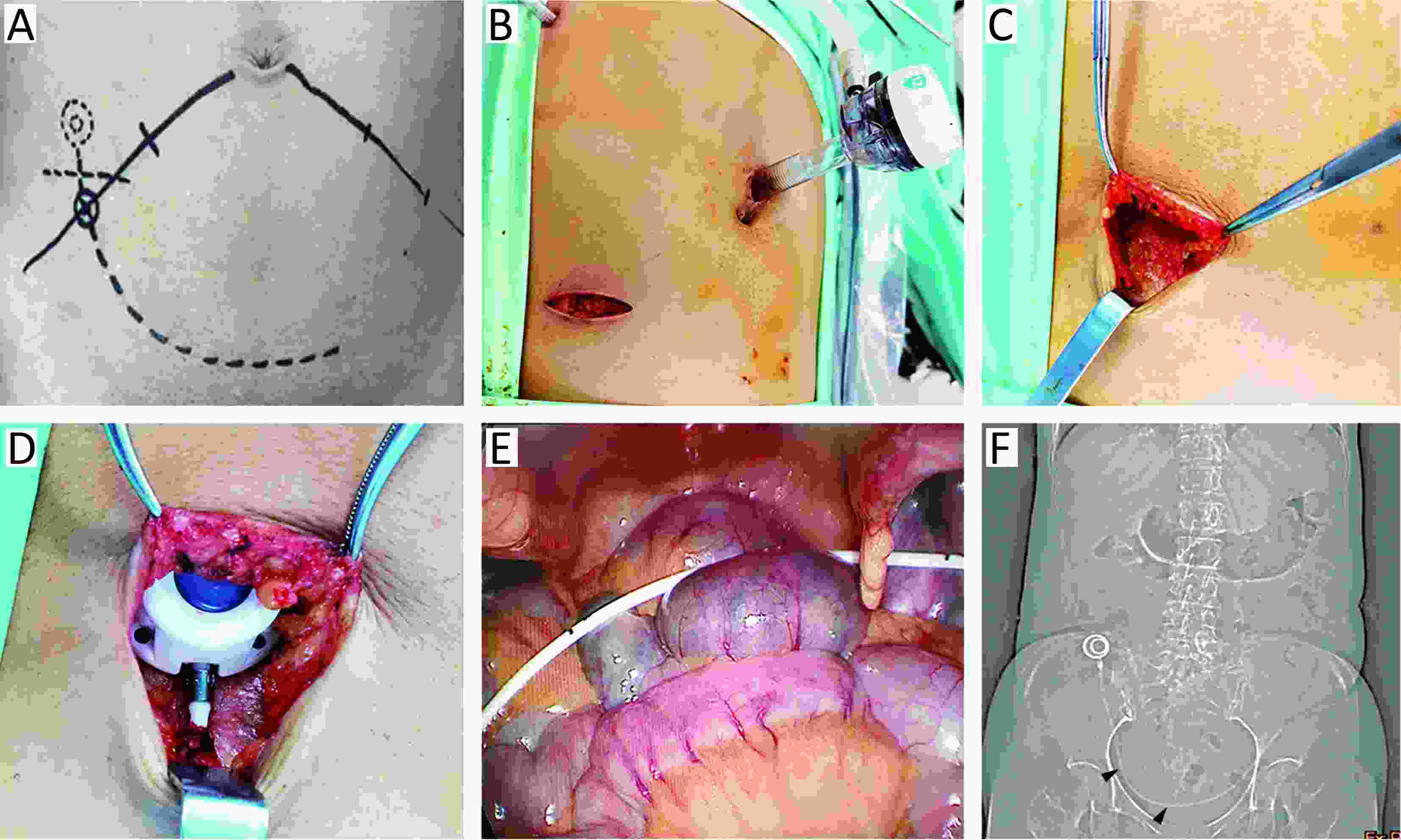
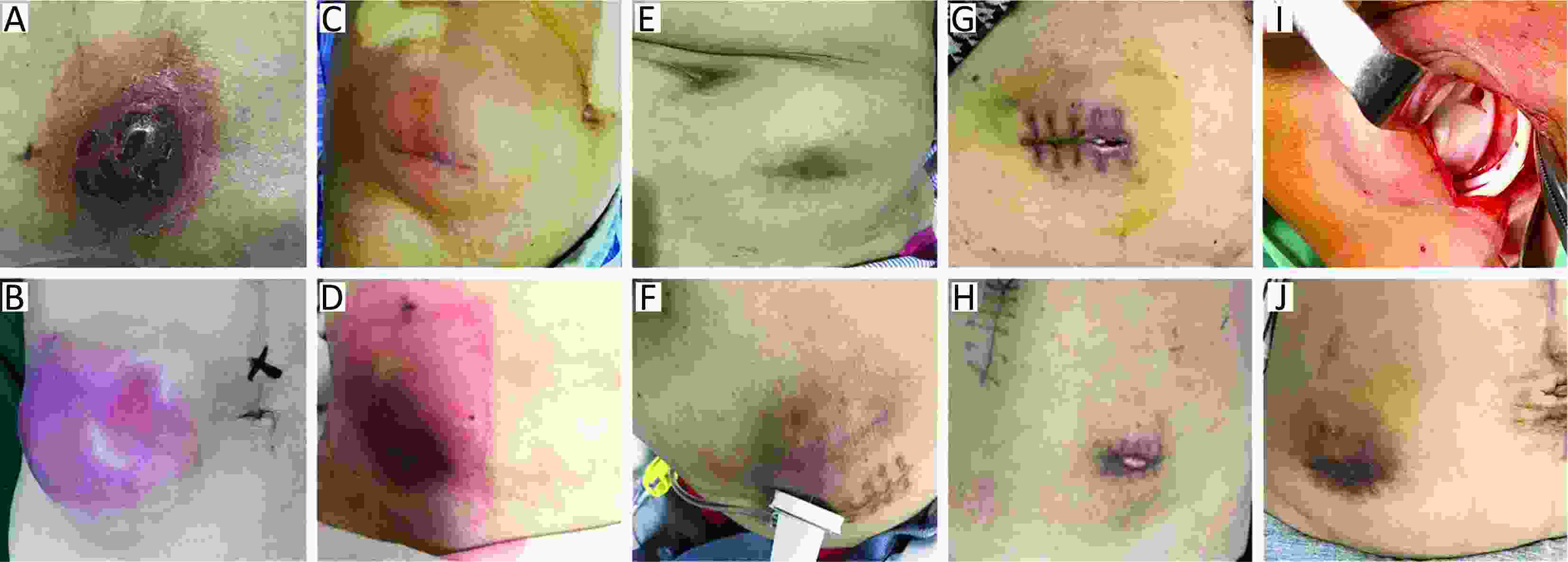
ObjectiveIntraperitoneal (IP) chemotherapy through subcutaneous port is an effective treatment for gastric cancer (GC) patients with peritoneal metastasis (PM). The objective of this study is to assess the port complications and risk factors for complications in GC patients with PM. MethodsIn retrospective screening of 301 patients with subcutaneous ports implantation, 249 GC patients with PM who received IP chemotherapy were screened out for analysis. Port complications and risk factors for complications were analyzed. ResultsOf the 249 analyzed patients, 57 (22.9%) experienced port complications. Subcutaneous liquid accumulation (42.1%) and infection (28.1%) were the main complications, and other complications included port rotation (14.1%), wound dehiscence (12.3%), inflow obstruction (1.7%) and subcutaneous metastasis (1.7%). The median interval between port implantation and occurrence of complications was 3.0 months. Eastern Cooperative Oncology Group (ECOG) performance status [odds ratio (OR), 1.74; 95% confidence interval (95% CI), 1.12−2.69], albumin (OR, 3.67; 95% CI, 1.96−6.86), implantation procedure optimization (OR, 0.33; 95% CI, 0.18−0.61) and implantation groups (OR, 0.37; 95% CI, 0.20−0.69) were independent risk factors for port complications (P<0.05). ECOG performance status was the only factor that related to the grades of port complications (P=0.016). ConclusionsPort complications in GC patients who received IP chemotherapy are manageable. ECOG performance status, albumin, implantation procedure and implantation group are independent risk factors for port complications in GC patients with PM.
Characteristics of cancer susceptibility genes mutations in 282 patients with gastric adenocarcinoma
2020, 32(4): 508-515.
doi: 10.21147/j.issn.1000-9604.2020.04.08
Abstract:
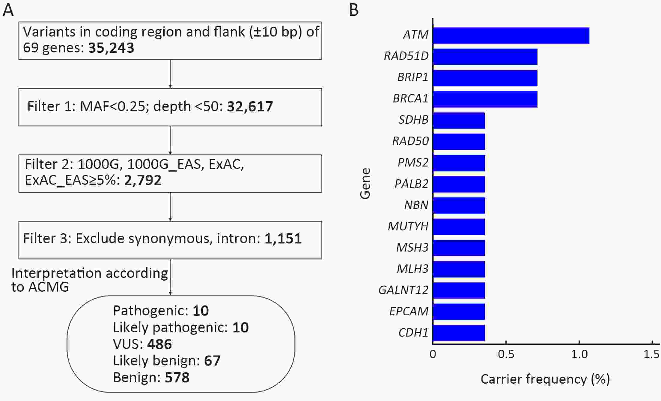
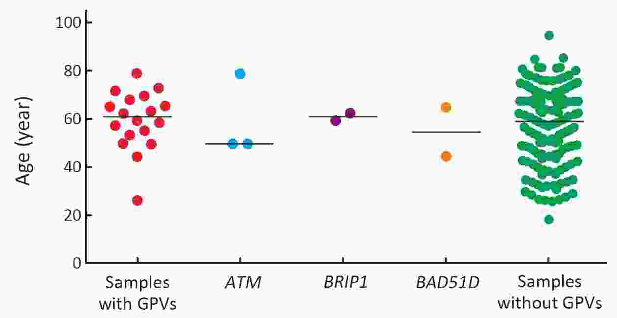
ObjectiveTo reveal the distribution signature of cancer susceptibility genes in patients with gastric adenocarcinoma, offering a diagnostic and prognostic surrogate for disease risk management and therapeutic decisions. MethodsA total of 282 patients with gastric adenocarcinoma (182 males and 100 females) were enrolled in this study, with peripheral blood genomic DNA extracted. Mutations of 69 canonical cancer susceptibility genes or presumably tumor-related genes were analyzed by targeted capture-based high-throughput sequencing. Candidate mutations were particularly selected for discussion on tumor pathogenesis according to the American College of Medical Genetics and Genomics (ACMG) guidelines. ResultsIn this study, 7.1% (20/282) of patients with gastric adenocarcinoma were found to harbor mutations of canonical or presumable cancer susceptibility genes. The detection rate in male patients (3.8%, 7/182) was significantly lower than that in female patients (13%, 13/100) (P=0.004). The most recurrent mutations were in A-T mutated (ATM) (1.1%, 3/282), followed by BRCA1, BRIP1 and RAD51D, all showed a detection rate of 0.7% (2/282). Mutations in three genes associated with hereditary gastric cancer syndromes were detected, namely, PMS2 and EPCAM associated with Lynch syndrome and CDH1 associated with hereditary diffuse gastric cancer. The detection frequencies were all 0.4% (1/282). Notwithstanding no significant difference observed, the age of patients with pathogenic mutations or likely pathogenic mutations is slightly younger than that of non-carriers (median age: 58.5 vs. 60.5 years old), while the age of patients with ATM mutations was the youngest overall (median age: 49.3 years old). ConclusionsOur study shed more light on the distribution signature and pathogenesis of mutations in gastric cancer susceptibility genes, and found the detection rate of pathogenic and likely pathogenic mutations in male patients was significantly lower than that in female patients. Some known and unidentified mutations were found in gastric cancer, which allowed us to gain more insight into the hereditary gastric cancer syndromes from the molecular perspective.
2020, 32(4): 516-529.
doi: 10.21147/j.issn.1000-9604.2020.04.09
Abstract:
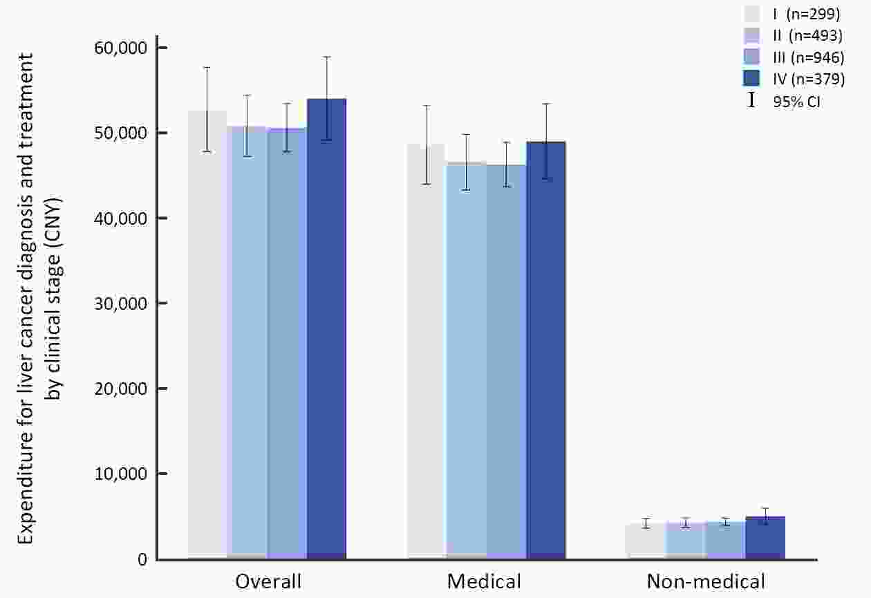
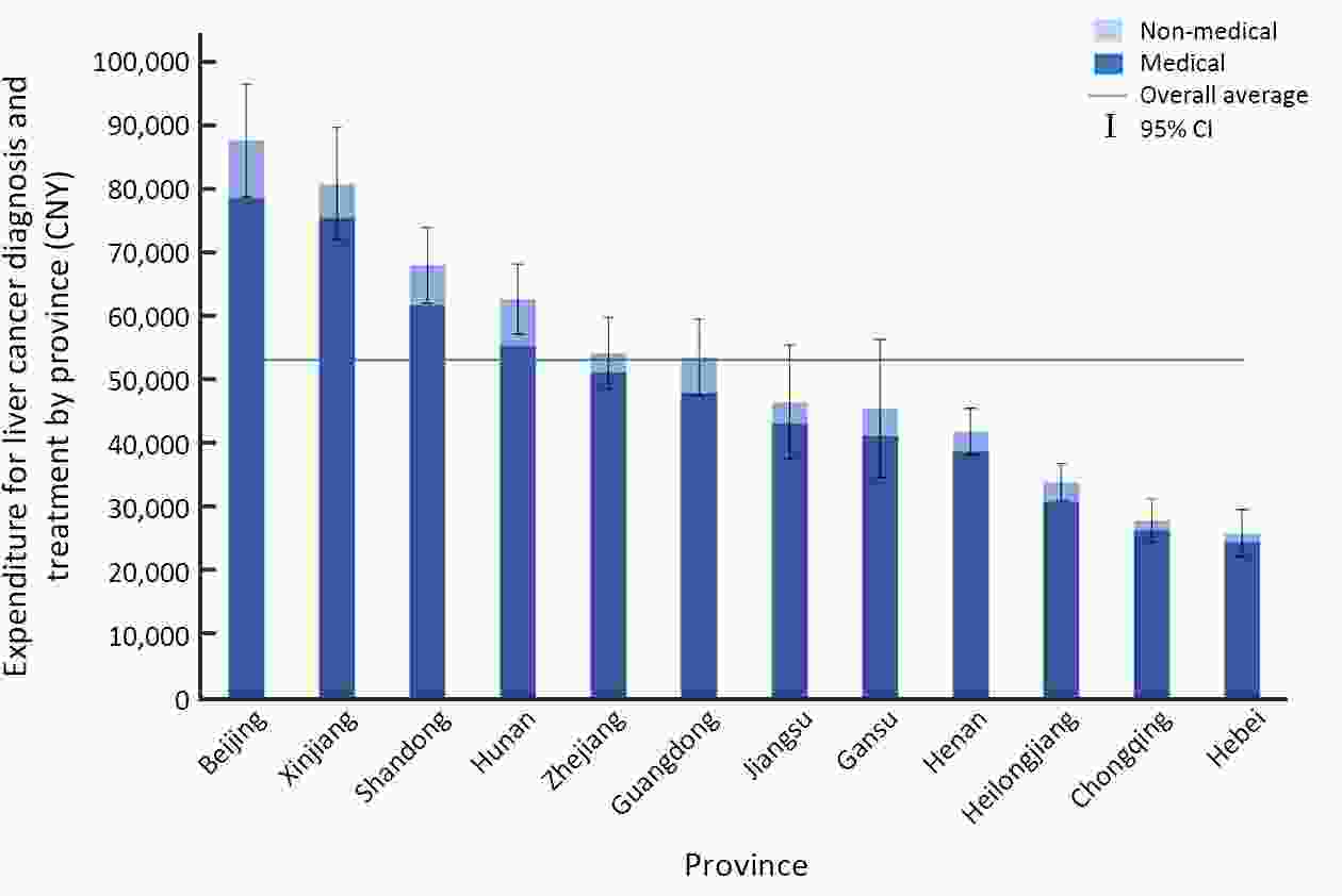
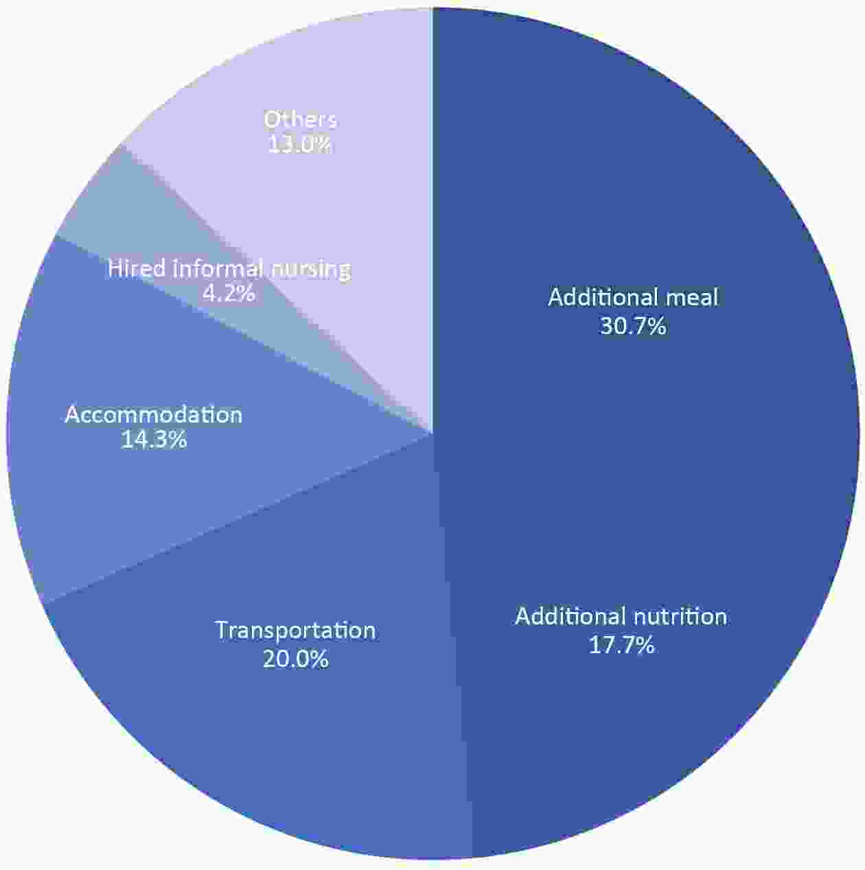
ObjectiveThe number of liver cancer patients in China accounts for more than half of the world. However, China currently lacks national, multicenter economic burden data, and meanwhile, measuring the differences among different subgroups will be informative to formulate corresponding policies in liver cancer control. Thus, the aim of the study was to measure the economic burden of liver cancer by various subgroups. MethodsA hospital-based, multicenter and cross-sectional survey was conducted during 2012-2014, covering 39 hospitals and 21 project sites in 13 provinces across China. The questionnaire covers clinical information, sociology, expenditure, and related variables. All expenditure data were reported in Chinese Yuan (CNY) using 2014 values. ResultsA total of 2,223 liver cancer patients were enrolled, of whom 59.61% were late-stage cases (III-IV), and 53.8% were hepatocellular carcinoma. The average total expenditure per liver cancer patient was estimated as 53,220 CNY, including 48,612 CNY of medical expenditures (91.3%) and 4,608 CNY of non-medical expenditures (8.7%). The average total expenditures in stage I, II, III and stage IV were 52,817 CNY, 50,877 CNY, 50,678 CNY and 54,089 CNY (P>0.05), respectively. Non-medical expenditures including additional meals, additional nutrition care, transportation, accommodation and hired informal nursing were 1,453 CNY, 839 CNY, 946 CNY, 679 CNY and 200 CNY, respectively. The one-year out-of-pocket expenditure of a newly diagnosed patient was 24,953 CNY, and 77.2% of the patients suffered an unmanageable financial burden. Multivariate analysis showed that overall expenditure differed in almost all subgroups (P<0.05), except for sex, clinical stage, and pathologic type. ConclusionsThere was no difference in treatment expenditure for liver cancer patients at different clinical stages, which suggests that maintaining efforts on treatment efficacy improvement is important but not enough. To furtherly reduce the overall economic burden from liver cancer, more effort should be given to primary and secondary prevention strategies.
2020, 32(4): 530-539.
doi: 10.21147/j.issn.1000-9604.2020.04.10
Abstract:
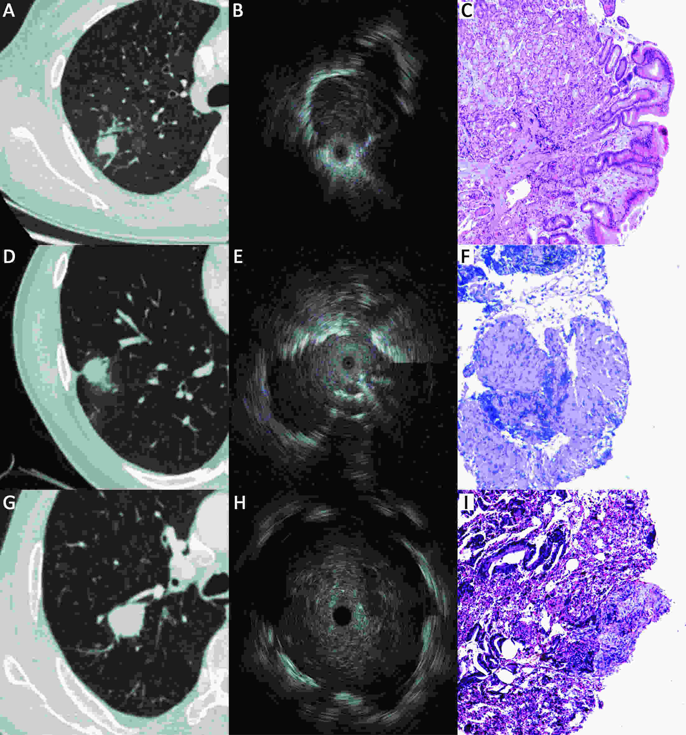
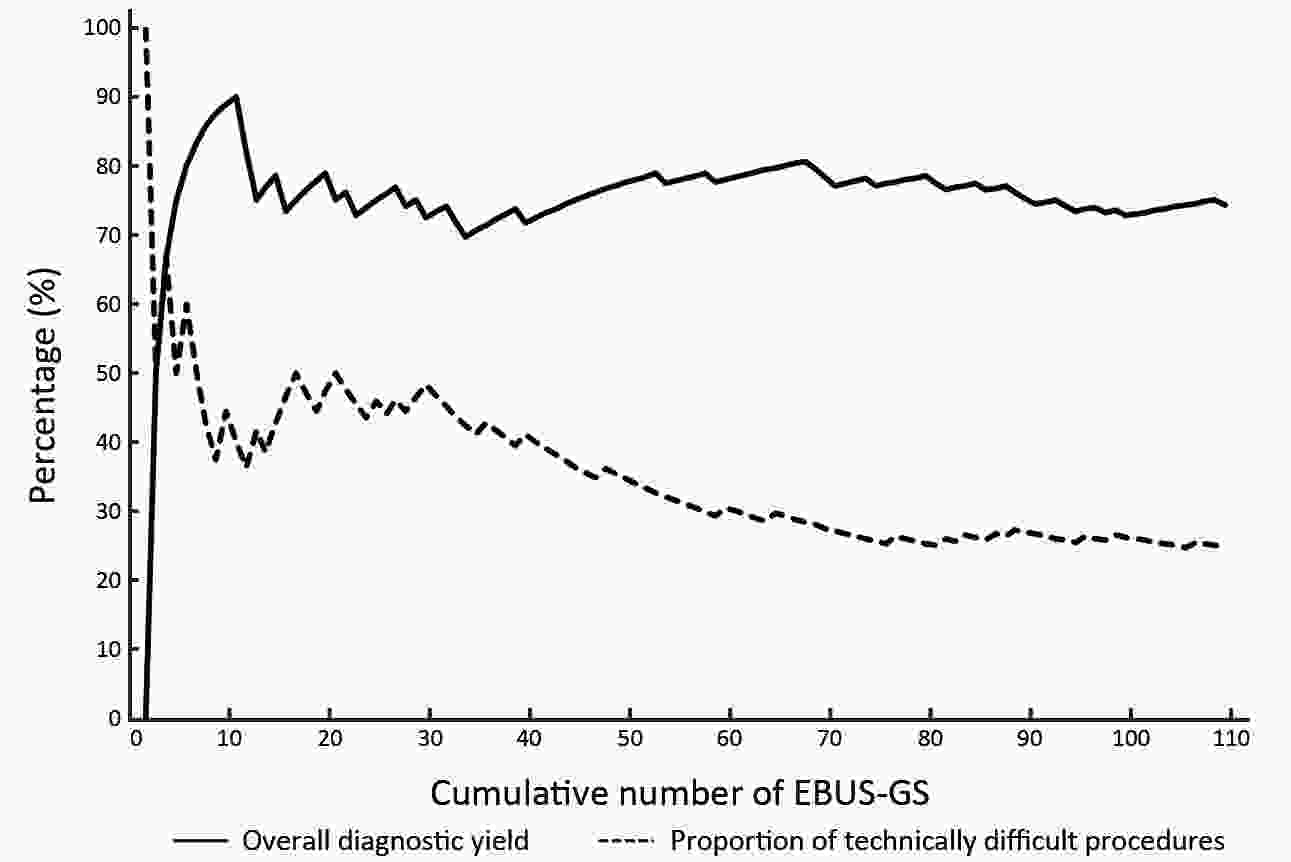
ObjectiveFluoroscopy guidance is generally required for endobronchial ultrasonography with guide sheath (EBUS-GS) in peripheral pulmonary lesions (PPLs). Virtual bronchoscopic navigation (VBN) can guide the bronchoscope by creating virtual images of the bronchial route to the lesion. The diagnostic yield and safety profiles of VBN without fluoroscopy for PPLs have not been evaluated in inexperienced pulmonologist performing EBUS-GS. MethodsBetween January 2016 and June 2017, consecutive patients with PPLs referred for EBUS-GS at a single cancer center were enrolled. The diagnostic yield as well as safety profiles was retrospectively analyzed, and our preliminary experience was shared. ResultsA total of 109 patients with 109 lesions were included, 99 (90.8%) lesions were visible on EBUS imaging. According to the procedure time needed to locate the lesion on EBUS, 24.8% (27/109) were deemed technically difficult procedures; however, no significant relationships were identified between candidate parameters and technically difficult procedures. The overall diagnosis yield was 74.3% (81/109), and the diagnostic yield of malignancy was 83.7% (77/92). Lesions larger than 20 mm [odds ratio (OR), 2.758; 95% confidence interval (95% CI), 1.077−7.062; P=0.034] and probe of within type (OR, 3.174; 95% CI, 1.151−8.757, P=0.026) were independent factors leading to a better diagnostic yield in multivariate analysis. About 30 practice procedures were needed to achieve a stable diagnostic yield, and the proportion of technically difficult procedures decreased and stabilized after 70 practice procedures. Regarding complications, one patient (0.9%) had intraoperative hemorrhage (100 mL) which was managed under endoscopy. ConclusionsVBN without fluoroscopy guidance is still useful and safe for PPLs diagnosis, especially for malignant diseases when performed by pulmonologist without previous experience of EBUS-GS. VBN may simplify the process of lesion positioning and further multi-center randomized studies are warranted.
2020, 32(4): 540-546.
doi: 10.21147/j.issn.1000-9604.2020.04.11
Abstract:
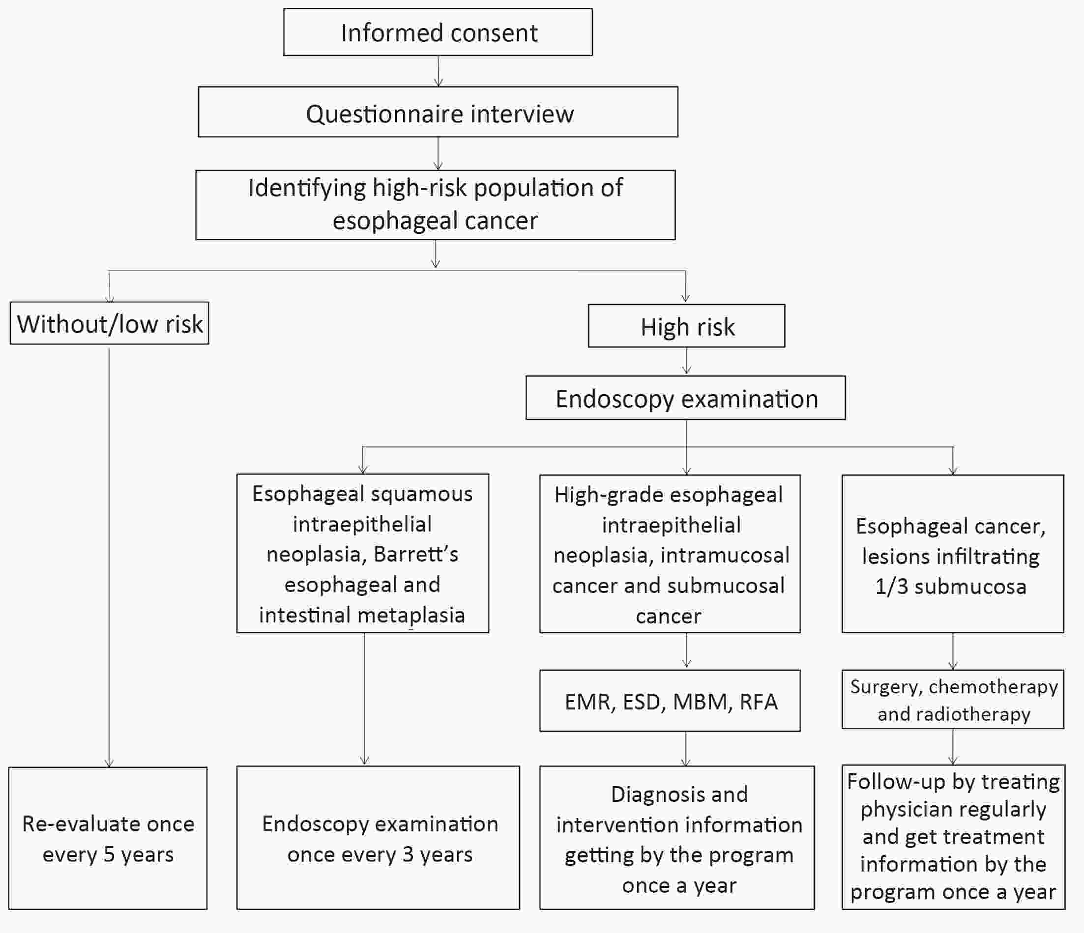
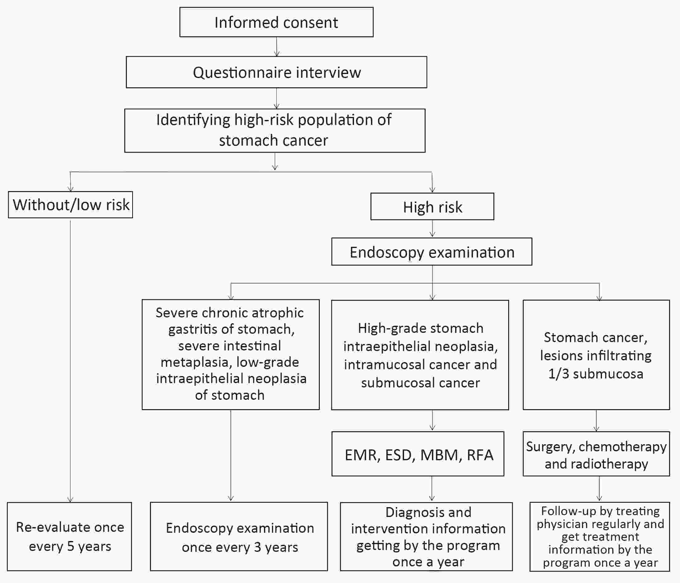
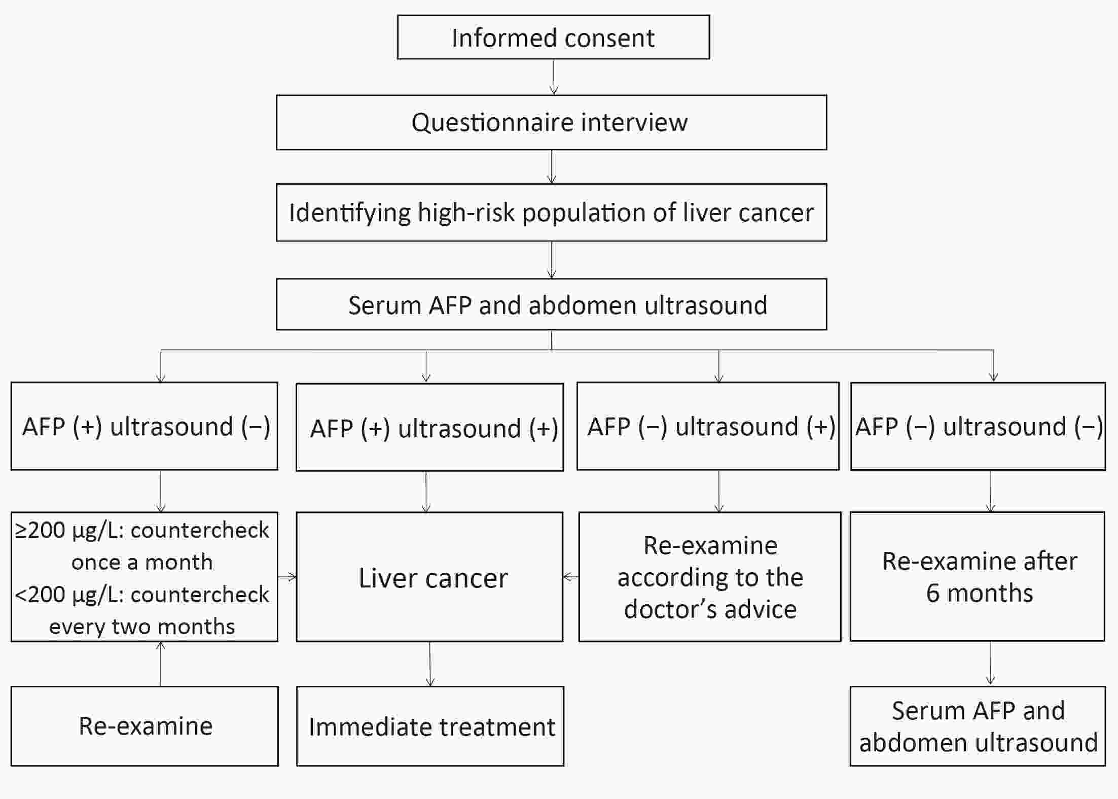
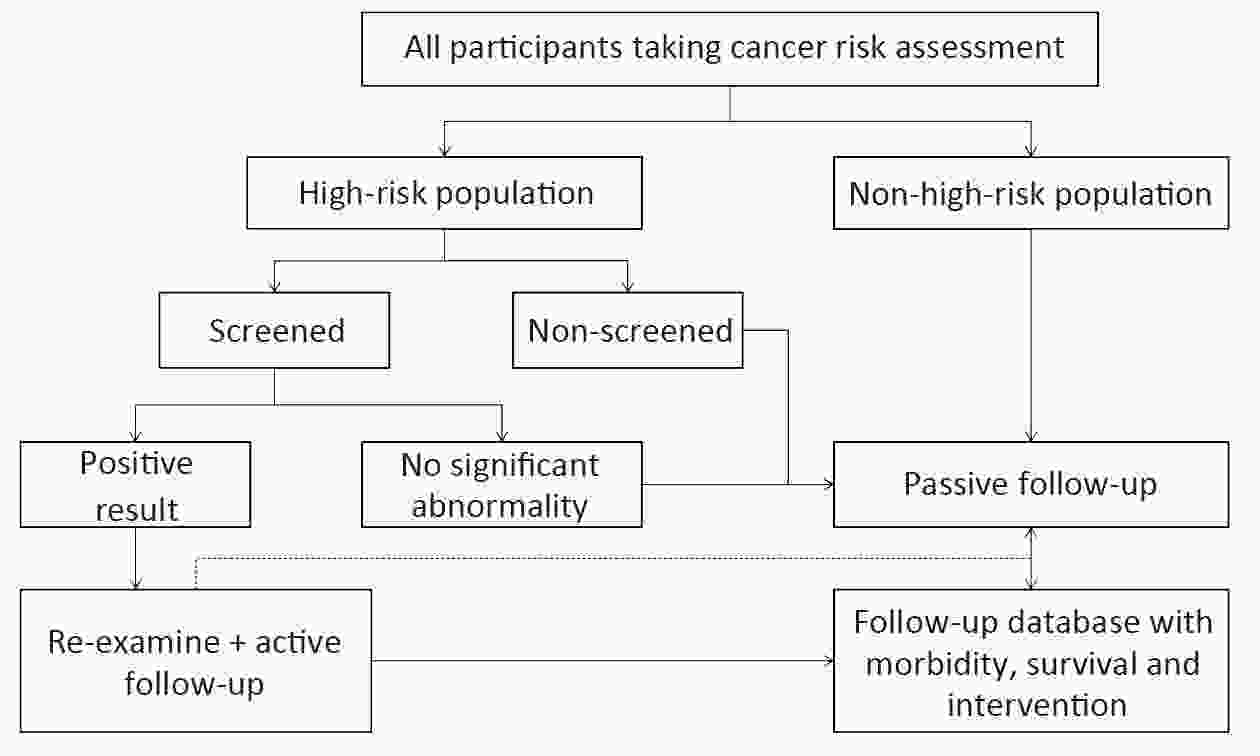
ObjectiveNational Health Commission of the People’s Republic of China collaborated with many ministries and commissions government and initiated a population-based cancer screening program in high-risk area of rural China, targeting three types of cancer that are most prevalent in these areas, including esophageal, stomach and liver cancer. This study protocol was reported to show the design and evaluate the effectiveness of cancer screening and appropriate screening strategies of three cancers in rural China. Methods and analysisA two-step design with cancer risk assessment based on questionnaire interview, Hepatitis B surface antigen (HBsAg) test strip and subsequent clinical intervention for high-risk populations was adopted free of charge at the local hospitals designated in the program. Ethic and disseminationThis study was approved by the Institutional Review Board of Cancer Hospital, Chinese Academy of Medical Sciences and Peking Union Medical College. The results will evaluate the effectiveness of cancer screening and appropriate screening strategies in rural China.

 Abstract
 FullText HTML
 PDF 764KB